Method and apparatus for minimally invasive delivery, tensioned deployment and fixation of secondary material prosthetic devices in patient body tissue, including hernia repair within the patient's herniation site

a technology of secondary material and prothesis, which is applied in the field of methods and equipment for minimally invasive delivery, tensioned deployment and fixation of secondary material, which can solve the problems of difficult to tension a planar mesh sheet across the patient, loose or flaccid mesh may not provide sufficient structural integrity, and the pliable body tissue does not always establish tension, so as to reduce the likelihood of future exfiltration

Inactive Publication Date: 2010-03-18
VENTRALFIX
View PDF72 Cites 133 Cited by
  • Summary
  • Abstract
  • Description
  • Claims
  • Application Information

AI Technical Summary

Benefits of technology

[0016]The present invention enables minimally invasive, selective orientation and tensioned deployment of a secondary material into a patient's body cavity. For example, a hernia can be repaired directly at the herniation site through a relatively small incision of approximately 0.2 inch (5 millimeters) to 2 inches (50 mm). Some embodiments of the apparatus instrumentation of the present invention and the inventive methods for their use may circumferentially pretension the secondary material, such as hernia repair mesh, into a relatively taught planar sheet that may be oriented and abutted against the target tissue, such as generally concave, three-dimensional abdominopelvic interior wall of a patient. The tensioned, abutting alignment of the secondary material increases likelihood of successful structurally sound marginal affixation of said material to the patient's target tissue. In the example of hernia repair, tensioned mesh provides additional structural integrity to the repaired herniation site, thereby reducing likelihood of future ex-filtration of the patient's abdominal organ tissue through the repaired site.

Problems solved by technology

Pushing the instrument distal tip laterally across a relatively confined body cavity space between the cavity walls and viscera and thereafter against resilient, pliable body tissue does not always establish tensioned, taut abutting contact between the prosthetic device and the tissue.
It is difficult to tension a sheet of planar mesh across the patient's generally cylindrical, concave inner abdominopelvic wall via laterally oriented access points.
Loose or flaccid mesh may not provide sufficient structural integrity for the hernia repair and may necessitate future remedial repair.
Mesh that is not properly tensioned over and affixed to the parietal peritoneum layer of the abdominal wall may not have sufficient structural integrity to inhibit ex-filtration of internal organ tissue through the existing herniation site or under the marginal edges of the mesh patch.
However, the umbrella-like ribs require relatively large free volumetric space between the viscera and body cavity walls so that the umbrella may deploy.
Hence, the ribs need considerable space to complete their motion from the pre-deployed to deployed states.
Such references do not address solutions for surgeons who want or need to implant a prosthesis device as part of a medical procedure.

Method used


Image

  • Method and apparatus for minimally invasive delivery, tensioned deployment and fixation of secondary material prosthetic devices in patient body tissue, including hernia repair within the patient's herniation site
  • Method and apparatus for minimally invasive delivery, tensioned deployment and fixation of secondary material prosthetic devices in patient body tissue, including hernia repair within the patient's herniation site
  • Method and apparatus for minimally invasive delivery, tensioned deployment and fixation of secondary material prosthetic devices in patient body tissue, including hernia repair within the patient's herniation site

Examples


first embodiment

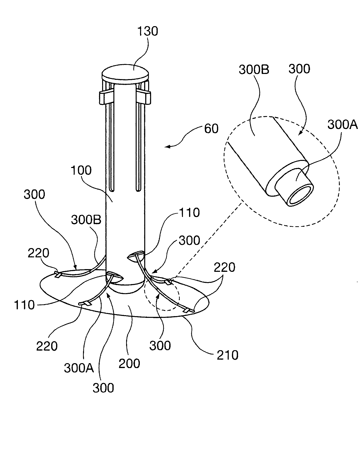
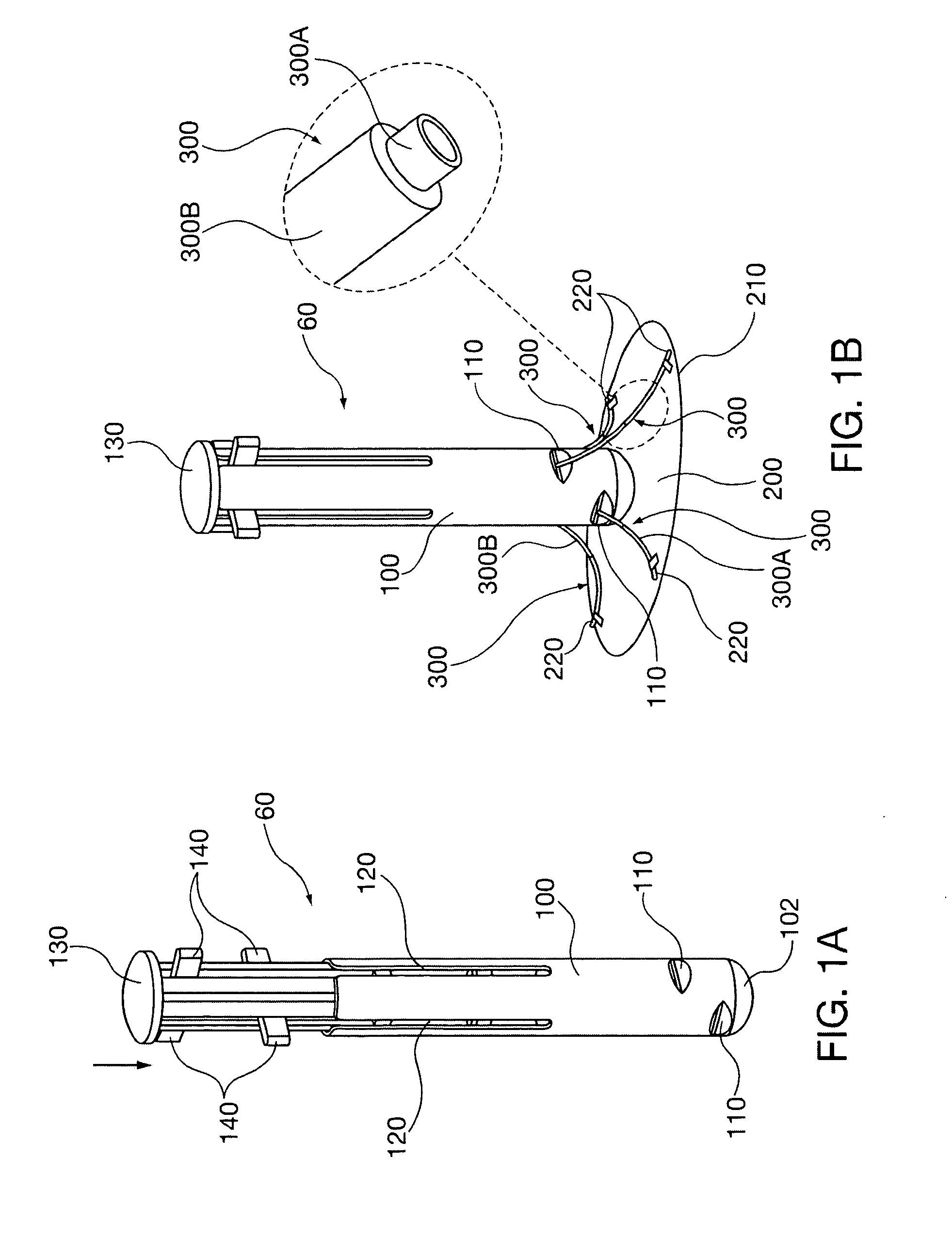
[0046]FIGS. 1A and 1B show the repair instrument 60 of the present invention. The instrument 60 has an outer tubular device body 100, preferably with a distal rounded end 102 for insertion into an incision formed at the hernia site, to be discussed below in connection with operational descriptions for use of the instrument. Near the distal end 102 the device body 100 defines two opposing pairs of ports 110 in its outer periphery, oriented radially every 90 degrees. The ports might be staggered axially, for example to accommodate different shapes and dimensions of mesh. The device body 100 also defines four needle advancement channels 120 that are in axial peripheral alignment with a corresponding port 110. It is envisioned that the device body 100 and other delivery cannulae described herein preferably will have an outer diameter of between 0.5 inch (approximately 12 mm) and 2 inches (approximately 50 mm).

[0047]A plunger 130 is slidably received within the device body 100. The plung...

embodiment 70

[0058]FIGS. 9-15 show a constant radius needle is instrumentation embodiment 70. This embodiment utilizes a cannula port 160 with insertion depth stop 161. The cannula port 160 has an inner bore that defines needle tracks 162 having a curvature radius and diameter adapted for receipt and guidance of constant radius cannulae 330, oriented at 90 degree circumferential positions. The cannula port 160 retains pre-deployment configuration repair mesh 200, shown schematically in FIG. 9. The mesh sutures 450 respectively are threaded into one of the corresponding constant radius cannulae 330. As was described with respect to prior embodiments, the distal end of each suture 450 is affixed to the mesh 200 circumference and the proximal end is coupled to a needle. In this embodiment constant radius needles 430 are utilized, having a profile conforming to the constant radius cannulae 330.

[0059]For illustrative purposes, two mesh deployment and primary fixation site alignment apparatus and meth...

embodiment 530

[0075]Another primary fixation fastener expanding braid anchor 540 is shown in FIGS. 38-39A. The general functional concepts of a radially outwardly bowed fastener and delivery instrument 536 that were employed in the anchor embodiment 530 of the immediately prior drawing descriptions are applicable to the braid anchor embodiment.

[0076]As described in connection with other fixation fastener embodiments, a needle and sutures 450 are advanced through the patient's abdominal tissue. The delivery instrument (not shown) captures braid fastener 540 between the front and compression collars.

[0077]Braid fastener 540 has a pair of braid tubes 542 that axially sandwich a length of coreless braided cable 544 between them. The braided cable 544 may be constructed of a polymer such as polyester or polypropylene. Relative compression of the pair of braid tubes 542 with the delivery instrument 536 in the manner described with respect to the expanding anchor fastener 530 radially bows out the braid...


PUM


Abstract

Apparatus and methods enable insertion and tensioned deployment of a secondary material prosthetic device into a body cavity or other tissue of a patient, such as for example hernia repair mesh into the abdominopelvic cavity of a patient through the hernia site. The present invention establishes fixation sites for the prosthetic device and tensions it against the body tissue. It may also be used implant fixation devices within the body tissue so that the prosthetic device is tensioned into firm abutting contact with the body tissue. Instrument deployment and fixation struts may be advanced in retrograde fashion in order to reduce needed deployment volume within the patient's body cavity. The prosthetic device advantageously may be flexibly coupled to the instrument via fixation devices such as sutures, so as to increase orientation flexibility.

Description

CLAIM TO PRIORITY[0001]This application claims priority under 35 U.S.C. §119 (e) to the following U.S. provisional applications: Ser. No. 61 / 192,208 filed on Sep. 16, 2008 and Ser. No. 61 / 214,316 filed on Apr. 21, 2009, each of which is hereby incorporated by reference in its entirety.BACKGROUND OF THE DISCLOSURE[0002]1. Field of the Invention[0003]The invention relates to methods and apparatus minimally invasive delivery, tensioned deployment and fixation of secondary material, including prosthetic devices, in a selected implantation site defined by a patient's body tissue. The tissue may include tissue within or defining a body cavity. An exemplary, non-limiting application of this invention is for repair of a herniation in a wall of the abdominopelvic cavity of a patient.[0004]2. Description of the Prior Art[0005]A hernia is a weakness or hole within a patient's abdominopelvic wall that may allow internal organ tissue, such as intestines or bowel, to ex-filtrate the abdominal cav...

Claims


Application Information

Patent Timeline
Patent Type & Authority Applications(United States)
IPC IPC(8): A61B17/08
CPCA61B17/0057A61B2017/00659A61F2002/0072A61B2017/047A61F2/0063A61B2017/00663
Inventor ROSLIN, MITCHELLSHAH, PARESH C.SHIKHMAN, OLEGFERREIRA, DANIAL P.RADZIUNAS, JEFFREY P.BATTLES, CHRISTOPHER A.
Owner VENTRALFIX
Why Eureka
Try Eureka