Standardized evaluation of therapeutic efficacy based on cellular biomarkers

a cellular biomarker and clinical trial technology, applied in the field of pharmaceutical therapies, can solve the problems of undesirable side effects, potentially a significant opportunity loss of therapy, and the addition of toxic side effects to all patients, so as to reduce the amount of a particular biomarker, increase fluorescence, and increase the effect of fluorescen

Inactive Publication Date: 2010-03-25
LESKO STEPHEN A +1
View PDF2 Cites 0 Cited by
  • Summary
  • Abstract
  • Description
  • Claims
  • Application Information

AI Technical Summary

Benefits of technology

[0011]In another aspect, the present invention provides a method of selecting a therapeutic agent for the treatment of cancer. Such a method may comprise obtaining a cell sample from a patient, wherein the sample comprises circulating cancer cells. Typically, said cancer cells may comprise one or more biomarkers. Cancer cells may then be contacted with a test compound (e.g., a targeting moiety coupled to a reporter moiety) that specifically binds to one or more biomarker. Test compounds typically comprise one or more reporter moieties, for example, a fluorescent moiety. The intensity of the fluorescence of the cells may be measured, for example, using a fluorescence microscope; and the intensity of the fluorescence of the cells may be compared to that of a reference standard. The fluorescent intensity of the cells is typically measured under standardized conditions (e.g., at the same time of exposure). Typically, the ratio of the intensity of the stained cells to the reference standard correlates to the effectiveness of the therapeutic agent against the cancer. The ratio may be conveniently expressed as a percent of the reference standard. For example, the presence and / or increased amount of a biomarker (indicated by increased fluorescence) may correlate to susceptibility of the cancer cell to the therapeutic reagent. Likewise the presence and / or increase amount of a particular biomarker may indicate resistance to the therapeutic. The absence and / or reduced amount of a particular biomarker may indicate susceptibility of the cancer to the therapeutic or the absence and / or reduced amount may indicate resistance to the therapeutic. Those skilled in the art will appreciate that the amount of fluorescence may be compared to a reference standard, for example, a known cancer cell of known type and susceptibility.
[0012]The present invention also provides kits for the practice of one or more methods of the invention. For example, the present invention provides a kit for determining the susceptibility of a cancer cell to a therapeutic agent, comprising a targeting moiety specific for a biomarker. A targeting moiety may be the therapeutic agent. A targeting moiety may be coupled to a fluorescent moiety. One skilled in the art will appreciate that a targeting moiety (e.g., a therapeutic agent) may be of any type known in the art, for example, small molecules, peptides, proteins, enzymes, monoclonal antibodies, oligonucleotides (DNA, RNA, mixed DNA and RNA, which may contain one or more non-naturally occurring nucleotide) and the like. Kits of the invention may comprise one or more therapeutic agents, which may be coupled to one or more fluorescent moieties. A targeting moiety and / or a therapeutic agent may comprise one or more antibodies, which may include one or more monoclonal antibodies. Kits of the invention may comprise at least one antibody, which may be a monoclonal antibody, which is specific for a cytokeratin. Antibodies of the kits of the invention may be polyclonal or monoclonal antibodies and may comprise a fluorescent moiety. Kits of the invention may comprise a reference standard. Such reference standards may comprise a known amount of a non-bleaching fluorescent moiety. One example of a suitable reference standard is a fluorescent microsphere. Kits of the invention may comprise one or more reagent selected from the group consisting of buffers, buffer salts, detergents, surfactants, fixatives, and the like. In a particular embodiment, kits of the invention may comprise a permeability buffer.

Problems solved by technology

However, the use of these drugs so far has been empirical, and not based on diagnostic drug-action response parameters, with overall modest benefit, approximately 20-30% favorable responses.
There is potentially a significant opportunity loss for therapy if the disease is still progressing (70-80% of the cases) with added toxic side effects to all the patients.
Trastuzumab is a very effective therapy, but it has undesirable side effects and only 30 to 35% of selected breast cancer patients respond to trastuzumab as a single agent.
Data on the clinical validity of such characterizations are, however, inconclusive, possibly because of the subjective nature of the determination, variability of test conditions, variability of different scorers' techniques or variability among laboratory equipment.

Method used


Image

  • Standardized evaluation of therapeutic efficacy based on cellular biomarkers
  • Standardized evaluation of therapeutic efficacy based on cellular biomarkers
  • Standardized evaluation of therapeutic efficacy based on cellular biomarkers

Examples


example 1

[0023]Initially, an attempt to obtain the desired results using the obvious approach, and the failure of that approach, will be described.

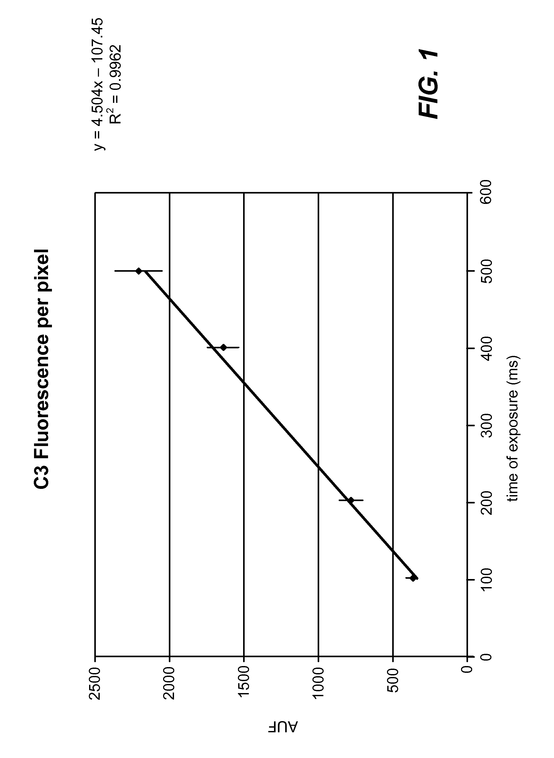
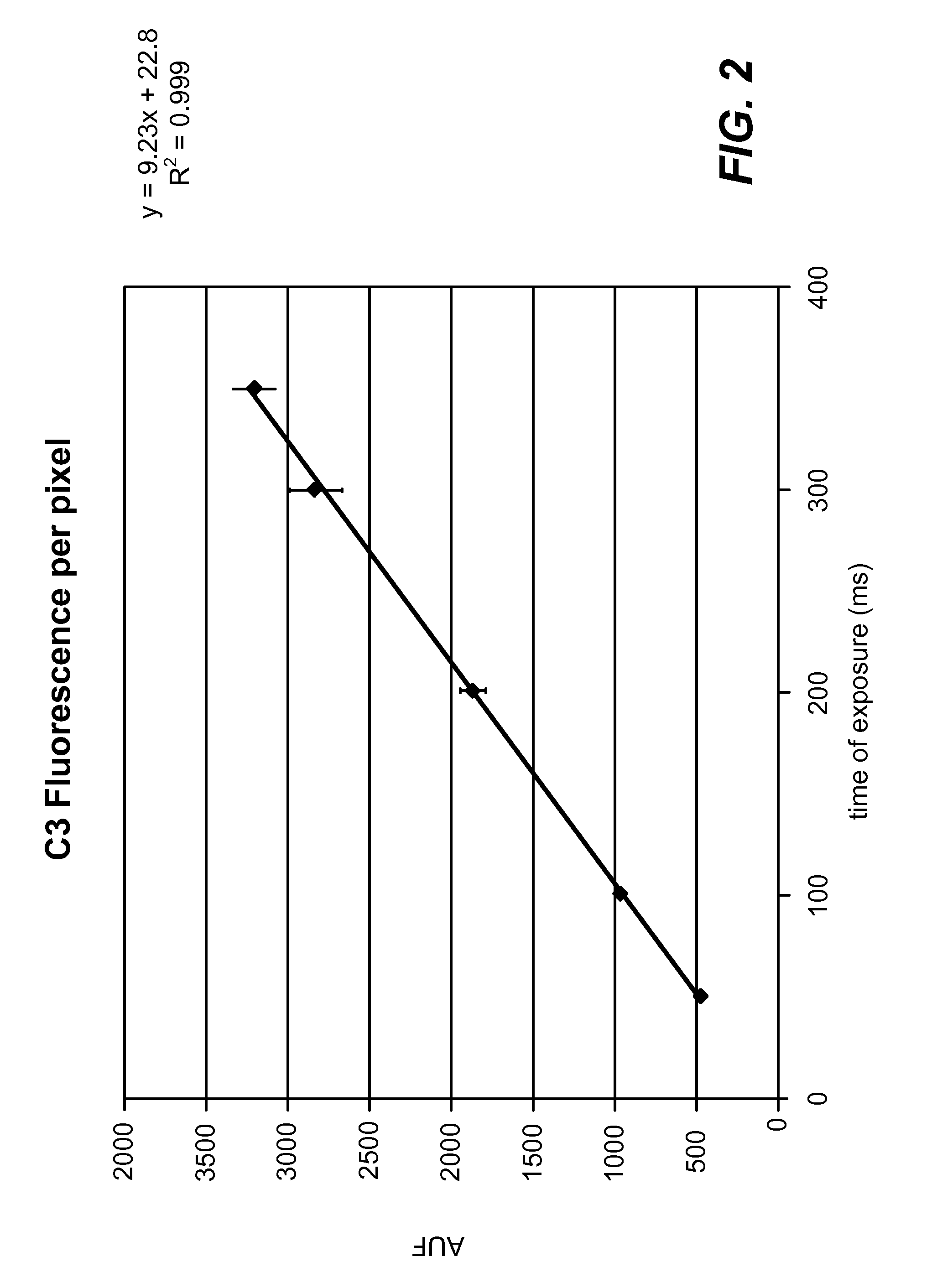
[0024]A fluorescence microscopy standard (4.0 rim-diameter microspheres, Kit M-7901 from Molecular Probes) was used to calibrate two different Leica microscopes. Standard curves of fluorescence per pixel (average of about 20 microspheres at each time point) versus exposure time in milliseconds were constructed (see FIG. 1 and FIG. 2). A linear response is observed with both microscopes over the range of exposure time used to acquire images.

[0025]A breast cancer cell line, HCC 2218 (positive for HER2 / neu expression) was stained simultaneously with anti-cytokeratin-FITC and anti-HER2 / neu-Alexa 532 (red fluorescence) antibodies. Digital images were acquired of identical fields using filter cubes that differentiate the two fluorescence signals. The HER2 / neu images were acquired using exposure times within the linear range of the standard curves, viz.,...

example 2

[0027]It was then determined that the desired consistency of results could be achieved by the following method.

[0028]An antibody specific for the receptor is labeled with fluorescent dye and a fluorescence standard has been obtained for the generation of standard curves to allow HER2 / neu fluorescence to be normalized as a percentage of the non-bleaching standard. HER2 / neu values can then be correlated from sample to sample and from laboratory to laboratory based on quantitative calibration on a universal fluorescence standard.

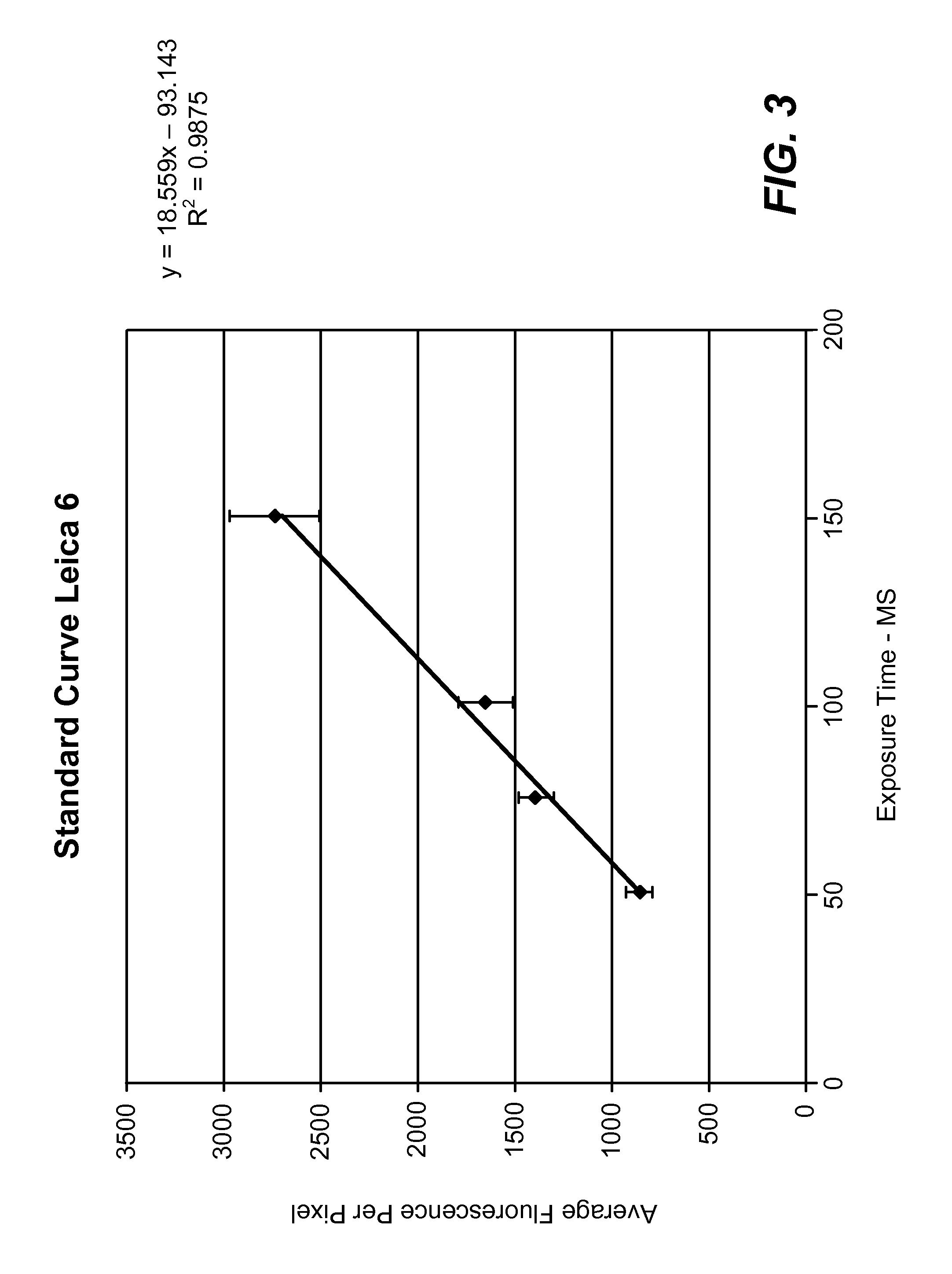
[0029]A fluorescence microscopy standard (4.0 um-diameter microspheres, Kit M-7901 from Molecular Probes) was used to calibrate two different Leica microscopes. Standard curves of fluorescence per pixel (average of about 20 microspheres at each time point) versus exposure time in milliseconds were constructed (see FIG. 3 and FIG. 4). A linear response is observed over the range of exposure time used to acquire images. From these linear curves, an exposure time ...

example 3

[0032]Cells: Six breast cancer cell lines were purchased from ATCC and grown in medium containing 10% fetal bovine serum. HCC2218, HCC38, HCC2O2 and T-47D were grown in RPMI 1640, MCF-7 was grown in EMEM and SK-BR-3 was grown in McCoy's. Exponentially growing cells were trypsinized and spun onto microscope slides from a megafunnel with a Cytospin 3 (Shandon) at 1000 rpm for 10 minutes and then air-dried for at least two hours, preferably overnight.

[0033]Reagents: An antibody cocktail for identifying epithelial cells containing monoclonal antibodies covalently labeled with FITC and which recognizes nine different cytokeratin peptides and a tumor-associated glycoprotein. Anti-ERCC-1 (sc-10785, Santa Cruz Biotechnology), anti-thymidylate synthase (clone TS 106, Exalpha Biologicals), anti-estrogen receptor (clone TE 111 .5D 11, Exalpha Biologicals) and Trastuzumab (Genentech) were conjugated to Alexa Fluorescent (AF) dyes AF 594, AF 647, AF 594 and AF 532 (Molecular Probes, Eugene, Oreg...


PUM

PropertyMeasurementUnit
exposure timeaaaaaaaaaa
fluorescent microscopeaaaaaaaaaa

Abstract

The present invention provides materials and methods for predicting the response of a disease state to a therapeutic agent. A targeting moiety specific for a biological marker is labeled with a reporter moiety and used to analyze cells characteristic of the disease state. The output of the reporter moiety, which may be fluorescence intensity, is compared to the output of reference standard analyzed under similar or identical conditions. The use of a reference standard allows biomarker reporting to be normalized. Biomarker values can then be correlated from sample to sample and from laboratory to laboratory based on quantitative calibration on a universal reference standard.

Description

CROSS REFERENCE TO RELATED APPLICATIONS[0001]This application is a continuation of U.S. patent application Ser. No. 10 / 787,585, filed Feb. 27, 2004, which claims the benefit of U.S. Provisional Application Nos. 60 / 451,050, filed Feb. 27, 2003 and 60 / 488,865, filed Jul. 21, 2003, the contents of each are specifically incorporated herein by reference in their entirety.FIELD OF THE INVENTION[0002]The present invention relates in general to pharmaceutical therapies, and more particularly to compounds and methods for predicting the efficacy of particular therapies for particular patients.BACKGROUND[0003]One of the most urgent needs for cancer patients is to find an effective drug, particularly for cancer patients with a metastatic disease after removal / destruction of the primary tumor in the original organ site by surgery or radiation. The goal of any therapy is to improve the life expectancy and quality of life in a cost-effective manner. Through much effort and expense, a number of new...

Claims


Application Information

Patent Timeline
Patent Type & Authority Applications(United States)
IPC IPC(8): G01N33/53G01NG01N1/00G01N33/50G01N33/574
CPCG01N33/5011G01N2800/52G01N33/5091
Inventor LESKO, STEPHEN A.TS'O, PAUL O.P.
Owner LESKO STEPHEN A
Why Eureka
Try Eureka