

Injection and hemostasis site
a technology which is applied in the field of injection and hemostasis site, can solve the problems of unsterile needles, a certain amount of bleeding that occurs when the needle is removed, and cannulation-induced infection of the cannulation tract and/or vessel
- Summary
- Abstract
- Description
- Claims
- Application Information
AI Technical Summary
Benefits of technology
Problems solved by technology
Method used
Image

Examples
Embodiment Construction
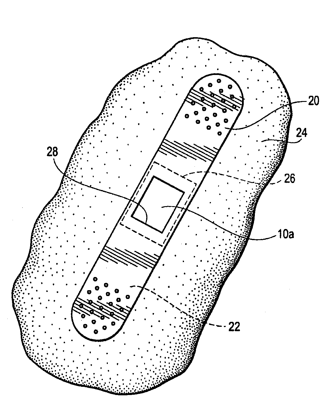
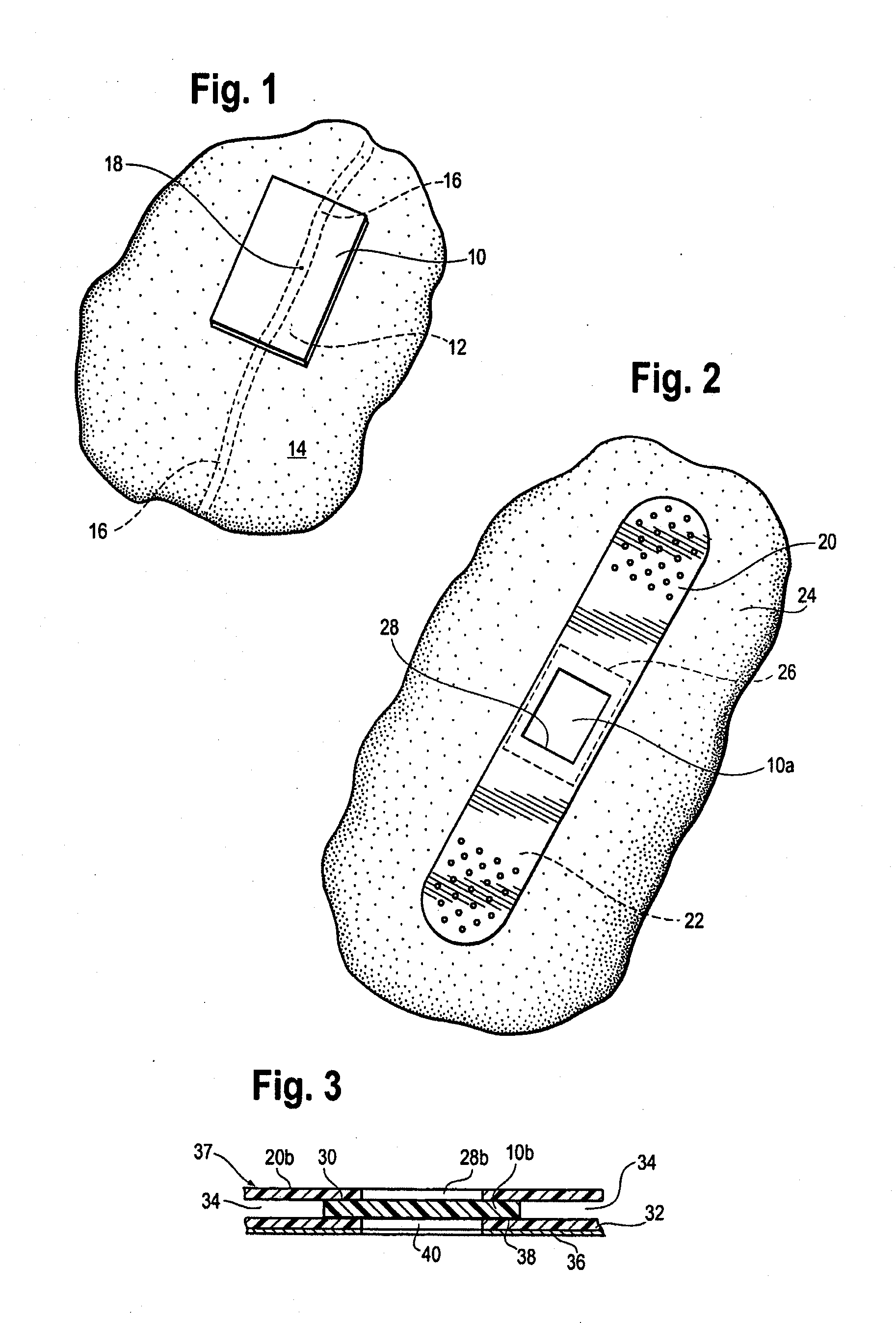
[0009]By this invention, a supersoft, typically elastomeric, pad or sheet is disclosed as an improved replacement for gauze squares, and other modes of achieving hemostasis on any bleeding site, whether by cannulation with needles of any size, or trauma from gunshot wounds and the like. Its action is the same. The typically substantially nonporous-surfaced pad stops the bleeding and promotes clotting, being a solid, pore free pad, or a porous pad typically having at least one, nonporous, skin like surface. The nonporous, skin like surface lies against the bleeding site, for hemostasis. The pores, if present, may be closed cells, or interconnected, open cells if desired, to contribute to softness of the pad. “Pores” are spaces in the pad which are large enough to pass blood or other medical fluid. The pad may be provided in a package in medically clean, or preferably sterile, condition. The pad may, in some embodiments, comprise a pair of opposed, parallel, equally sized major faces,...
PUM
 Abstract
Description
Claims
Application Information