Fluorescent detection of proteins in polyacrylamide gels

a polyacrylamide gel and protein technology, applied in the direction of fluid pressure measurement, liquid/fluent solid measurement, peptide measurement, etc., can solve the problem of irradiation-induced production of long wavelength emitting substances, and achieve the effect of not having to laborious labeling or staining steps

Inactive Publication Date: 2010-04-15
UNIV TECH INT +1
View PDF3 Cites 30 Cited by
  • Summary
  • Abstract
  • Description
  • Claims
  • Application Information

AI Technical Summary

Benefits of technology

[0008]The applicants have recently determined that a UV-light-dependent reaction between Trp in the presence of trichloro compounds such as trichloroacetic acid yields fluorescent products that emit in the visible spectrum. This reaction is similar to the reaction between Trp and chloroform described in the prior art. This knowledge permitted the development of a procedure for visualizing proteins after SDS-PAGE which allowed fluorescent visualization of proteins shortly after completion of electrophoresis and without laborious labeling or staining steps.

Problems solved by technology

This generation of blue fluorescence in biological tissue samples is used to monitor aging and lipid peroxidation; but the use of chloroform / methanol in the extraction of tissues and subsequent production of long wavelength emitting substances upon irradiation has been noted as a complication in these studies.

Method used


Image

  • Fluorescent detection of proteins in polyacrylamide gels
  • Fluorescent detection of proteins in polyacrylamide gels
  • Fluorescent detection of proteins in polyacrylamide gels

Examples


example 1

Post-Electrophoresis Incorporation—Materials and Methods

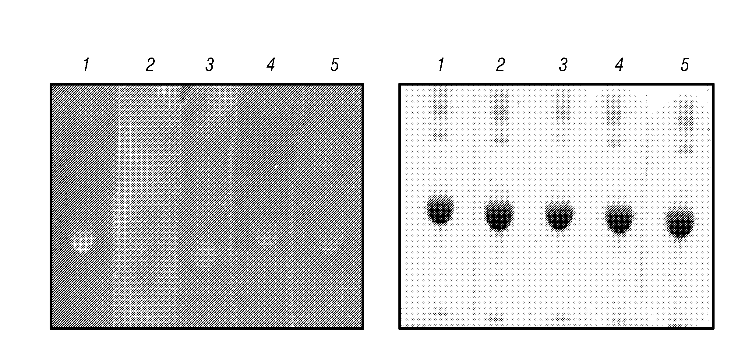
[0042]Electrophoresis was performed in each case using an electrophoresis apparatus from Owl Scientific (Portsmouth, N.H.) with 15 3 20 3 0.15-cm gel cassettes or a Mini Protean II™ system from Bio-Rad (La Jolla, Calif.). Upon completion of electrophoresis, the gel was removed from the cassette and placed in the haloalkane solution. After soaking from 5 to 10 minutes, the haloalkane solution was decanted, and the gel was rinsed several times with tap water to remove residual solution and then placed in water to prevent drying. If trichloroacetic acid (TCA) is used, it is important to rinse the gel to remove excess TCA prior to protein visualization since TCA is corrosive and will damage the UV transilluminator. To visualize proteins, the gel was subjected to UV illumination using a standard UV box. During the course of UV irradiation, resolved proteins became visible as bluish-green bands against the background of pale blue gel...

example 2

Comparison of Fluorescent Detection with CBB Staining

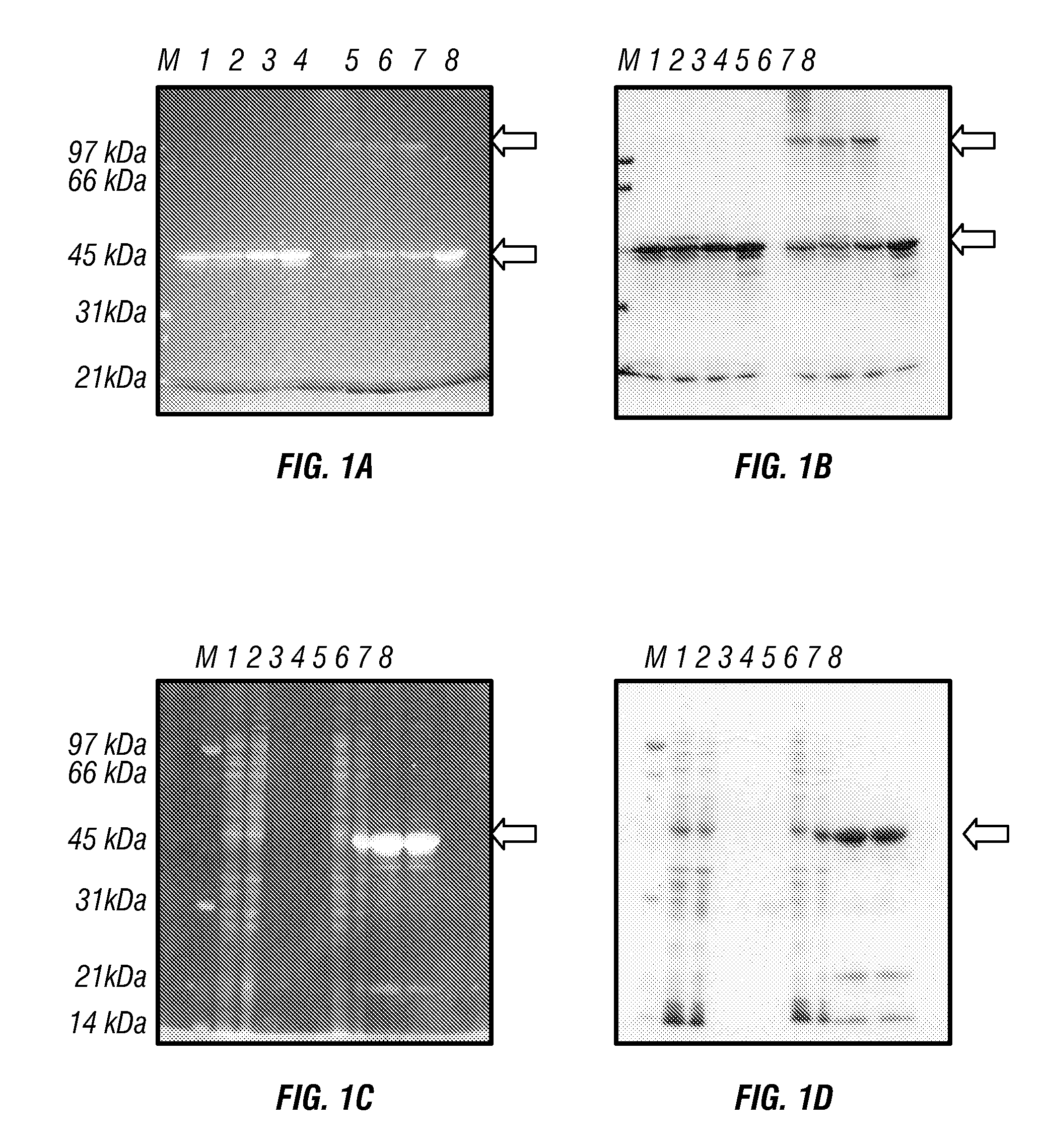
[0044]FIG. 1 shows results which validate the utility of this method. In FIGS. 1A and 1B, approximately 20 μg of a recombinant protein and three of its mutants, all with electrophoretic mobility corresponding to 46 kDa, were loaded on the gel in sample buffer either with or without added β-mercaptoethanol (BME). FIG. 1A is a picture of the gel soaked in 10% TCA for 5 minutes and rinsed three times with tap water. FIG. 1B is the same gel after conventional CBB staining. It is evident that wild-type (wt), as well as mutants 1 and 2, form aggregates of a higher molecular weight in the absence of BME, while mutant 3 does not. These proteins could be visualized by UV illumination 10 min after the completion of the PAGE run. In FIGS. 1C (TCA) and 1D (CBB), an expression of a recombinant protein by two bacterial clones and its purification were followed. The purifications were run in duplicate. The gel demonstrates that clone 1 does not ...

example 4

Pre-Electrophoretic Incorporation: Optimization and Sensitivity

[0058]In optimization studies 0.02, 0.05, 0.1, 0.2, 0.5, 1.0 and 2.0% TCE (vol / vol) were added to the gel before polymerization. For the TCE staining the 12% SDS-PAGE was run and then soaked in 10% TCE (v / v) in water:methanol (1:1) for ten minutes. Then the gel was washed in water and visualized as described above.

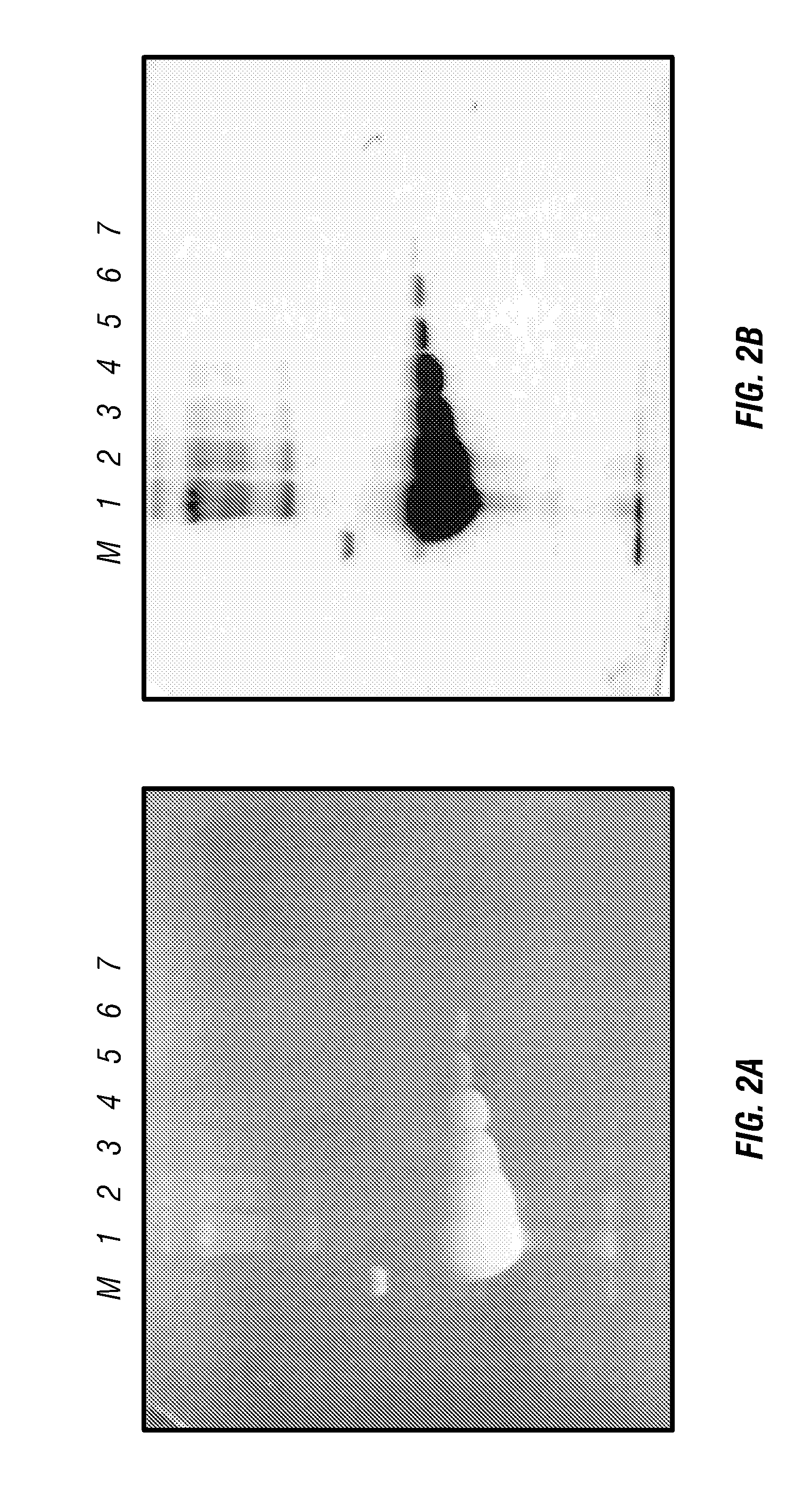
[0059]Calculating the intensities for FIGS. 2 and 3A, the sum of the intensity of phosphorylase b, albumin, ovalbumin and trypsin bands was used. These were chosen because they contain percentages of tryptophan near the average for soluble proteins. Four sets of 0.25 μg and 0.5 μg bands were used to calculate intensity per μg.

[0060]FIG. 8 shows that maximum intensity is reached with a TCE concentration of 0.5% and that lower concentrations have a lower intensity while higher concentrations do not have higher intensities.

[0061]FIG. 3A shows that intensity increases nearly linearly with the amount of protein up t...


PUM

PropertyMeasurementUnit
wavelengthaaaaaaaaaa
temperatureaaaaaaaaaa
wavelengthaaaaaaaaaa

Abstract

The mechanism of the UV light-induced reaction between the indole moiety of tryptophan and chloroform, and the structure of the modified tryptophan and polypeptides including such modified tryptophan residues. The excited indole moiety, which is formed upon UV light irradiation, emits a solvated electron which initiates a series of events that yield fluorescent derivatives that have CHO group covalently bound to the indole moiety. These derivatives are herein referred to as formyltryptophan, and are relatively stable. Similar reactions are observed when 5-hydroxytryptophan, 5-fluorotryptophan, or N-methylindolacetate are used in place of tryptophan, or when other haloalkanes, such as trichloracetic acid, trichlorethanol, trichlorethane, bromoform, and iodoactetate are used in place of chloroform. The derivatives can be used in a variety of applications in fluorescence spectroscopy, and for nuclear magnetic resonance, X-ray crystallography, infra-red spectroscopy, circular dicroism and mass spectroscopy. Additionally, the UV light-induced reaction between the indole moiety of tryptophan and haloalkanes can be used to prepare derivatives of tryptophan for chemical cross-linking studies of proteins and peptides.

Description

CROSS REFERENCE TO RELATED APPLICATIONS[0001]This application claims the priority benefit of U.S. Provisional Patent Application No. 60 / 319,099 filed Jan. 25, 2002; No. 60 / 352,225 filed Jan. 29, 2002; and No. 60 / 319,810 filed Dec. 24, 2002; the contents of each of which are incorporated by reference.BACKGROUND OF INVENTION[0002]This invention relates to polyacrylamide gel electrophoresis and in particular, to a method of visualizing proteins in a polyacrylamide gel.[0003]Polyacrylamide gel electrophoresis is a well-known technique for determining the molecular weight of a protein and for separating proteins on the basis of their molecular weight. Electrophoresis in the absence of any denaturing reagent (native-PAGE) results in separation on the basis of charge and size. It gives an estimation of the size of the folded protein by reference to proteins of known size. In order to determine the molecular weight of the polypeptide chain it is necessary to carry out the electrophoresis in...

Claims


Application Information

Patent Timeline
Patent Type & Authority Applications(United States)
IPC IPC(8): C07K1/26G01N33/559G01N33/68
CPCG01N33/6827
Inventor EDWARDS, ROBERT A.TURNER, RAYMOND J.LADNER, CAROLSTARKEY, JEANLARSON, ERICKAZMIN, DMITRI
Owner UNIV TECH INT
Why Eureka
Try Eureka