Method for double staining in immunohistochemistry

a technology of immunohistochemistry and double staining, which is applied in the field of immunohistochemistry, can solve the problems of inability to exclude, undesirable flow cytometry, and insufficient equipment for diagnostic or pathological laboratories, and achieve the effect of a measurably different imag

Active Publication Date: 2010-06-17
CORNELL UNIVERSITY
View PDF6 Cites 20 Cited by
  • Summary
  • Abstract
  • Description
  • Claims
  • Application Information

AI Technical Summary

Benefits of technology

[0017]This invention provides a method for double staining tissue samples, e.g., using methods typical of IHC but employing two different chromogens, each producing a distinctly colored product, and which when co-localized those products form a distinct color that is different than either isolated color. These differences in color are detectable by naked eye and are measurably different by computer scanning. While some light reactive IHC co-localization studies have been successful in showing two proteins in the same cell (e.g., one protein in the nucleus and another in the cell membrane), none have shown co-localization in the same cellular structure as in the present invention. In the presen

Problems solved by technology

However, non-cancer cells expressing the protein indicative of cell proliferation must be excluded from the analysis; such exclusion is not possible using other methods.
Flow cytometry is undesirable because it does not allow for any visualization or morphologic assessment.
For these reasons, most diagnostic or pathology laboratories are not equipped for IF.
For these reasons, IHC is the diagnostic standard widely used in pathology laboratories and ind

Method used


Image

  • Method for double staining in immunohistochemistry
  • Method for double staining in immunohistochemistry
  • Method for double staining in immunohistochemistry

Examples


example 1

Bone Marrow Specimen Procurement

[0063]Bone marrow specimens were obtained from volunteers without hematological disease at the Hospital for Special Surgery and from multiple myeloma patients at the New York-Presbyterian Hospital under informed consent as part of an Institutional Review Board approved study. The posterior and superior iliac spines were identified before beginning the procedure. The patient was placed in a ventral or lateral decubitus position. The field was cleaned with Betadine and the area of interest was anesthetized with 1% Lidocaine. A bone marrow aspirate and biopsy was performed unilaterally using a modified Jamshidi bone marrow aspiration / biopsy needle, 11 gauge×4 inch. Several 5-7 cc aspirates were obtained by relocating the needle in each aspirate to obtain a total of approximately 30 cc of marrow. The core biopsy was then obtained. The bone marrow core biopsies were fixed in 10% neutral-buffered formalin and paraffin-embedded.

example 2

Immunohistochemistry Using Two Nuclear Biomarkers

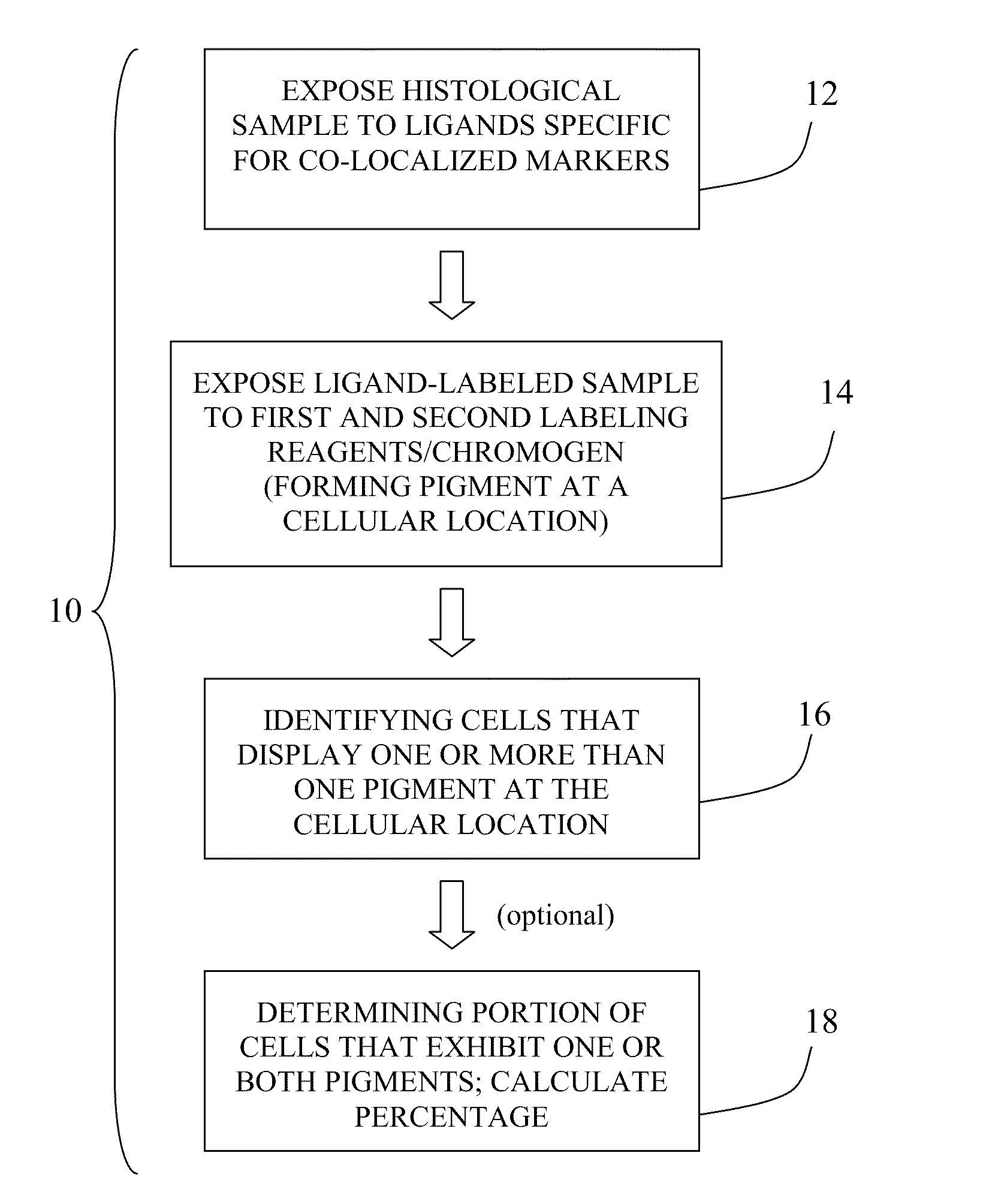
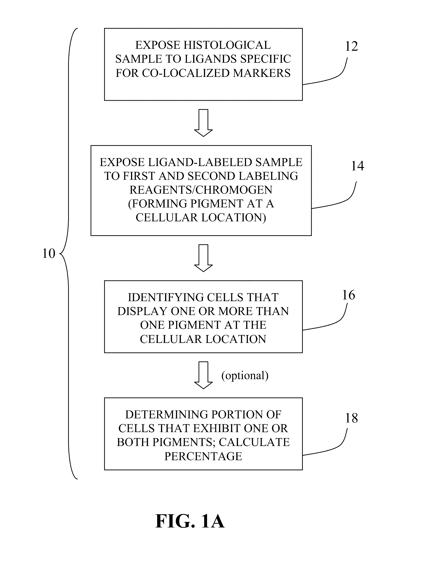
[0064]Because cell cycle molecules are not unique to cancer cells and because some tumors are composed of cancer cells that coexist with large populations of non-cancer cells, in such tumors cell cycle analysis requires co-localization, detecting the cell cycle molecule in the cancer cells while excluding non-cancer cells from the analysis. For example, some tumors contain large numbers of actively proliferating immune cells; if they are not excluded from the analysis, assessment of proliferation may yield falsely high data.

[0065]In hematopoietic cancer biopsies, the non-cancer hematopoietic and immune cells must be excluded. In this example, multiple myeloma (MM) cells were identified in biopsy samples with an anti-MUM1 antibody and a detection system yielding a red signal. Specificity of MUM1 for myeloma cells in MM biopsies is estimated at ˜99%, thus excluding non-MM cells from the analysis. Double IHC was performed to detect cell ...

example 3

Automated Image Analysis

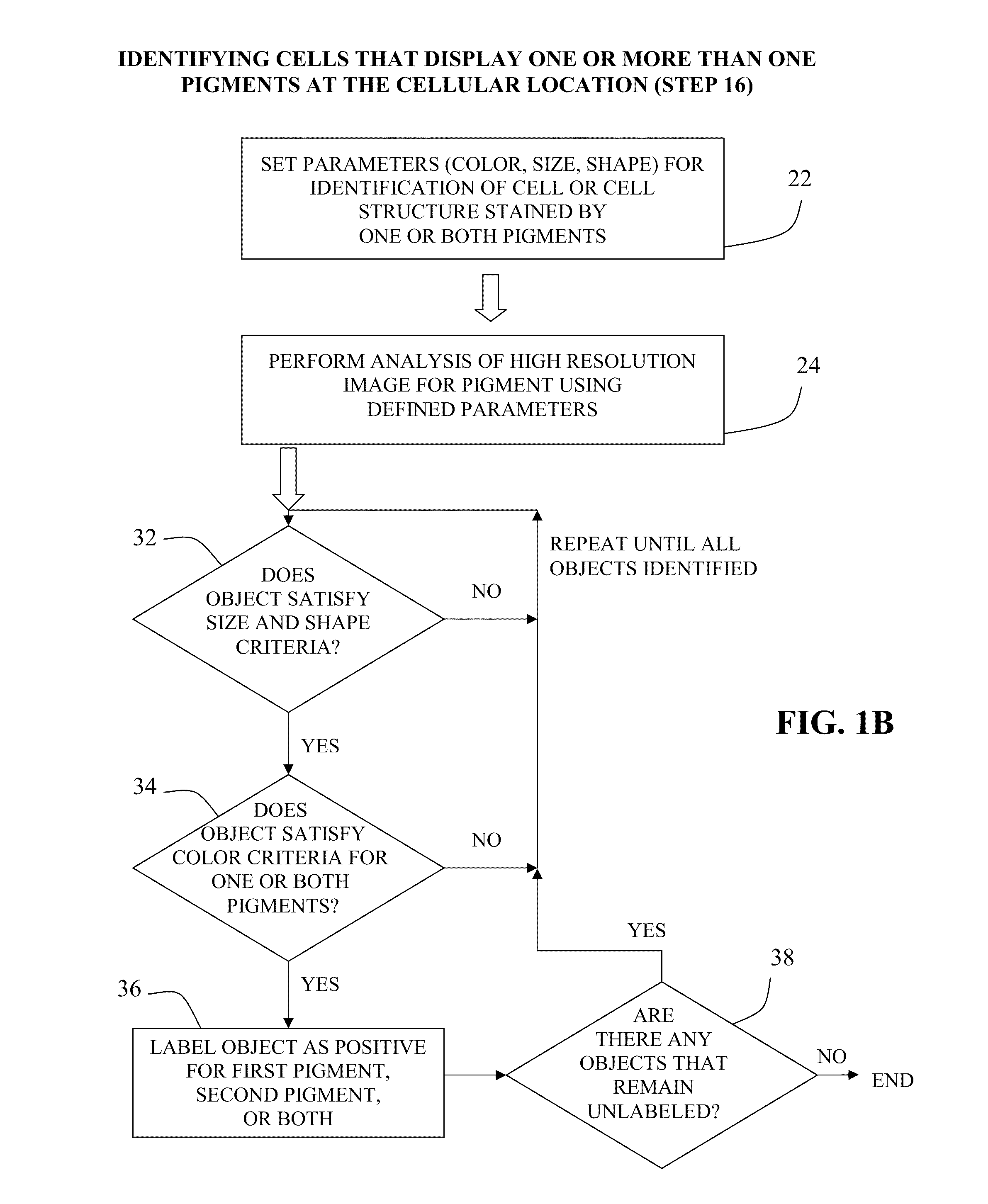
[0073]The double stained IHC slides were accessioned into the image analysis system and then loaded onto the machine in batches of 60. A robotic arm picked up each slide, one at a time, and loaded it onto the microscope. Via a motorized stage, the microscope moves and scans the slide, looking for tissue, the location of which varies slightly from slide to slide. Once it has found the tissue, the device scans the tissue and the camera records low resolution images. Using the scanned low resolution images, the inventor selected ten good areas for subsequent high resolution scanning and analysis. Numerous data points were then calculated for each of the ten areas per slide, and those data were averaged.

[0074]Since no counterstain was used in these slides, all identifiable cells are positive for at least one marker (e.g., in FIG. 3A, cells that express neither MUM1 nor pSRb are not visible). The characteristics used for the analysis (“classifiers”) were determine...


PUM


Abstract

The present invention relates to kits and methods for performing dual-staining immunohistochemistry (IHC) for the detection of specific cell populations in tissue samples containing heterogeneous populations of cells, which can be observed by a light microscope for co-localization of distinct pigments. The method includes providing a tissue sample comprising fixed cells; exposing the sample to first and second ligands that recognize different marker proteins found at the same cellular location, thereby forming a ligand-labeled sample; exposing the ligand-labeled sample to first and second labeling reagents, the first labeling reagent binding to the first ligand and the second labeling reagent binding to the second ligand, the first and second labeling reagents each forming distinct pigments; and identifying the number of cells that display only one particular pigment, or more than one pigment, by the different coloration of the cellular location labeled by the distinct pigment.

Description

[0001]This application claims the priority benefit of U.S. Provisional Patent Application Ser. No. 61 / 138,422, filed Dec. 17, 2008, which is hereby incorporated by reference in its entirety.FIELD OF THE INVENTION[0002]The present invention relates generally to immunohistochemistry (“IHC”) methods for functional assessment of specific cell types in fixed tissue samples. More specifically, the invention relates to kits and methods that employ two distinct chromogens that localize to the same cell structure and their resulting pigments can be observed by a light microscope. Co-localization of the pigments can be analyzed manually or by automatic scanning of the tissue section.BACKGROUND OF THE INVENTION[0003]Traditional methods of detecting gene expression detect or measure protein or RNA extracted from whole tissue. However, the observation of gene expression within specific cell types is critical in many clinical scenarios. For example, in observing or diagnosing bone marrow cancers ...

Claims


Application Information

Patent Timeline
IPC IPC(8): C12Q1/70G01N33/53
CPCG01N33/581G01N33/574
Inventor ELY, SCOTT A.
Owner CORNELL UNIVERSITY
Why Eureka
Try Eureka