Cellular Libraries of Peptide Sequences (CLiPS) and Methods of Using the Same
- Summary
- Abstract
- Description
- Claims
- Application Information
AI Technical Summary
Benefits of technology
Problems solved by technology
Method used
Image
Examples
example 1
Development of a Whole-Cell Assay for Peptidase Activity
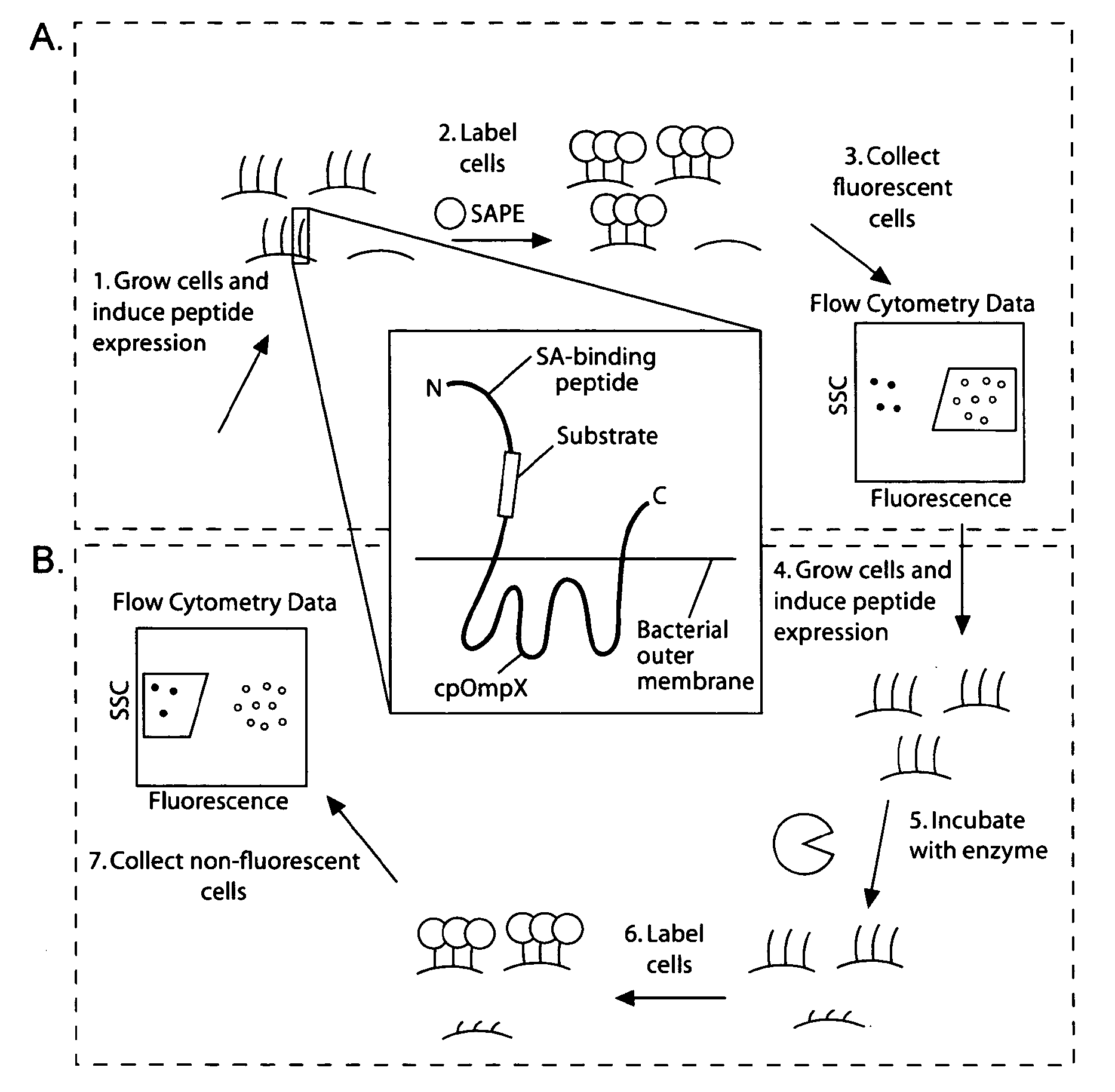
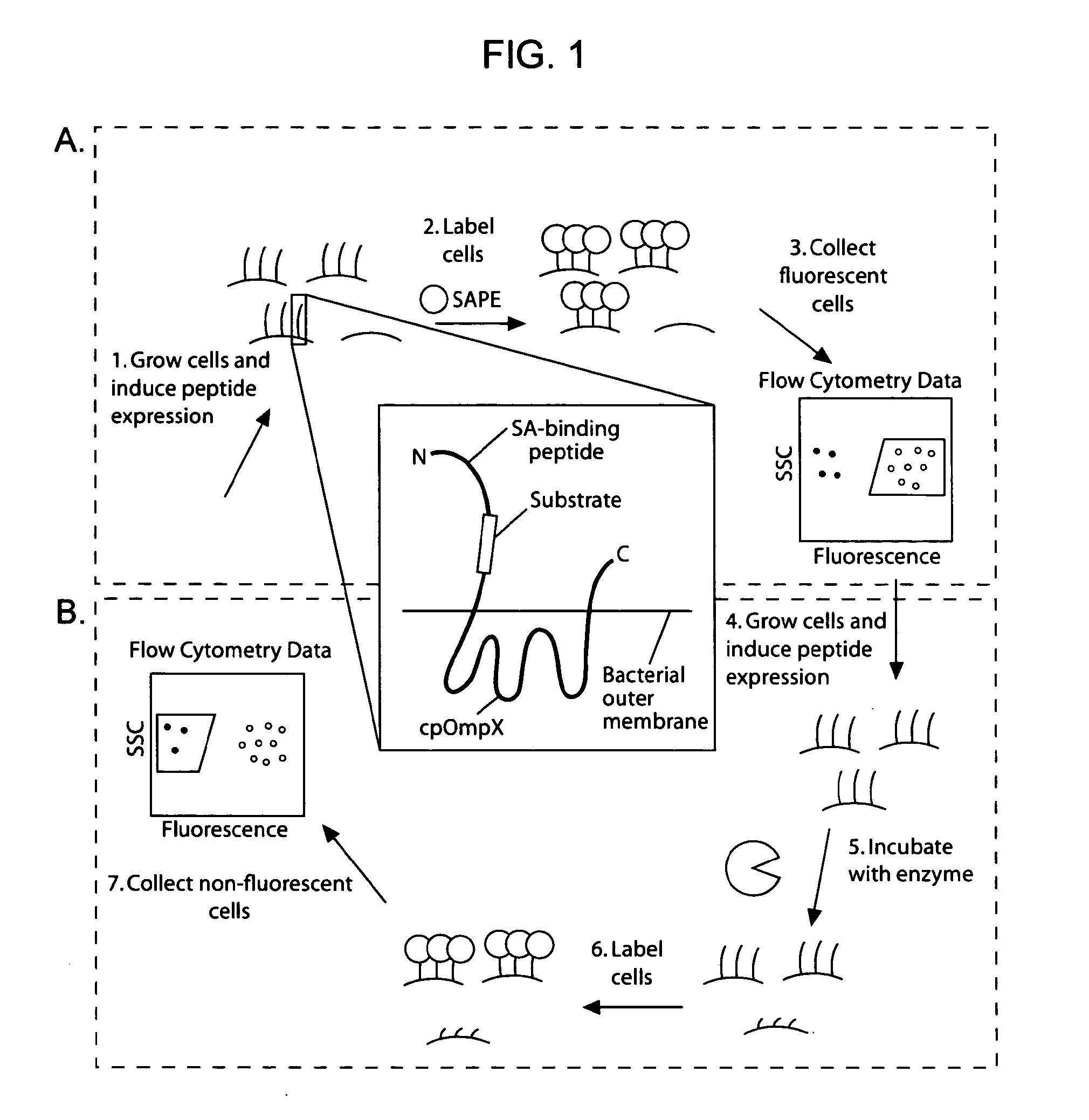
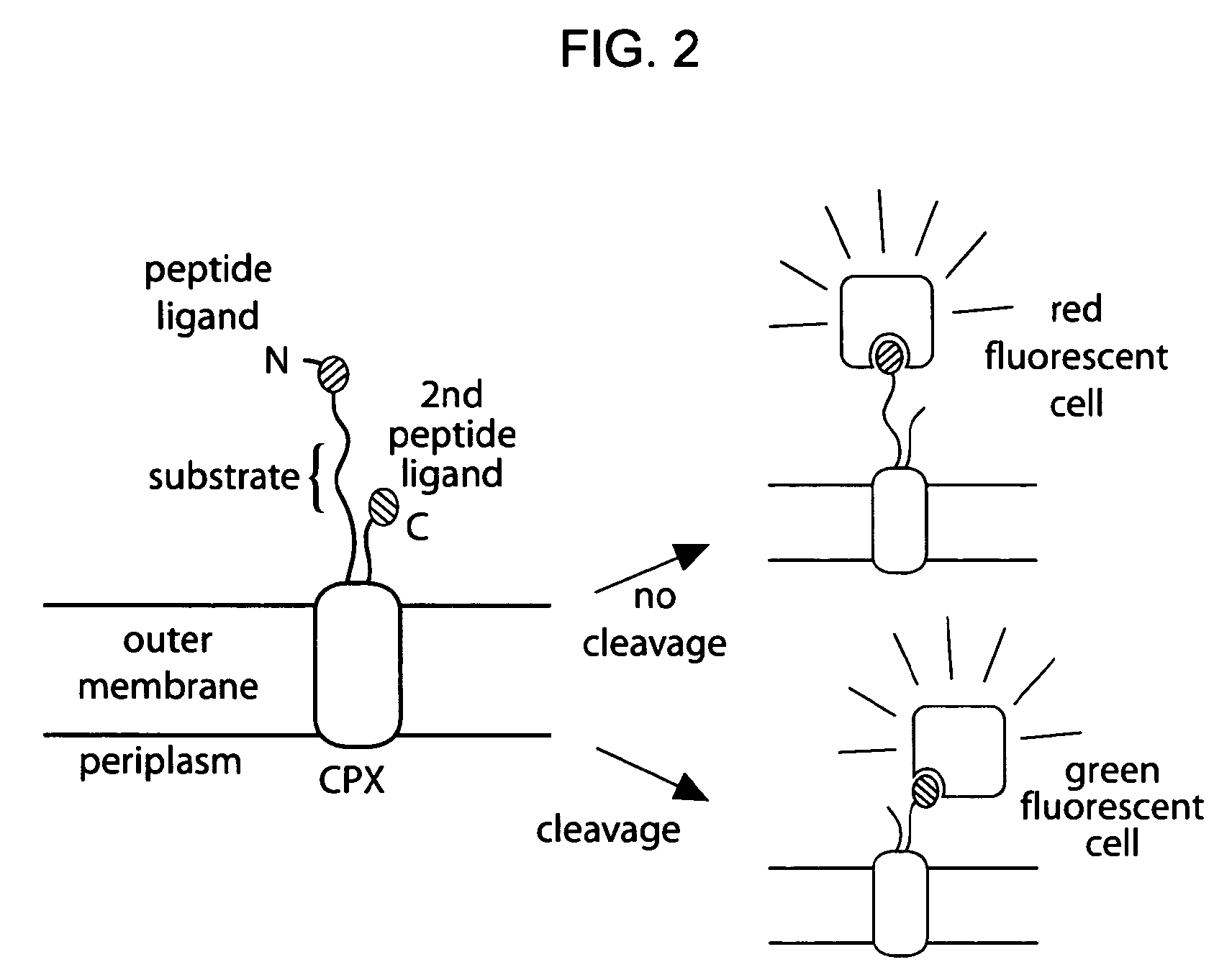
[0163]Given the utility of fluorescence-activated cell sorting (FACS) as a quantitative library screening tool, peptidase activity was assayed by displaying reporter-substrates on the surface of Esherichia coli (FIG. 1, panel A). Reporter-substrates were designed consisting of a peptide ligand (D) that binds the fluorescent probe streptavidin-R-phycoerythrin (SA-PE) and a peptide substrate oriented such that cleavage removes the SA-PE-binding ligand from the cell surface. In this way, protease activity towards a given substrate would be detectable by monitoring whole-cell fluorescence using FACS. Reporter-substrates were displayed on E. coli using circularly permutated outer membrane protein X (CPX), which presents both N- and C-termini on the cell surface, enabling presentation of candidate peptides as non-constrained, terminal fusions (Rice et al., Protein Sci. 15(4):825-36 (2006)). As a control, a substrate-reporter display ...
example 2
Determination of Enteropeptidase and Caspase-3 Specificity Using CLiPS
[0165]To demonstrate the general utility of CLiPS, the 6-mer substrate library was screened to identify optimal substrates for two unrelated proteases: caspase-3 and enteropeptidase. These proteases recognize the canonical substrates DEVD↓ (SEQ ID NO:16) (Barrett et al., Handbook of proteolytic enzymes (Academic Press, San Diego) (2004)) and DDDDK↓ (SEQ ID NO:02) (Bricteux-Gregoire et al., Comp Biochem Physiol B 42:23-39 (1972)), respectively. Caspase-3 was chosen to validate CLiPS, since specificity has been investigated extensively using both substrate phage and fluorogenic substrates (Stennicke et al., Biochem J350 Pt 2, 563-8 (2000); Lien et al., Protein J23:413-25 (2004)). In contrast, enteropeptidase specificity is less well characterized and has been investigated primarily using individually synthesized, fluorogenic substrate variants (Likhareva et al., Letters in Peptide Science 9:71-76 (2002)). For each p...
example 3
Characterization of Substrate Cleavage Kinetics
[0168]The use of multi-copy substrate display on whole cells enabled simple and direct quantitative characterization of cleavage kinetics. Consequently, flow cytometry was used to rank individual isolated clones on the basis of substrate conversion, and those clones exhibiting more than 50% conversion were identified by DNA sequencing (Tables 3 and 4). Substrates efficiently cleaved by caspase-3 revealed a strong substrate consensus of DxVDG (SEQ ID NO:45) (Table 3), in agreement with the known specificity of caspase-3. The substrates identified for enteropeptidase shared a consensus sequence of D / ERM, indicating a substrate preference at the P1′ position (Table 3). Interestingly, enteropeptidase substrates identified by CLiPS were cleaved more rapidly than the canonical sequence, DDDDK (SEQ ID NO:02) (Table 4). Four isolated clones with high conversion were investigated further to quantify cleavage kinetics. Clones exhibiting multiple ...
PUM
 Abstract
Description
Claims
Application Information
 - R&D
- Intellectual Property
- Life Sciences
- Materials
- Tech Scout
- Unparalleled Data Quality
- Higher Quality Content
- 60% Fewer Hallucinations
Browse by: Latest US Patents, China's latest patents, Technical Efficacy Thesaurus, Application Domain, Technology Topic, Popular Technical Reports.
© 2025 PatSnap. All rights reserved.Legal|Privacy policy|Modern Slavery Act Transparency Statement|Sitemap|About US| Contact US: help@patsnap.com


