Methods and Systems for Performing Submucosal Medical Procedures

a technology of submucosal and tunneling, applied in the direction of surgical forceps, catheters, applications, etc., can solve the problems of en bloc removal of large flat mucosal lesions, numerous problems for current endoscopic tools and techniques, and inability to perform full thickness biopsies

Active Publication Date: 2010-07-08
APOLLO ENDOSURGERY INC
View PDF101 Cites 36 Cited by
  • Summary
  • Abstract
  • Description
  • Claims
  • Application Information

AI Technical Summary

Benefits of technology

[0042]In accordance with a further aspect of the present invention the tubular member of the submucosal biopsy instrument includes a second lumen. The distal end of the second lumen is connected to an extendable bellows member having a first contracted configuration and a second extended configuration. The bellows member is positioned within the first lumen at the distal end of the tubular member and connected to the distal end of the tissue cutter. When a pressure source is coupled to the proximal end of the second lumen, pressure within the bellows member transitions the bellows member from the first contracted configuration to the second extended configuration, thereby causing the tissue cutter to move from a first position distal to the window member to a second position proximal to the window member.
[0043]In accordance with yet a further aspect of the present invention the tubular member of the submucosal biopsy instrument includes a second lumen. The distal end of the second lumen is positioned proximal to the proximal end of the window member. An elongate member having proximal and distal ends is slidably disposed within the second lumen where the distal end is connected to the tissue cutter. The tissue cutter is configured to encircle the distal end of the tubular member. Movement of the proximal end of the elongate member in a proximal direction relative to the proximal end of the tubular member causes the tissue cutter to move from a first position distal to the window member to a second position proximal to the window member. The tissue cutter may take the form of an electrosurgical cutter in which tissue suctioned into the window member may be severed, when high frequency energy is supplied to the tissue cutter and the tissue cutter is moved from the first position to the second position.

Problems solved by technology

While many of these devices and techniques have been useful in identifying and removing some neoplastic lesions of the mucosal layer as well as providing access to general locations within the digestive tract for the placement of submucosal implants, there are some lesions and areas of the digestive tract which are extremely difficult to resect or access.
For example, the en bloc removal of large flat mucosal lesions presents numerous problems for current endoscopic tools and techniques.
Currently, access to these types of specimens requires full thickness biopsies which can be particularly difficult from an endoscopic approach requiring extremely skilled closure techniques.
While EMR techniques have been shown to be effective in treating some flat neoplastic lesions there are limitations and complications associated with these techniques.
A major limitation associated with this technique is the size of the lesion that can be resected.
While larger or irregular shaped lesions may be resected in a piecemeal fashion, this is undesirable since small portions of the lesion may remain.
Another limitation of these techniques includes uncertainty of the area being resected.
One complication associated with these EMR techniques is in relation to the use of the needle injection system.
Manipulating the injection catheter to position the needle through the mucosal layer into the submucosal layer can ultimately result in puncturing the muscular wall of the digestive tract which may lead to infection or peritonitis.
Another complication associated with EMR techniques is damage to the underlying muscular layer.
Saline and other non-viscous fluids used to elevate the lesion dissipate relatively quickly after injection into the submucosal layer, such that portions of the underlying muscular layer may be included in the suctioned tissue and inadvertently damaged when using the electrosurgical tool for resection.
While this procedure allows the physician to resect large, irregular shaped lesions en bloc, it requires a high degree of skill on the part of the physician and is still subject to the complications associated with needle perforations and muscular layer injury.
During application of this pushing force there is a risk that the tip of the instrument may injure or perforate the muscular wall or the mucosal layer.
During the second hole formation and subsequent holes, without visual confirmation of the orientation of the high frequency knife there is a risk of perforating the muscular wall or mucosal layer.

Method used


Image

  • Methods and Systems for Performing Submucosal Medical Procedures
  • Methods and Systems for Performing Submucosal Medical Procedures
  • Methods and Systems for Performing Submucosal Medical Procedures

Examples


Embodiment Construction

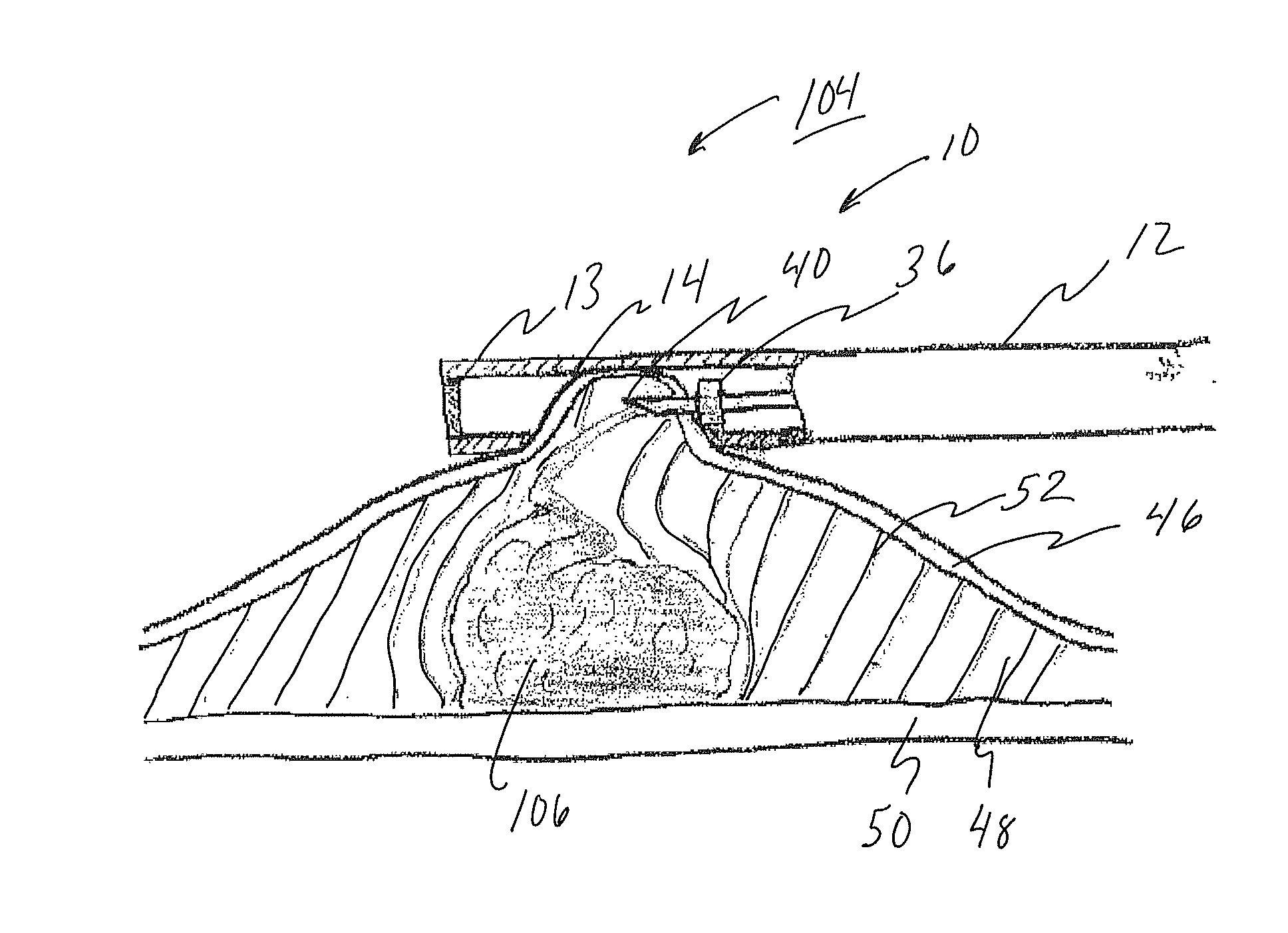
[0092]Methods and devices for performing submucosal medical procedures in a desired area of the digestive tract using an endoscope are described.

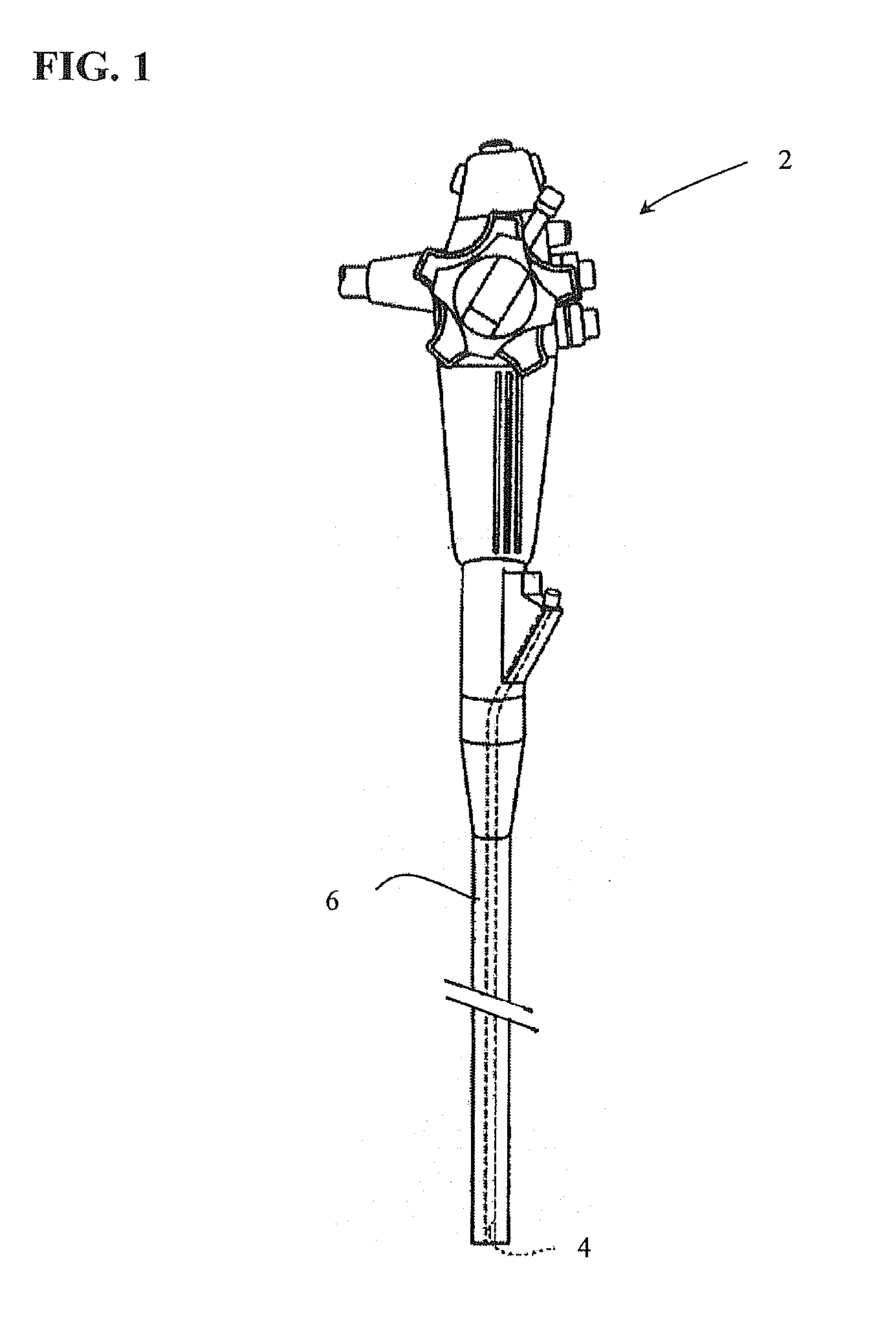
[0093]FIG. 1 illustrates an endoscope 2 of the type used in endoscopic procedures and suitable for use with embodiments of the present invention. The endoscope 2 has a working channel 4 extending from a proximal portion of the endoscope to the distal end of the endoscope. The endoscope 2 also has an insertion section 6 which enters the body of a patient passing through a natural orifice such as the mouth or rectum. The insertion section 6 is generally navigated to a position with the digestive tract when performing a submucosal medical procedure. Devices for use in performing submucosal medical procedures are preferably delivered through working channel 4 of the endoscope 2; however devices may be delivered along side the insertion section 6 of the endoscope.

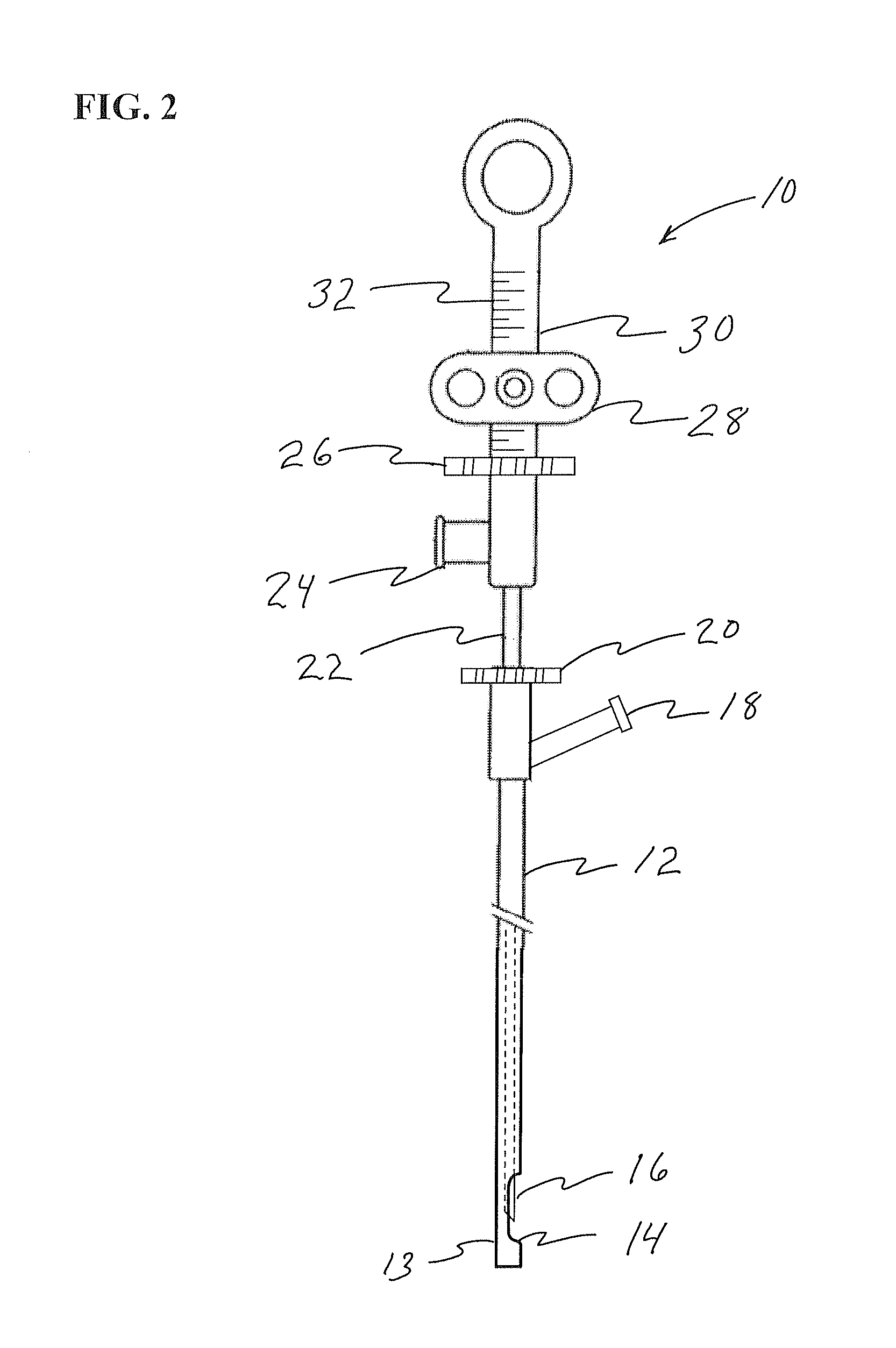
[0094]FIG. 2 illustrates a safe access needle injection instrument 10 which is used...


PUM


Abstract

Instruments, systems and methods are provided for performing submucosal medical procedures in a desired area of the digestive tract using endoscopy. Instruments include safe access needle injection instruments incorporating electrosurgical capability, a submucosal tunneling instrument, a submucosal dissection instrument, a mucosal resection device. Systems include a combination of one or more of such instruments with or without injectable agents. Embodiments of various methods for performing the procedures are also provided.

Description

CROSS REFERENCE TO RELATED APPLICATIONS[0001]This application claims the benefit of U.S. Ser. No. 61 / 144,805, filed Jan. 15, 2009, and is a continuation-in-part of U.S. Ser. No. 11 / 775,996, filed Jul. 11, 2007, both of which are hereby incorporated by reference herein in their entireties.FIELD OF THE INVENTION[0002]The present invention relates to a safe access needle injection instrument, a submucosal tunneling instrument, a submucosal dissection instrument, a mucosal resection device, a system and a method for performing submucosal medical procedures in a desired area of the digestive tract using an endoscope.BACKGROUND OF THE INVENTION[0003]The field of gastrointestinal endoscopy has for many years focused on diagnostic and therapeutic techniques to observe, modify and remove tissues located in the digestive tract. General endoscopic procedural techniques such as visualizing, dilating, cutting and manipulating tissue have been accomplished using flexible devices such as endoscope...

Claims


Application Information

Patent Timeline
Patent Type & Authority Applications(United States)
IPC IPC(8): A61B17/34A61B17/00
CPCA61B10/0283A61B17/22031A61B17/32056A61B17/320783A61B17/3478A61B18/1477A61B18/1492A61B2017/00269A61B2017/00539A61B2017/2937A61B2017/306A61B2017/320048A61B2017/320064A61B2017/320791A61B2017/3488A61B2018/00291A61B2018/1407A61B2018/1425A61M25/0084A61B18/12
Inventor MITELBERG, VLADIMIRJONES, DONALD K.
Owner APOLLO ENDOSURGERY INC
Why Eureka
Try Eureka