

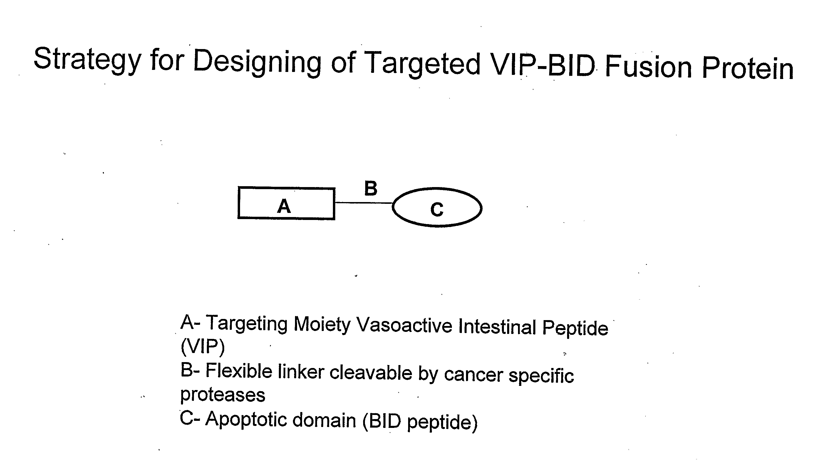
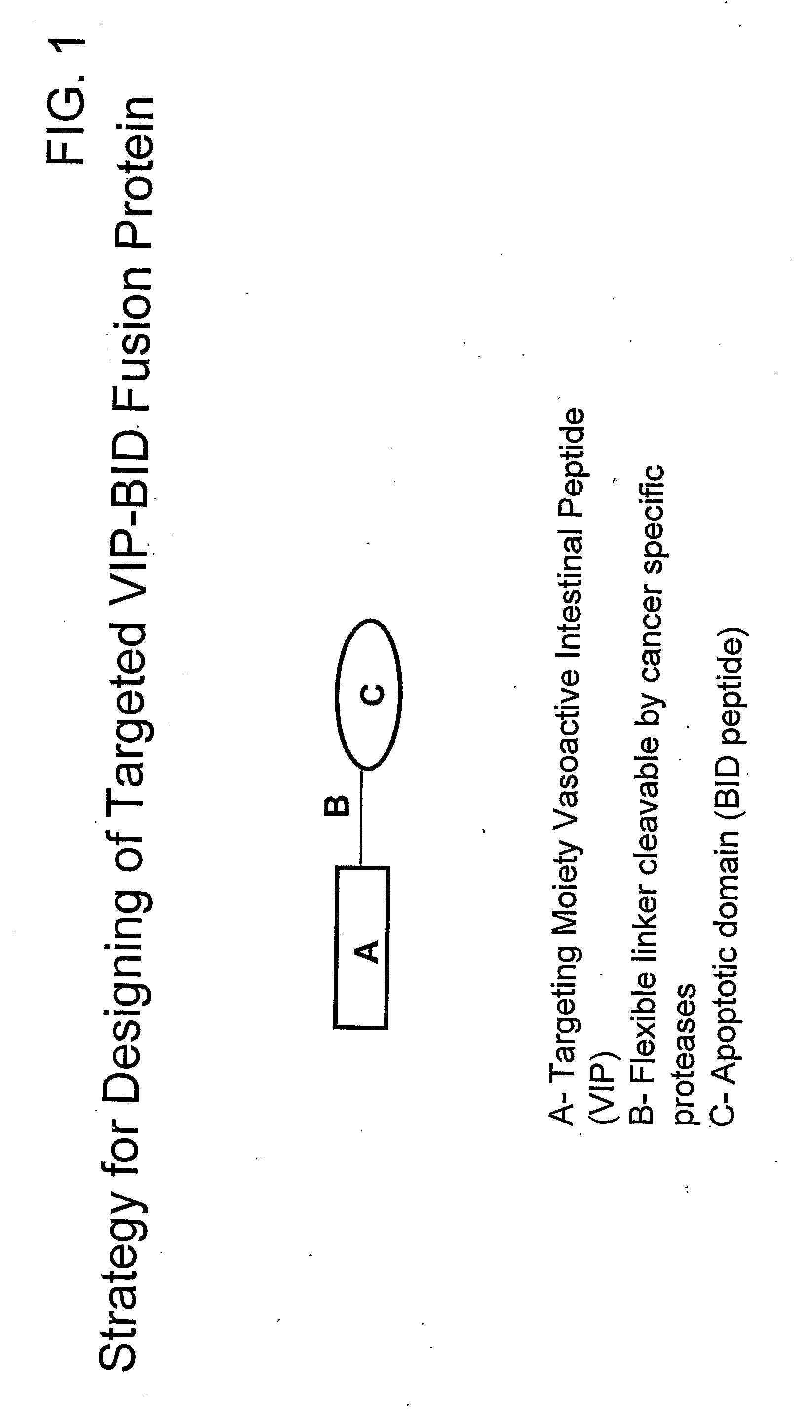
Targeted fusion proteins for cancer therapy
a cancer therapy and fusion protein technology, applied in the field of cancer therapy targeted fusion proteins, can solve the problems of limited practical use of bh3 peptides, trigger cell death, and -bak bh3 peptides, and achieve the effect of decreasing the mitochondrial membrane potential
- Summary
- Abstract
- Description
- Claims
- Application Information
AI Technical Summary
Benefits of technology
Problems solved by technology
Method used
Image

Examples
example 1
EXAMPLE 1
Construction of VIP-BH3 Bid Coding Sequence
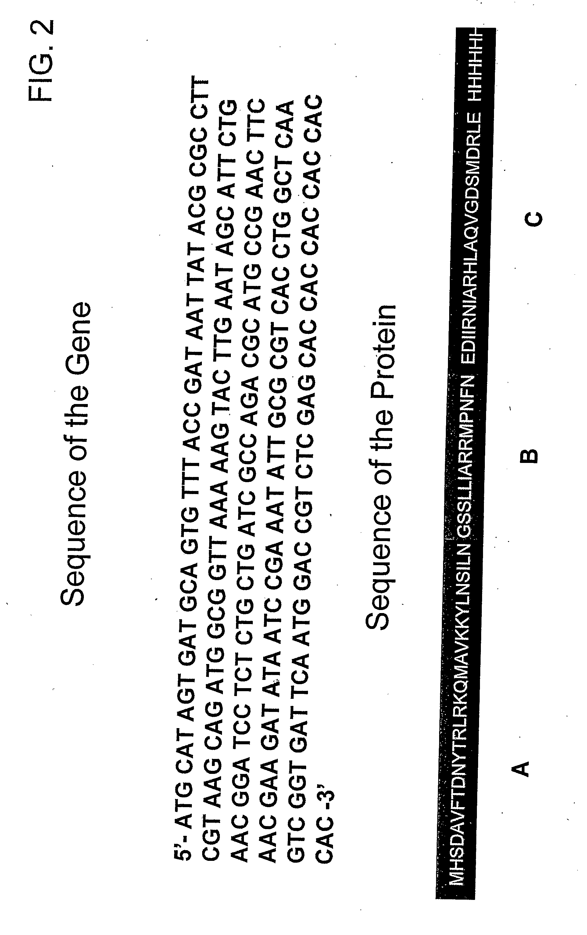
[0106]A plasmid for the expression of VIP-BH3 BID fusion protein under the control of the T7 promoter was constructed as shown in FIG. 3, which carried the fusion gene (SEQ ID: 5 of Table I). [Fishman et al., Biochem, 1994, 33, 6235-6243]. A cDNA encoding human VIP and BH3 BID / BAK was obtained by reverse transcription polymerase chain reaction (RT-PCR), using RNA isolated from HT-29 cells grown to logarithmic phase in presence of 10% Fetal Bovine Serum (FBS). Total RNA was isolated and was reverse transcribed into first strand cDNA, using the reverse transcription system (Promega, USA) under conditions recommended by the manufacturer. The cDNA was diluted to a total volume of 1 ml with 10 mM Tris-HCl pH 7.6, 1 mM EDTA and stored at 4° C.
[0107]An oligonucleotide encoding human BH3 BID / VIP was also synthesized chemically and purified by Poly acrylamide gel Electrophoresis (PAGE). The VIP encoding fragment was amplified using sense ...
example 2
EXAMPLE 2
Protein Expression and Purification
[0108]The p-fusion protein plasmid containing the fused coding sequences was transformed into E. coli strain BL21 (DE3) and the fusion protein (SEQ ID NO: 10) was expressed. A pellet of expressing cells was suspended in 10 mM Tris-HCl (pH 8.0) buffer containing Protease inhibitor cocktail (Sigma Chemical Co., St. Louis, Mo., USA), sonicated (six 45-s bursts) and centrifuged at 13,000 rpm for 10 min. The supernatant (soluble fraction) was removed and kept for analysis. The pellet was denatured in extraction buffer: 10 mM Tris-HCl-sodium chloride buffer (pH 8.0) containing 6M Guanidium chloride, and stirred for 30 min. at 4° C. The suspension was cleared by centrifugation at 13,000 rpm for 10 min. and the pellet discarded. The supernatant was incubated with NiNTA resin (Qiagen, Hilden, Germany) and loaded on to a mini column. After washing the resin with 5 column volumes of wash buffer, the bound protein was eluted as per manufacturer's in...
example 3
EXAMPLE 3
Western Blot Analysis
[0109]The electrophoresis samples were transferred onto nitro-cellulose and immunoblotted. The blot was probed with Anti-human VIP, anti-human BID, and anti-Hexahistidine tag at dilutions of 1:1500, 1:3000, 1:3000, respectively. Anti-human VIP, anti-human BID was obtained from Santacruz (Santa Cruz Biotechnology, Inc, Santa Cruz, Calif., USA.) and anti-Hexahistidine tag was obtained from Sigma (Sigma Chemical Co., St. Louis, Mo., USA.).
PUM
| Property | Measurement | Unit |
|---|---|---|
| Composition | aaaaa | aaaaa |
| Chemotherapeutic properties | aaaaa | aaaaa |
Abstract
Description
Claims
Application Information