Hypoxia-inducible protein 2 (hig 2), a diagnostic marker for clear cell renal cell carcinoma
a technology of hyperoxia and renal cell carcinoma, which is applied in the field of hyperoxia-inducible protein 2 (hig 2), can solve the problems of increasing the proliferation and tumor formation of renal cells, and achieve the effects of reducing the symptom of renal cell carcinoma, preventing rcc in subjects, and reducing the length
- Summary
- Abstract
- Description
- Claims
- Application Information
AI Technical Summary
Benefits of technology
Problems solved by technology
Method used
Image
Examples
example 1
General Methods
Clinical Samples
[0198]RCC tumor and surrounding normal kidney tissues used for cDNA microarray and immunohistochemical analyses were obtained with informed consent from 10 patients who underwent surgical resection. Clinicopathological data for each case is summarized in Table 1. All were diagnosed as being at stage I or II. The paired samples were snap-frozen in liquid nitrogen immediately after surgical resection and stored at −80° C. At least 90% of the viable cells in each specimen were identified as tumor cells; accordingly, contamination with non-cancerous cells was considered to be minimal. From among these tissues, cell populations with sufficient amount and quality of RNA were selected for the microarray analysis. Plasma samples from RCC and chronic glomerulonephritis patients (CGN) used for the ELISA analyses were also obtained with informed consent. In addition, 20 plasma samples from normal healthy volunteers were also obtained with informed consent.
TABLE 1...
example 2
Identification of Genes with a Clinically Relevant Expression Pattern in Renal Cell Carcinoma Cells
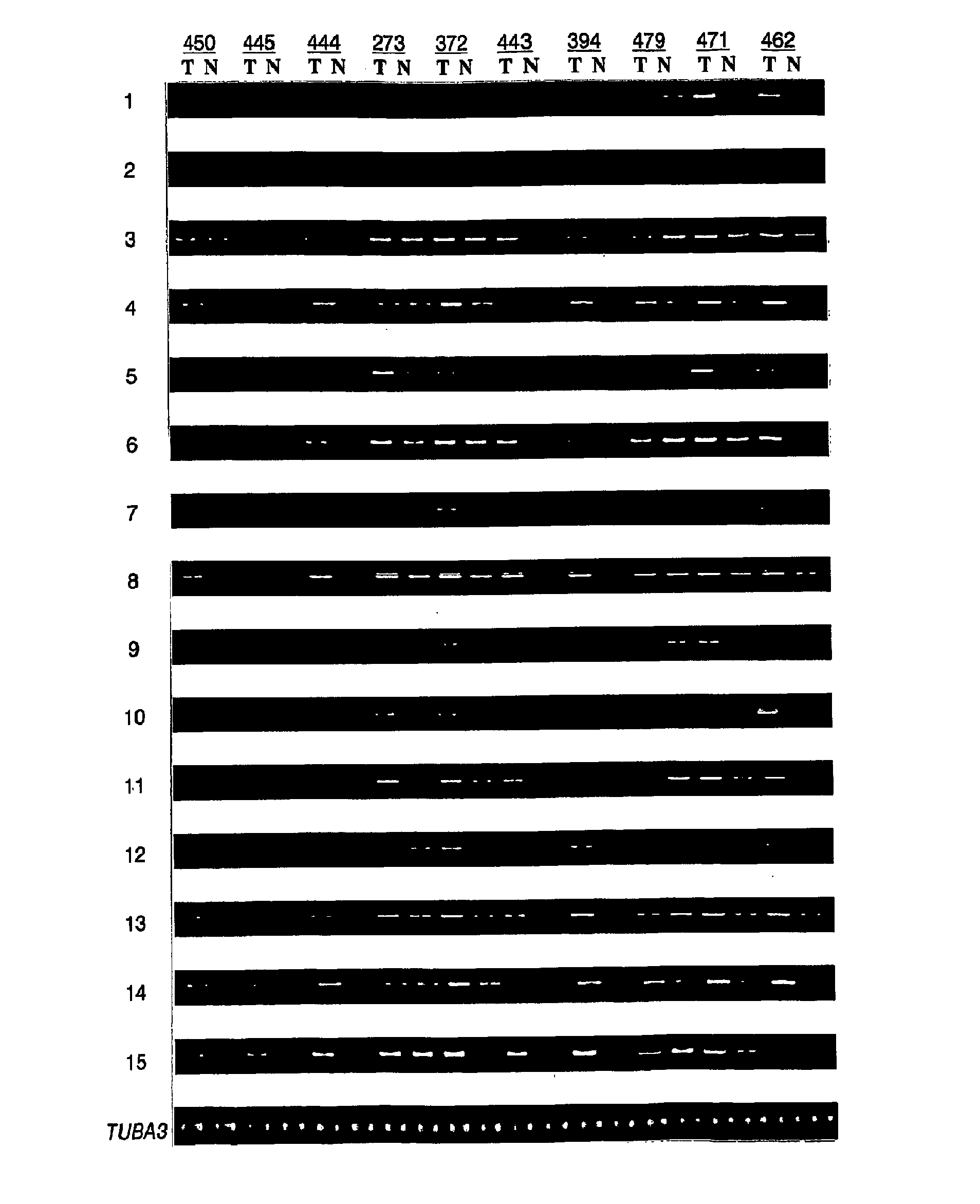
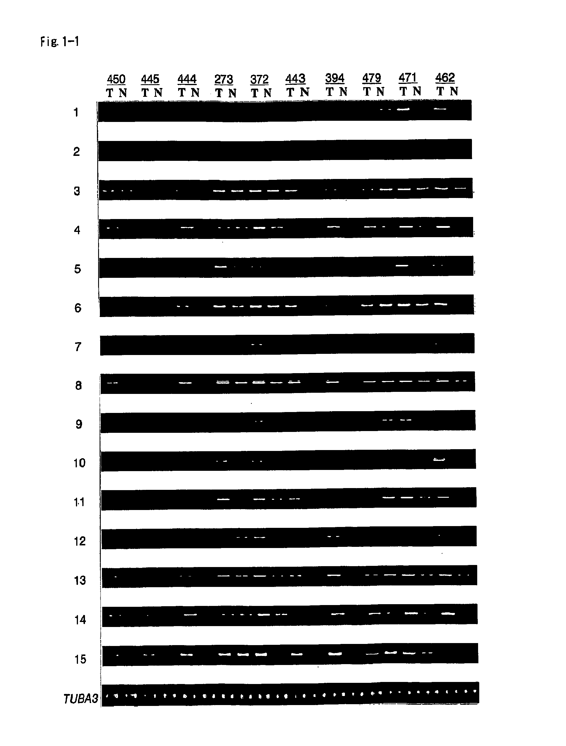
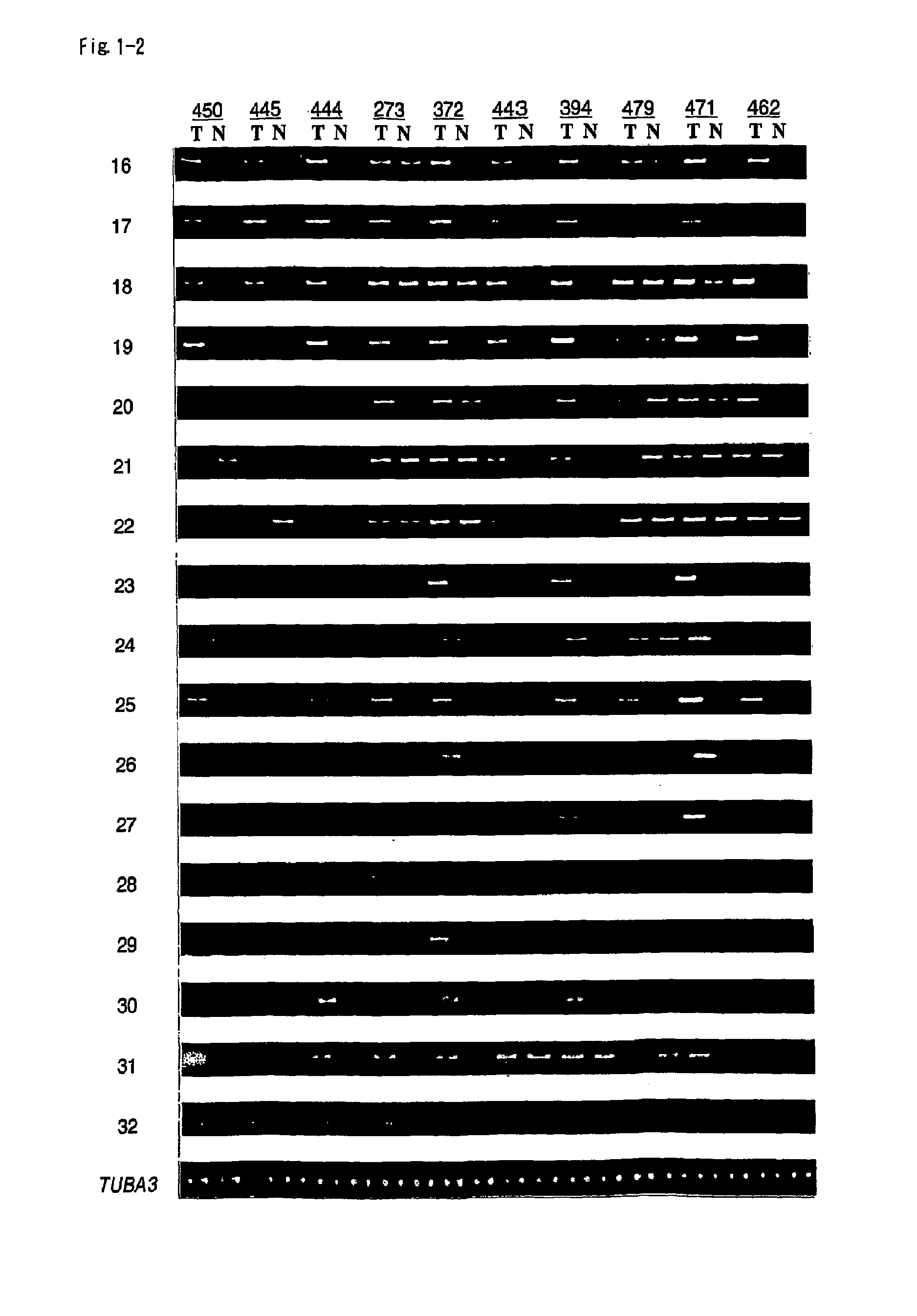
[0214]To clarify mechanisms underlying carcinogenesis of renal cell carcinoma, genes that were commonly up-regulated in this type of tumor were searched. A cDNA microarray analysis of more than 20,000 genes in 10 tumors identified 32 genes that were up-regulated in more than 50% of the cases examined (Table 3). Up-regulation of these 32 genes was reconfirmed by semi-quantitative RT-PCR analysis (FIG. 1).
TABLE 332 up-regulated genes in RCCRCCXAssignmentAccessionSymbolGene Name1AI088196ESTs2AA010816NM_031310ESTs, Weakly similar to S57447 HPBRII-7 protein[H. sapiens]3U62317NM_152299384D8-2hypothetical protein 384D8_64AA844401NM_138786ESTs, Weakly similar to T4S1_HUMANTRANSMEMBRANE 4 SUPERFAMILY,MEMBER 1 [H. sapiens]5AA156269NM_207310Homo sapiens mRNA; cDNA DKFZp434E2321(from clone DKFZp434E2321); partial cds6AI245600ESTs7AA844729ESTs8AA436623ESTs9AA903456NM_025085Homo sapiens cDNA FLJ1332...
example 3
Characterization of HIG2
[0215]HIG2 was up-regulated in 9 of 10 RCCs examined (FIG. 1, 2a). In contrast, HIG2 expression was hardly detectable in any of the other 29 organs examined, with the exception of fetal kidney (Saito-Hisaminato A., Katagiri T., Kakiuchi S., Nakamura T., Tsunoda T., Nakamura Y. Genome-wide profiling of gene expression in 29 normal human tissues with a cDNA microarray. DNA Res. 9, 35-45 (2002)). Multi-tissue northern (MTN) blot analysis also demonstrated that HIG2 expression was uniformly low level, confined to trachea, spinal cord and thyroid (FIG. 2b).
[0216]To further examine the expression level of HIG2 in tumors other than renal cell carcinomas, RT-PCR analysis was performed using cancer cell lines derived from various tissues. HIG2 was expressed strongly in RCC cell lines, but at very low levels in cancer cell lines derived from other organs, including colon cancer (CC), breast cancer (BC), and hepatocellular carcinoma (HCC) cell lines (FIG. 2c).
[0217]To c...
PUM
 Abstract
Description
Claims
Application Information
 - R&D
- Intellectual Property
- Life Sciences
- Materials
- Tech Scout
- Unparalleled Data Quality
- Higher Quality Content
- 60% Fewer Hallucinations
Browse by: Latest US Patents, China's latest patents, Technical Efficacy Thesaurus, Application Domain, Technology Topic, Popular Technical Reports.
© 2025 PatSnap. All rights reserved.Legal|Privacy policy|Modern Slavery Act Transparency Statement|Sitemap|About US| Contact US: help@patsnap.com


