Method for Automatic Segmentation of Images

a technology of automatic segmentation and image acquisition, applied in image data processing, character and pattern recognition, instruments, etc., can solve the problems of complicated training and variable intensity level of left ventricle, and achieve the effect of simplifying the segmentation of epicardial contour, accurate segmentation of papillary, and simplifying the detection of epicardial contour

Inactive Publication Date: 2010-08-26
SUNNYBROOK HEALTH SCI CENT
View PDF4 Cites 90 Cited by
  • Summary
  • Abstract
  • Description
  • Claims
  • Application Information

AI Technical Summary

Benefits of technology

[0017]It is an aspect of the invention to provide a method that simplifies the segmentation of the epicardial contour by mapping the pixels from Cartesian to approximately polar coordinates. By mapping the pixels from Cartesian to these “pseudo-polar” coordinates, the irregular, ring-shaped regions-of-interest are transformed to rectangular images or so-called “pseudo-polar maps”. In this way, the epicardial contour detection problem is simplified.
[0018]It is another aspect of the invention to provide a method for accurate segmentation of papillary and trabecular muscles, as well as endocardial and epicardial contours in all the phases. Clinical studies have employed different quantification methods for calculation of left ventricle volume, mass and ejection fraction by including or excluding papillary muscles and trabeculations in the ventricular cavity. Recent studies have shown that the papillary muscles and trabeculations have a significant impact on calculation of left ventricle volume and mass and ejection fraction; therefore, the method provides additional important options for daily

Problems solved by technology

Most model-based techniques require training with many manually drawn contours that are difficult to obtain.
In addition, the variability of the shap

Method used


Image

  • Method for Automatic Segmentation of Images
  • Method for Automatic Segmentation of Images
  • Method for Automatic Segmentation of Images

Examples


Embodiment Construction

Magnetic Resonance Imaging System

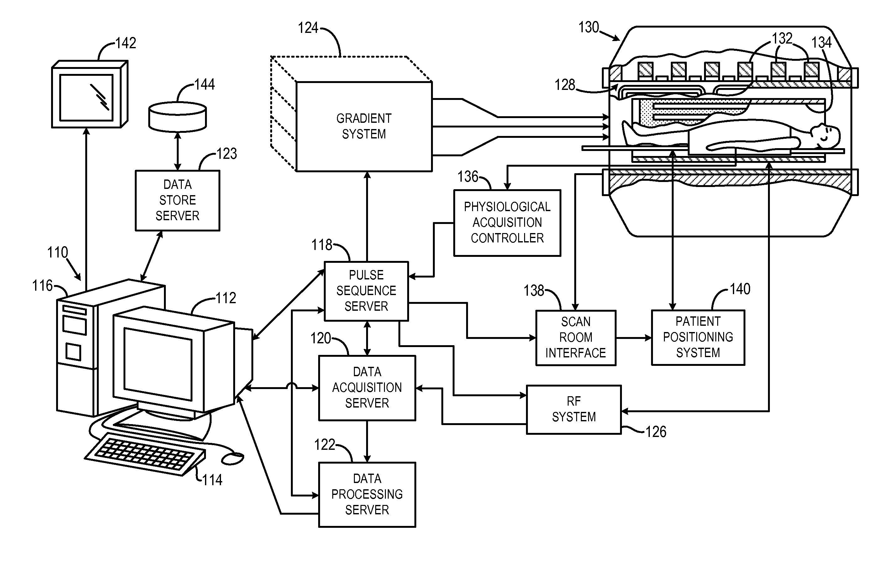
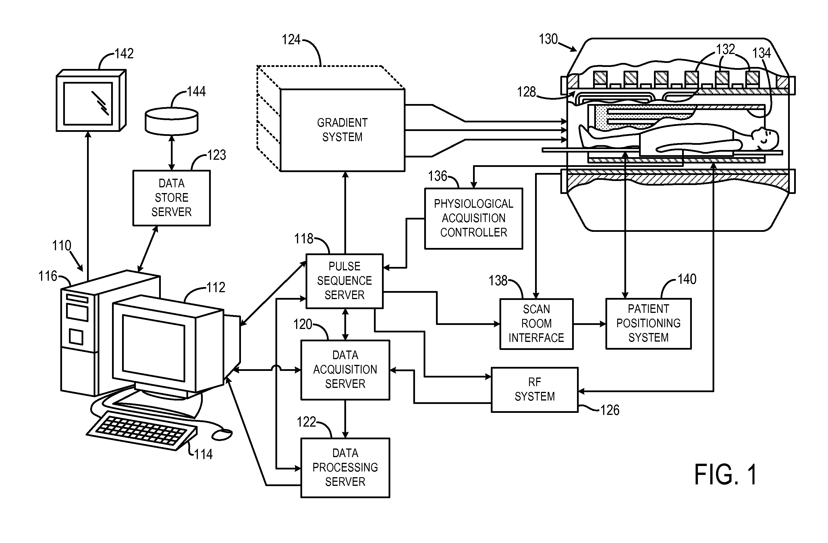
[0039]Referring particularly to FIG. 1, the preferred embodiment of the invention is employed in an MRI system. The MRI system includes a workstation 110 having a display 112 and a keyboard 114. The workstation 110 includes a processor 116 that is a commercially available programmable machine running a commercially available operating system. The workstation 110 provides the operator interface that enables scan prescriptions to be entered into the MRI system. The workstation 110 is coupled to four servers: a pulse sequence server 118; a data acquisition server 120; a data processing server 122, and a data store server 123. The workstation 110 and each server 118, 120, 122 and 123 are connected to communicate with each other.

[0040]The pulse sequence server 118 functions in response to instructions downloaded from the workstation 110 to operate a gradient system 124 and an RF system 126. Gradient waveforms necessary to perform the prescribed scan are p...


PUM


Abstract

A method for automatic left ventricle segmentation of cine short-axis magnetic resonance (MR) images that does not require manually drawn initial contours, trained statistical shape models, or gray-level appearance models is provided. More specifically, the method employs a roundness metric to automatically locate the left ventricle. Epicardial contour segmentation is simplified by mapping the pixels from Cartesian to approximately polar coordinates. Furthermore, region growing is utilized by distributing seed points around the endocardial contour to find the LV myocardium and, thus, the epicardial contour. This is a robust technique for images where the epicardial edge has poor contrast. A fast Fourier transform (FFT) is utilized to smooth both the determined endocardial and epicardial contours. In addition to determining endocardial and epicardial contours, the method also determines the contours of papillary muscles and trabeculations.

Description

CROSS-REFERENCE TO RELATED APPLICATIONS[0001]This application claims the benefit of U.S. Provisional patent application Ser. No. 61 / 154,556 filed on Feb. 23, 2009, and entitled “Method for Automatic Segmentation of Images.”BACKGROUND OF THE INVENTION[0002]The field of the invention is medical imaging methods and systems. More particularly, the invention relates to the segmentation of images acquired with a medical imaging system such as a magnetic resonance imaging (“MRI”) system, an x-ray computed tomography (“CT”) imaging system, or an ultrasound (“US”) imaging system.[0003]When a substance such as human tissue is subjected to a uniform magnetic field (polarizing field B0), the individual magnetic moments of the nuclei in the tissue attempt to align with this polarizing field, but precess about it in random order at their characteristic Larmor frequency. If the substance, or tissue, is subjected to a magnetic field (excitation field B1) that is in the x-y plane and that is near th...

Claims


Application Information

Patent Timeline
IPC IPC(8): G06K9/00
CPCG06T7/0083G06T2207/10081G06T2207/30048G06T2207/10132G06T2207/20168G06T2207/10088G06T7/12
Inventor LU, YINGLIRADAU, PERRYWRIGHT, GRAHAM A.
Owner SUNNYBROOK HEALTH SCI CENT
Why Eureka
Try Eureka