Eureka AIR delivers breakthrough ideas for toughest innovation challenges, trusted by R&D personnel around the world.

Automated fiber tracking of human brain white matter using diffusion tensor imaging

a technology of diffusion tensor imaging and human brain white matter, which is applied in the direction of image enhancement, measurement using nmr, instruments, etc., can solve the problems of inability to achieve the effect of unprocessed, inconvenient to operate, and insufficient processing value of unprocessed brain white matter

Active Publication Date: 2010-09-30
THE JOHN HOPKINS UNIV SCHOOL OF MEDICINE
View PDF8 Cites 30 Cited by
  • Summary
  • Abstract
  • Description
  • Claims
  • Application Information

AI Technical Summary

Benefits of technology

[0014]Another embodiment of the current invention provides a computer-readable medium comprising software, which when executed by a computer system, causes the computer system process at least one magnetic resonance image from a human subject showing a soft tissue region having at least one soft tissue substructure. The software includes: ...

Problems solved by technology

However, existing methods of fiber reconstruction require a substantial amount of anatomical knowledge of the brain white matter in order to extract only the tract regions of interest.
Because a straightforward reconstruction of all white matter yields a huge amount of fiber tracts entangled inside the brain, it does not have practical value in such an unprocessed form.
However, experienced operators with detailed knowledge of brain white matter anatomy may not be readily available, which may present a bottleneck in the application of fiber reconstruction technology in research and clinical studies.
Further, suboptimal quality of diffusion tensor imaging data may hamper the ability of even the experienced operators in using existing methods.

Method used


Image

  • Automated fiber tracking of human brain white matter using diffusion tensor imaging
  • Automated fiber tracking of human brain white matter using diffusion tensor imaging
  • Automated fiber tracking of human brain white matter using diffusion tensor imaging

Examples


Embodiment Construction

[0038]Some embodiments of the current invention are discussed in detail below. In describing embodiments, specific terminology is employed for the sake of clarity. However, the invention is not intended to be limited to the specific terminology so selected. A person skilled in the relevant art will recognize that other equivalent components can be employed and other methods developed without departing from the broad concepts of the current invention. All references cited herein are incorporated by reference as if each had been individually incorporated.

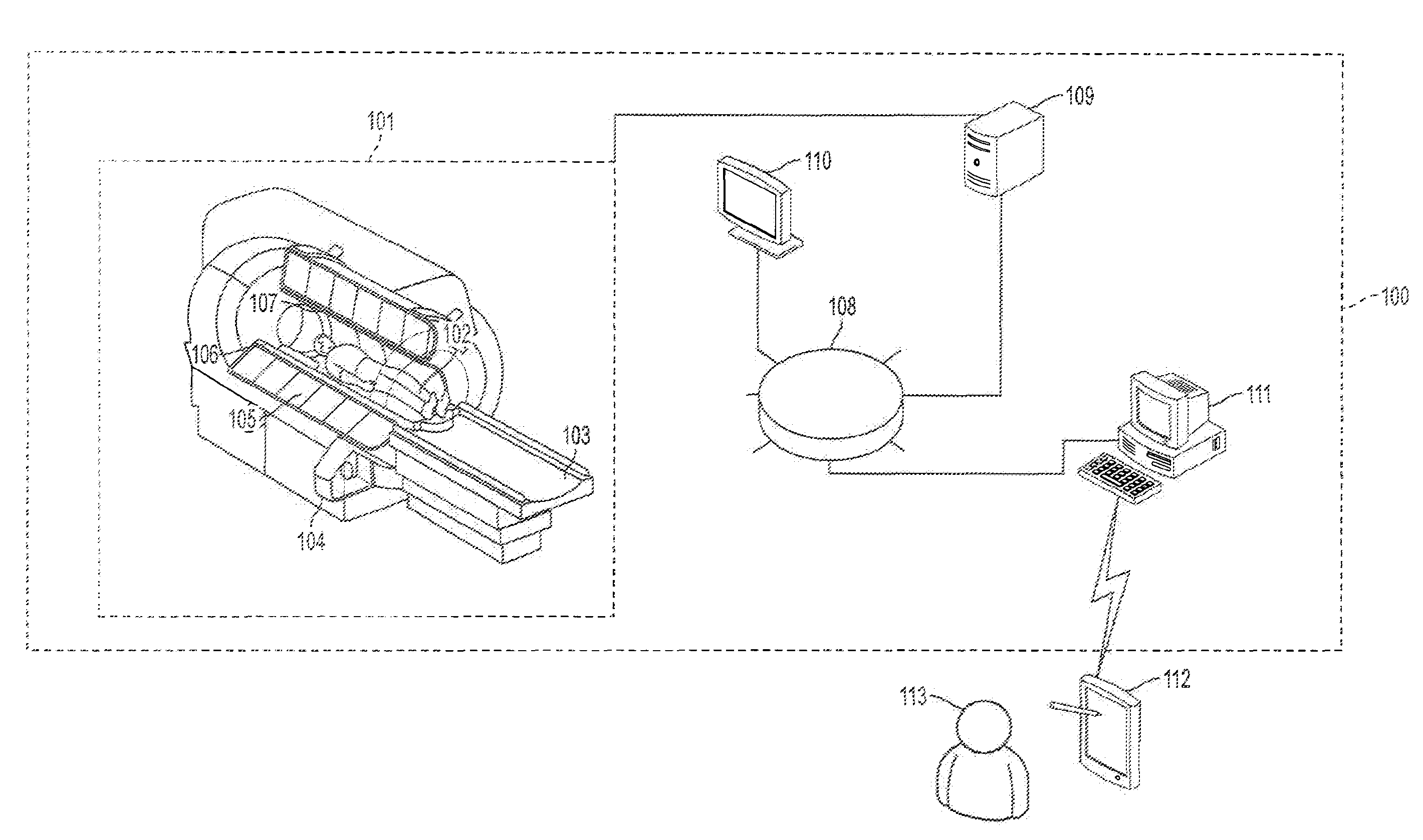
[0039]FIG. 1 is a schematic illustration of a magnetic resonance imaging (MRI) system 100 according to an embodiment of the current invention. The MRI system 100 has a magnetic resonance scanner 101, a data storage unit 108, and a signal processing unit 109. Magnetic resonance scanner 101 has a main magnet 105 providing a substantially uniform main magnetic field B0 for a subject 102 under observation on scanner bed 103, a gradient sy...


PUM


Abstract

A magnetic resonance imaging (MRI) system, comprising: a MRI scanner; a signal processing system in communication with the magnetic resonance imaging scanner to receive magnetic resonance (MR) signals for forming magnetic resonance images of a subject under observations; a data storage unit in communication with the signal processing system, wherein the data storage unit contains database data corresponding to a soft tissue region of the subject under observation. The database data includes information identifying at least one soft tissue substructure encompassed by the soft tissue region of the subject under observation. The signal processing system is adapted to process MR signals received from the MRI scanner to automatically identify at least one soft tissue substructure encompassed by the soft tissue region of the subject under observation.

Description

CROSS-REFERENCE OF RELATED APPLICATION[0001]This application claims priority to U.S. Provisional Application No. 61 / 009,913 filed Jan. 3, 2008, the entire contents of which are hereby incorporated by reference.[0002]The U.S. Government has a paid-up license in this invention and the right in limited circumstances to require the patent owner to license others on reasonable terms as provided for by the terms of Grant No.: R01AG20012 and P41RR15241, awarded by the National Institutes of Health.BACKGROUND[0003]1. Field of Invention[0004]The current invention relates to automated identification of soft tissue substructures in a soft tissue region of a human or animal subject in a non-invasive manner.[0005]2. Discussion of Related Art[0006]It is highly desirable to have automated identification of soft tissue substructures in soft tissue regions of a human and possibly animal subject in a non-invasive manner. For example, diagnosing brain pathologies associated with white matter brain tis...

Claims


Application Information

Patent Timeline
IPC IPC(8): G01R33/44
CPCA61B5/055G01R33/56341G06T2207/30016G06T7/0081G06T2207/10092G06T7/0032G06T7/344G06T7/11
Inventor MORI, SUSUMUZHANG, JIANGYANGHUA, KEGANG
Owner THE JOHN HOPKINS UNIV SCHOOL OF MEDICINE
Why Eureka
Eureka Blog
Learn More