Method for image data acquisition with a magnetic resonance device

a magnetic resonance device and image data technology, applied in the direction of magnetic measurements, measurement devices, instruments, etc., can solve the problems of difficult to repeat the acquisition, the number of artifacts known that can occur, and the difficulty of obtaining images

Active Publication Date: 2010-10-07
SIEMENS HEALTHCARE GMBH
View PDF14 Cites 15 Cited by
  • Summary
  • Abstract
  • Description
  • Claims
  • Application Information

AI Technical Summary

Benefits of technology

[0026]The method according to the invention can also be advantageously applied in regard to a technique what is known as the “phase oversampling method”. Phase oversampling means that a field of view is used that encompasses the entire subject so that there is no aliasing, with data outside of a narrower field of view being discarded after the acquisition. In the framework of the method according to the inve

Problems solved by technology

In the acquisition of image data with a magnetic resonance device, a number of artifacts are known that can occur when the image acquisition parameters are not ideally selected.
Such artifacts can occur in the partition direction even if no slice selection pulse is used, but is barely possible in practice.
Uncertainties and consequently measurements of images with aliasing in regions to be diagnosed can occur in the case of less well-trained personnel or less experienced personnel, such that the image acquisitions must be repeated.
This therefore leads to the situation that the user is not expected to input the field of vie

Method used


Image

  • Method for image data acquisition with a magnetic resonance device
  • Method for image data acquisition with a magnetic resonance device
  • Method for image data acquisition with a magnetic resonance device

Examples


Embodiment Construction

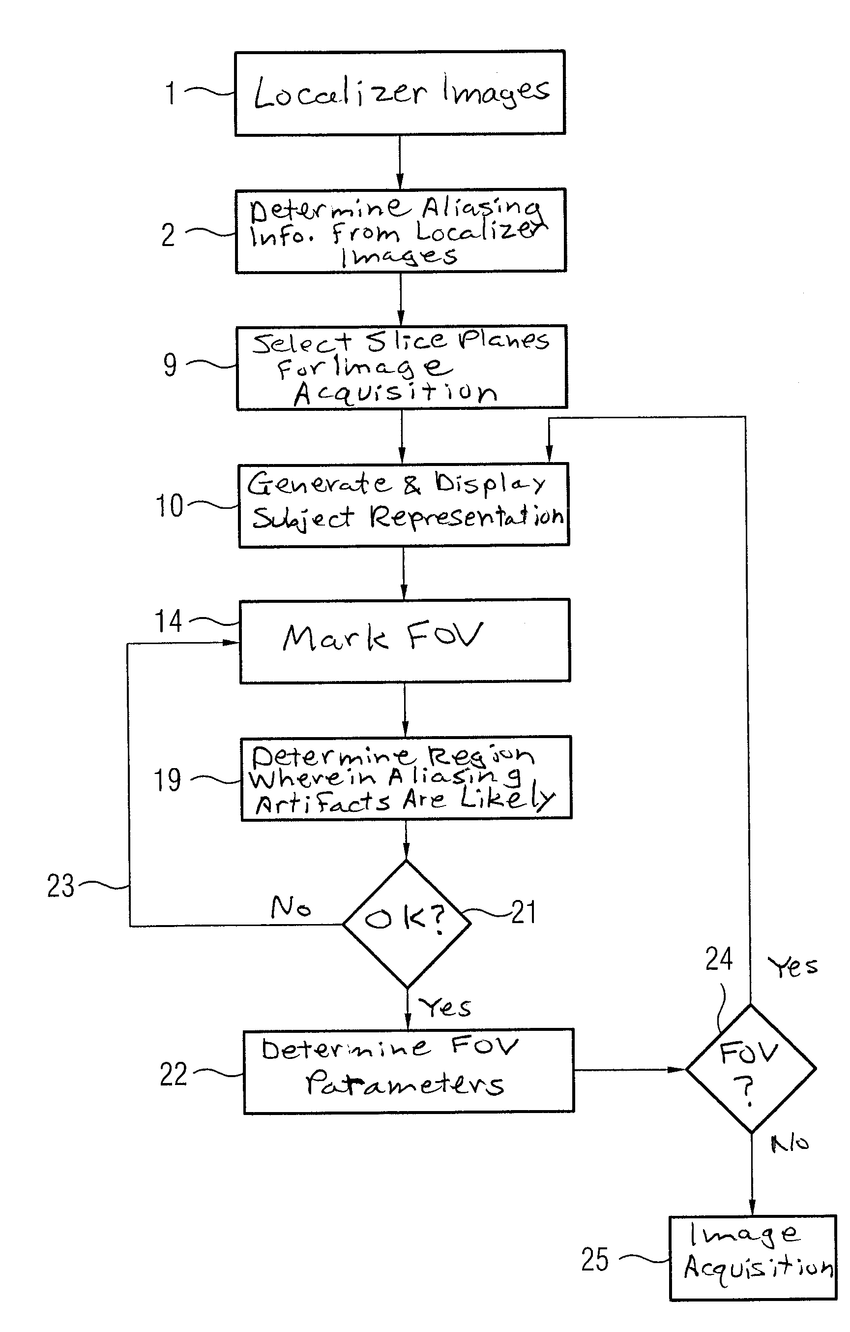
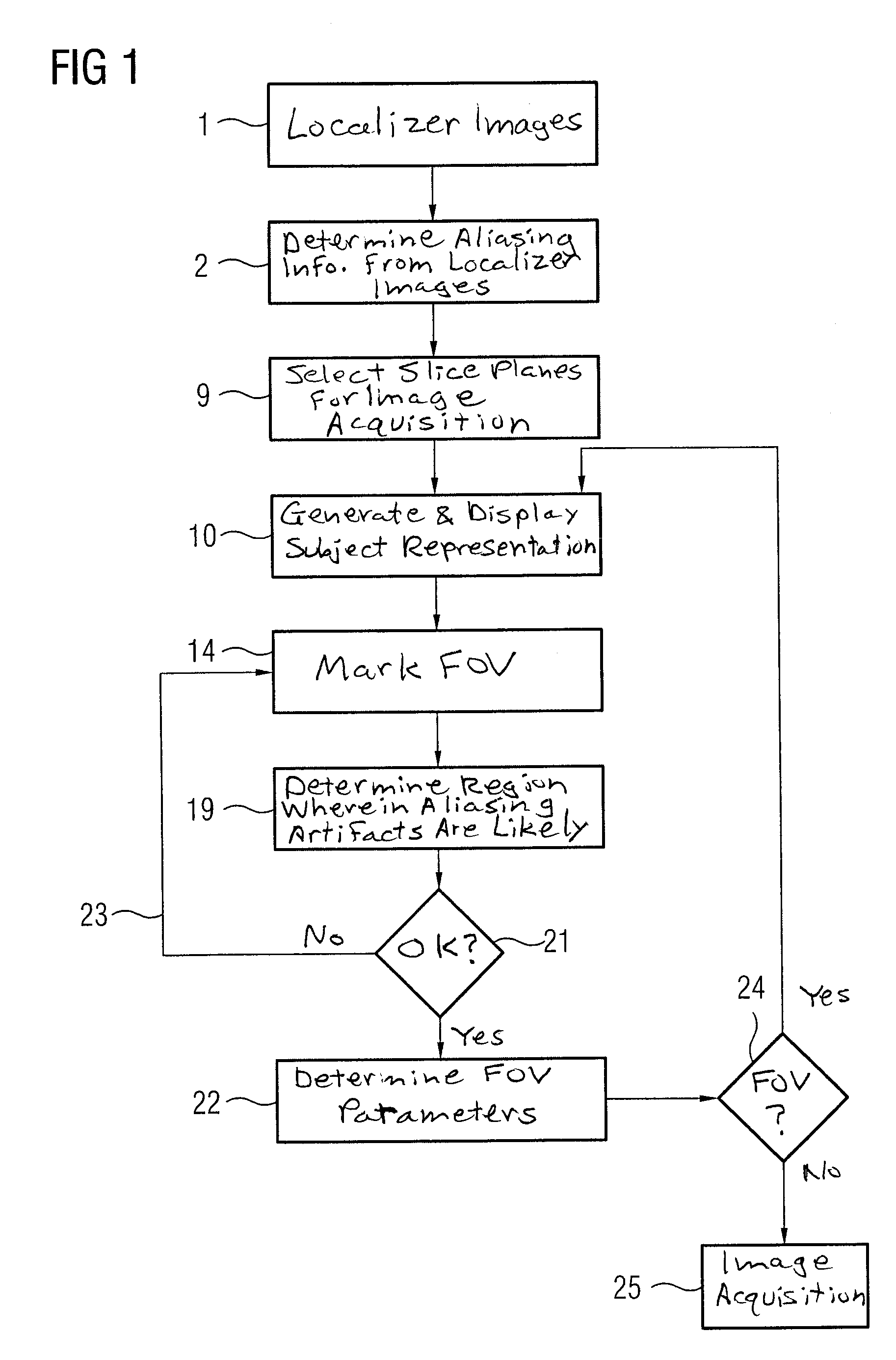
[0033]FIG. 1 shows a flowchart of a first embodiment of the method according to the invention. In a first step 1, a set of localizer exposures of a subject, which should later be the subject matter of an image acquisition with a magnetic resonance device, is initially generated, wherein in particular a region of interest should be acquired. Localizer exposures are already well known and do not need to be explained in detail herein.

[0034]In step 2 of the method according to the invention, aliasing information is determined from the localizer exposures. This information essentially reflects which dimensions the subject has and, correspondingly, how and where aliasing is to be expected. The aliasing information is thus subject-specific and is stored in the magnetic resonance device for the duration of the examination after its determination. In principle, two variants for the determination of said information are conceivable in the framework of the method according to the invention.

[00...


PUM


Abstract

In a method for image data acquisition of a region of interest in a subject with a magnetic resonance device, wherein, to establish the field of view, a minimal geometric shape encompassing the subject to be acquired and/or the surface of the subject is determined automatically from previously acquired localizer exposures as aliasing information for each exposure, at least one slice plane is determined for the acquisition of the region, and the phase coding direction and/or the extent of the field of view in the phase coding direction is determined for every slice plane using the aliasing information.

Description

BACKGROUND OF THE INVENTION[0001]1. Field of the Invention[0002]The present invention concerns a method for image data acquisition of a region of interest in a subject with a magnetic resonance device.[0003]2. Description of the Prior Art[0004]In the acquisition of image data with a magnetic resonance device, a number of artifacts are known that can occur when the image acquisition parameters are not ideally selected. In particular, an artifact known as aliasing can occur with regard to the determination of a field of view (FoV). This occurs when a field of view was selected in the phase coding direction so that a portion of the subject to be acquired (for example a patient body) is arranged outside of the field of view. Such artifacts can occur in the partition direction even if no slice selection pulse is used, but is barely possible in practice. The field of view in the phase coding direction (and possibly in the partition direction) is accordingly not selected based on the diagn...

Claims


Application Information

Patent Timeline
IPC IPC(8): G01V3/14
CPCG01R33/4833G01R33/543G01R33/5608
Inventor GRIMME, ANDREASGUEHRING, JENSJOLLY, MARIE-PIERESCHMIDT, MICHAELASPEIER, PETER
Owner SIEMENS HEALTHCARE GMBH
Why Eureka
Try Eureka