Animal Model Based on Targeted Apoptosis for the Study of Genetic Diseases Such as Parkinson's Disease
a genetic disease and animal model technology, applied in the field of animal models, can solve the problems of affecting the effectiveness of patients' movements, affecting the effect of patients' movements,
- Summary
- Abstract
- Description
- Claims
- Application Information
AI Technical Summary
Benefits of technology
Problems solved by technology
Method used
Image
Examples
example 2
Ablation of TIF-IA in Neural Stem Cells
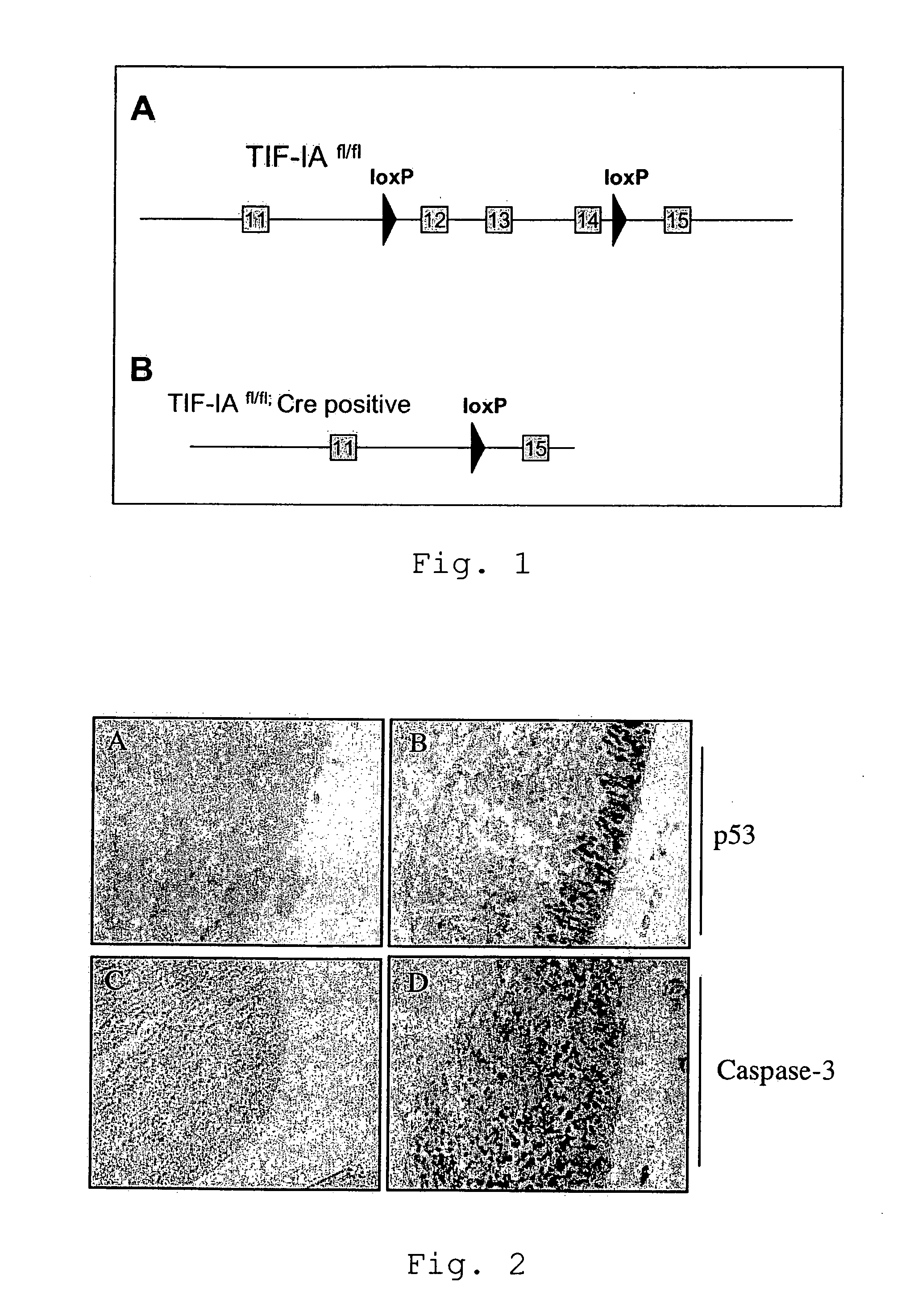
[0059]We induced the specific deletion of the TIF-IA gene in neural stem cells in the developing brain (TIF-IANesCre mutants). The NestinCre line has been previously described (Tronche et al., 1999). These mutants develop normally, but they are perinatally lethal; indeed, at birth they lack virtually all the neurons in the CNS. The deletion of the TIF-IA gene results in progressive loss of neural stem cells by activation of the apoptotic machinery mediated by p53 upregulation (FIG. 2).
example 3
Loss of the TIF-IA Gene Restricted to Dopaminergic Neurons
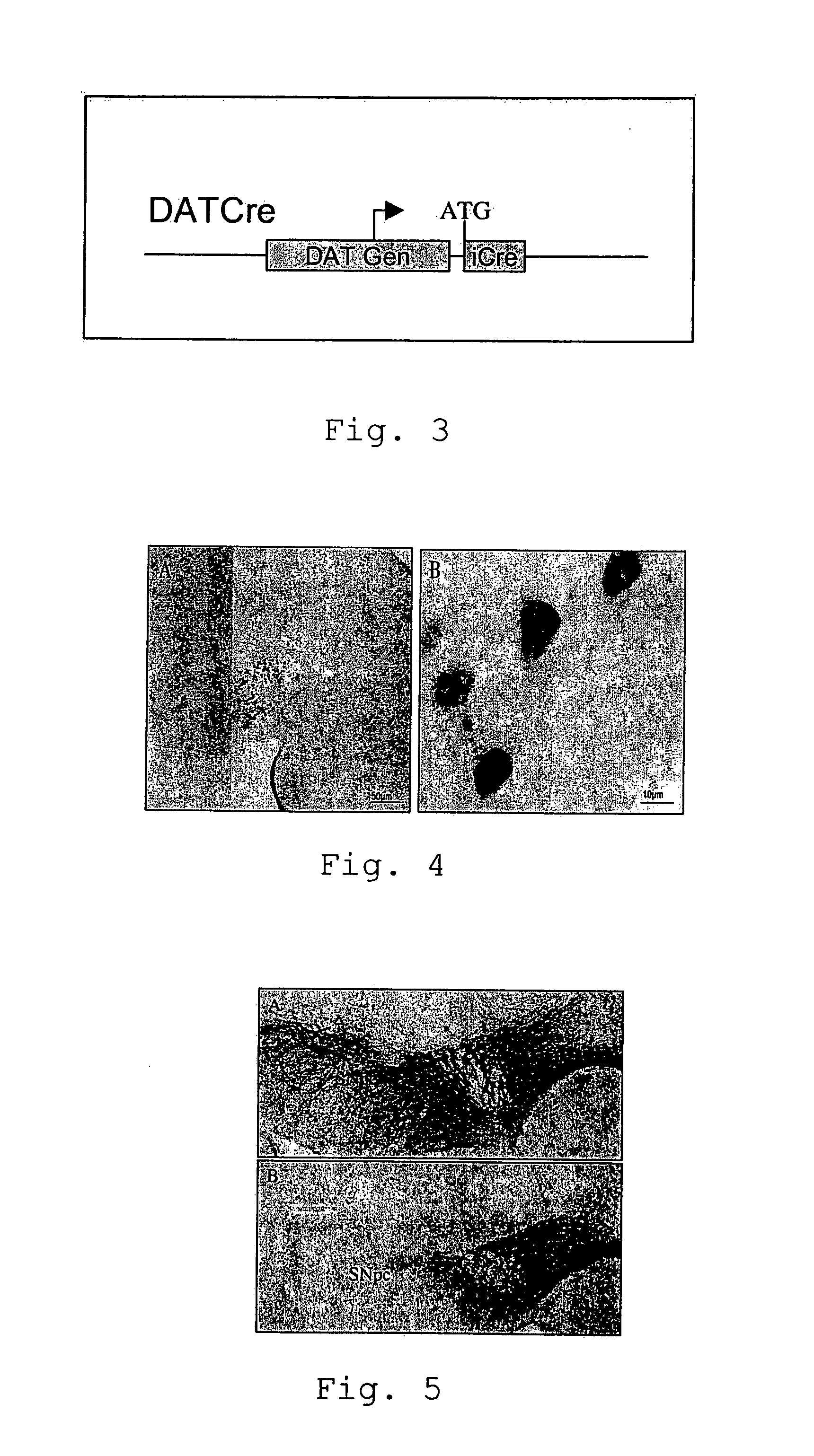
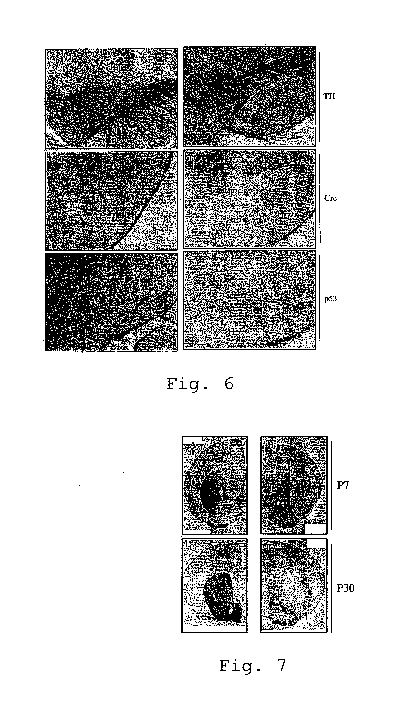
[0060]Parkinson's disease is a slowly progressing neurological disease affecting particular areas of the brain (the basal ganglions) which are involved in control of voluntary and involuntary movement. The slow degeneration of cells of the substantia nigra leads to a lack of the neurotransmitter dopamine. As a consequence, the classical symptoms of the disease are observed (main symptoms: akinesia, rigor, passive tremor and postural instability). As already mentioned earlier, so far, the experimental study of Parkinsons's disease was mainly based on model system using the neurotoxin MPTP (1-methyl-4-phenyl-1,2,3,6-tetrahydropyridine) which generates severe and irreversible symptoms in humans, primates and other mammals. However, in contrast to the MPTP based model which results in rapid apoptosis, the present mouse model shows slowly progressing degeneration of dopaminergic neurons by upregulation of the expression of the p53...
example 4
Immunohistochemical Analyses
[0065]Animals were sacrificed by use of CO2, brains were taken and incubated in cold 4% Paraformaldehyde (PFA) for 48 hours. The brains were stored in 0.4% PFA at 4° C. Coronal sections (50 μm) were prepared by use of a Vibratome (Leica).
[0066]Embryos were incubated in cold 4% PFA over night and then embedded in paraffin. Sagittal sections were prepared by use of a microtome (Leica). The sections were processed for immunohistochemical detection by using the VECTASTAIN ABC system (Vector Laboratories) and diaminobenzidine (Sigma) incubation. We used the following primary antibodies: anti-Cre, 1:3000 (Mantamadiotis et al, 2002); anti-caspase-3, 1:1000 (Cell Signaling, #9661); anti-p53, 1:1000 (anti-tyrosine-hydroxylase, 1:2000 (Chemicon, AB1542). In situ hybridization was performed as previously described (Nat. Neurosci. 2005 June; 8(6):759-67).
PUM
| Property | Measurement | Unit |
|---|---|---|
| Time | aaaaa | aaaaa |
| Therapeutic | aaaaa | aaaaa |
| Recombination enthalpy | aaaaa | aaaaa |
Abstract
Description
Claims
Application Information
 - R&D
- Intellectual Property
- Life Sciences
- Materials
- Tech Scout
- Unparalleled Data Quality
- Higher Quality Content
- 60% Fewer Hallucinations
Browse by: Latest US Patents, China's latest patents, Technical Efficacy Thesaurus, Application Domain, Technology Topic, Popular Technical Reports.
© 2025 PatSnap. All rights reserved.Legal|Privacy policy|Modern Slavery Act Transparency Statement|Sitemap|About US| Contact US: help@patsnap.com


