Marker-free chromosome screening

a chromosome and marker-free technology, applied in the field of chromosome analysis, can solve the problems of high disturbance resistance, time-consuming execution and analysis of methods, and high difficulty in detecting disturbances

Inactive Publication Date: 2011-03-10
HOCHSCHULE REUTLINGEN
View PDF6 Cites 7 Cited by
  • Summary
  • Abstract
  • Description
  • Claims
  • Application Information

AI Technical Summary

Problems solved by technology

A further disadvantage of the staining techniques is the time intensive nature of their execution and analysis which require special methodical skill and a precise knowledge of the banding of chromosomes.
Technically this method is extremely demanding and highly sensitive to disturbance by vibration, heat and static loading.
Furthermore it exhibits system generated faults so that rather than producing an image of the actual surface of the sample an image of the folding of the geometry of the needle point with the structure of the surface is produced.
Until now, in the prior art a chromosome analysis is carried out solely by disturbance-prone, elaborate atomic force techniques or by staining techniques which are time consuming and depend upon staining quality, degree of condensation and spreading of the chromosomes.

Method used


Image

  • Marker-free chromosome screening
  • Marker-free chromosome screening
  • Marker-free chromosome screening

Examples


examples

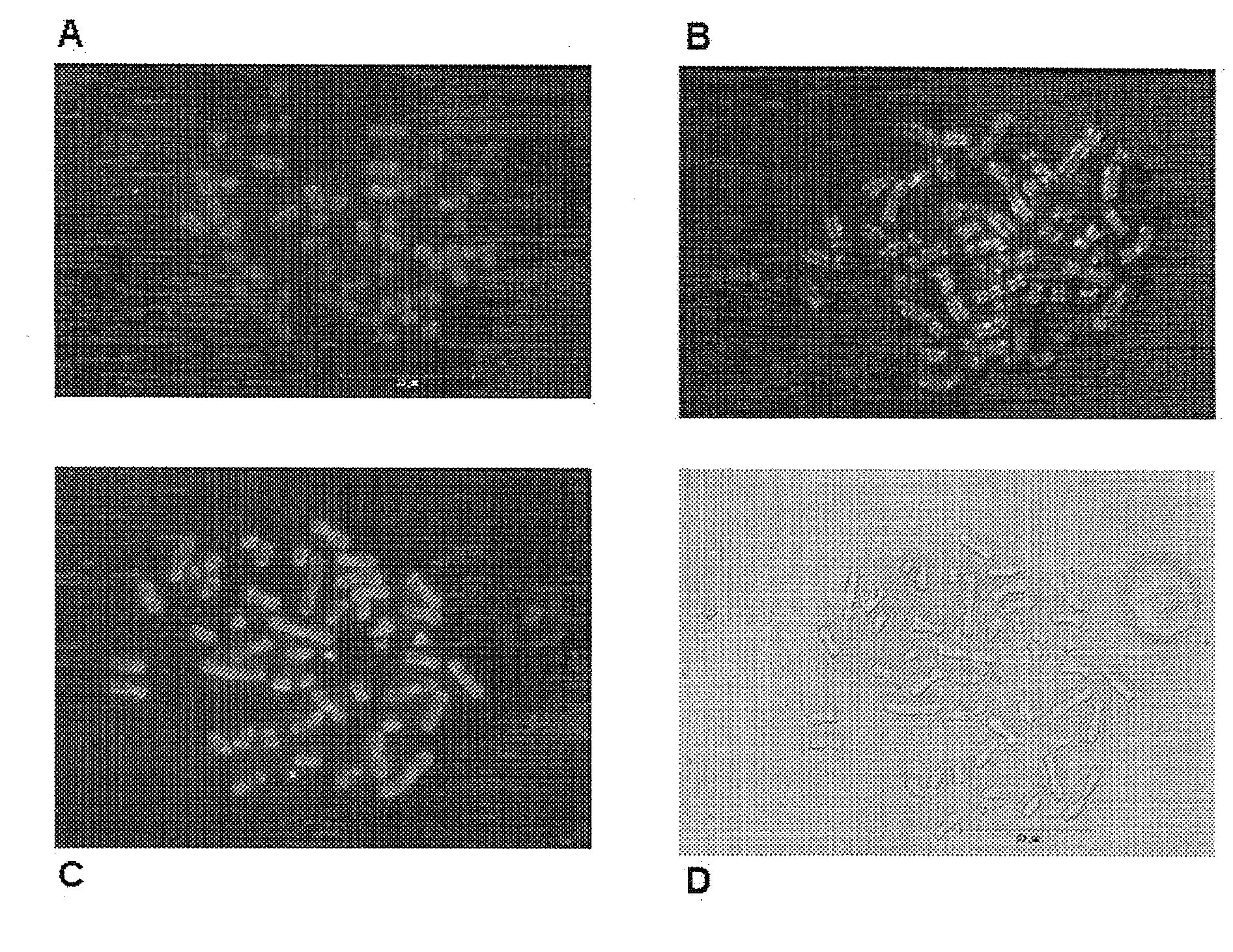
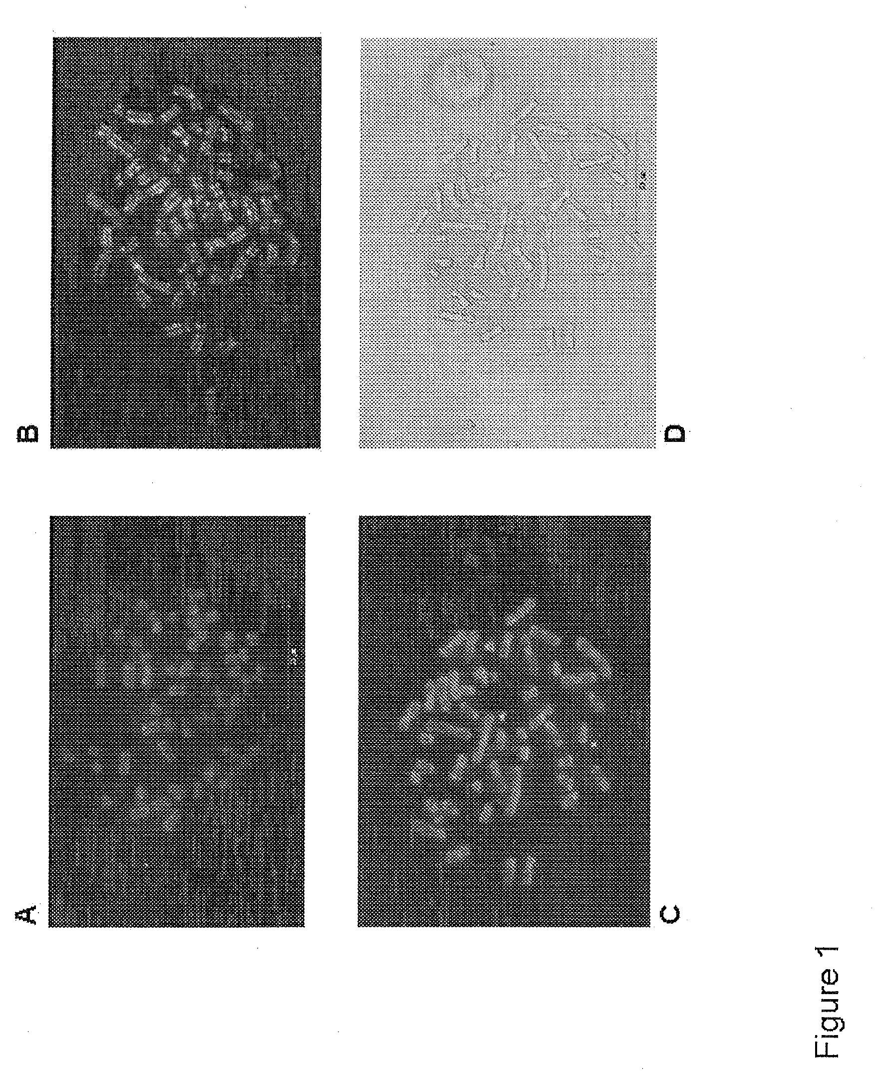
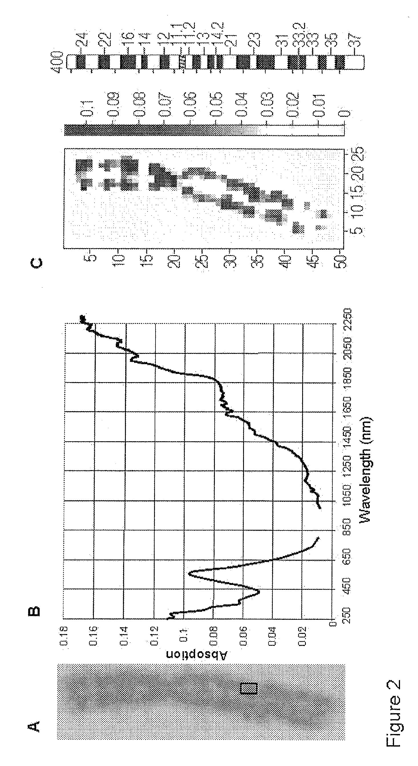
1. Preparation of the Metaphase Chromosomes

Blood lymphocytes taken from men and women whose karyotype had previously been shown to be normal 46, XX or XY were used for the manufacture of chromosome preparations. The sample of the blood culture, as well as the preparation of the blood culture, was carried out in accordance with the standardized methods and protocols of human cytogenetics.

2. Sample of Blood Culture

Venous blood was transferred to a small sterile tube wetted with heparin. The full blood was transferred under sterile conditions into culture flasks and mixed with chromosome medium B (Biochrom AG, Catalogue No. F5023). The T-lymphocytes were stimulated during a cultivation time of 72 hours at 37° C. in an incubator to undergo cell division.

3. Processing of the Blood Culture

Colcemid was added to the blood culture, incubated at 37° C. for 30 minutes and then centrifuged for 6-7 minutes. The supernatant was removed by suction down to the lower cone. The sediment was then shak...


PUM


Abstract

The present invention relates to a method for analyzing chromosomes by preparing a chromosome preparation, measuring at least one interference property of the chromosome preparation and characterizing at least one chromosome structure by way of the interference property. Also, the invention relates to the use of a near field microscope for analyzing unstained chromosomes.

Description

FIELD OF THE INVENTIONThe invention relates to a method for analyzing chromosomes and to the use of a near field microscope for the analysis of unstained chromosomes.BACKGROUND OF THE INVENTIONChromosomes are structures, which contain genes and, thereby, hereditary information. They contain DNA which is packed with proteins and are to be found in the nuclei of eukaryotic cells.In the middle of the 20th century techniques were developed in order to analyze the number and structure of cells which were metaphasing. In a metaphase preparation the chromosomes of a cell lie spread out beside one another, i.e. spread out on a microscope slide so that they can be counted under the microscope and compared with one another. In good preparations the individual chromosomes have the frequently displayed X-like shape.A classic staining technique to display metaphase chromosomes is the Giesma staining. After the chromosomes have been treated with the enzyme trypsin the chromatin complex of DNA and...

Claims


Application Information

Patent Timeline
Patent Type & Authority Applications(United States)
IPC IPC(8): G01J3/45G01Q60/00
CPCG01N21/45G01N21/31
Inventor KESSLER, RUDOLFMERZ, TOBIASREBNER, KARSTEN
Owner HOCHSCHULE REUTLINGEN
Why Eureka
Try Eureka