System for orientation assistance and display of an instrument in an object under examination particularly for use in human body

a technology of orientation assistance and display, which is applied in the field of endoscopes, can solve the problems of high radiation exposure of patients and operating room personnel, inability to display the entire length of the instrument, and only being poorly captured, and achieves improved distal end area of the endoscope, improved operator orientation, and reliable information.

Active Publication Date: 2011-03-24
KARL STORZ GMBH & CO KG
View PDF12 Cites 36 Cited by
  • Summary
  • Abstract
  • Description
  • Claims
  • Application Information

AI Technical Summary

Benefits of technology

[0016]In another preferred embodiment of the system, the at least one virtual image of the instrument, in particular of an endoscope, is projected onto a projection surface in the examination room. In this case the projection surface can in particular be a wall of an operating room and therefore inside the sterile zone, while the projection unit is positioned outside the sterile zone, set off from it by a glass partition. This makes efficient sterilization of the system possible, without affecting the improved orientation capability.
[0032]While advancing the instrument in the body, paying attention precisely either directly to the body of the patient as projected, or as merged in an extracorporeal body image of the patient captured on a monitor, the user thus receives visual information on the current placement / position of the instrument in the patient's body. The user thus receives a secure, rapid system, which causes significantly less radiation impact on the patient.

Problems solved by technology

The disadvantage, however, is that the entire length of the instrument is not displayed and can only be poorly captured.
The disadvantage of these processes is that in order to be able to construct an augmented-reality application, it is necessary at first, preoperatively, to provide an entire system, which is expensive as well as complex to operate, consisting of video camera, tracking devices, nuclear spin tomographs, support software, and so on.
In addition, the aforementioned methods have the great disadvantage that, before and sometime even during a single procedure, a number of x-ray images are required to produce the virtual image of the body interior by means of a computer tomograph (CT).
This means a high radiation exposure for the patient as well as for the operating room personnel.
In addition, in the event of an emergency intervention, there is often insufficient time to be able to provide the corresponding ED image data from computer tomography preoperatively.
The result is that the physician in these procedures is deprived of important information for the spatial orientation.

Method used


Image

  • System for orientation assistance and display of an instrument in an object under examination particularly for use in human body
  • System for orientation assistance and display of an instrument in an object under examination particularly for use in human body
  • System for orientation assistance and display of an instrument in an object under examination particularly for use in human body

Examples


Embodiment Construction

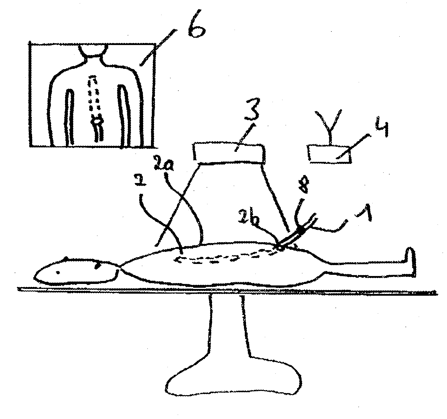
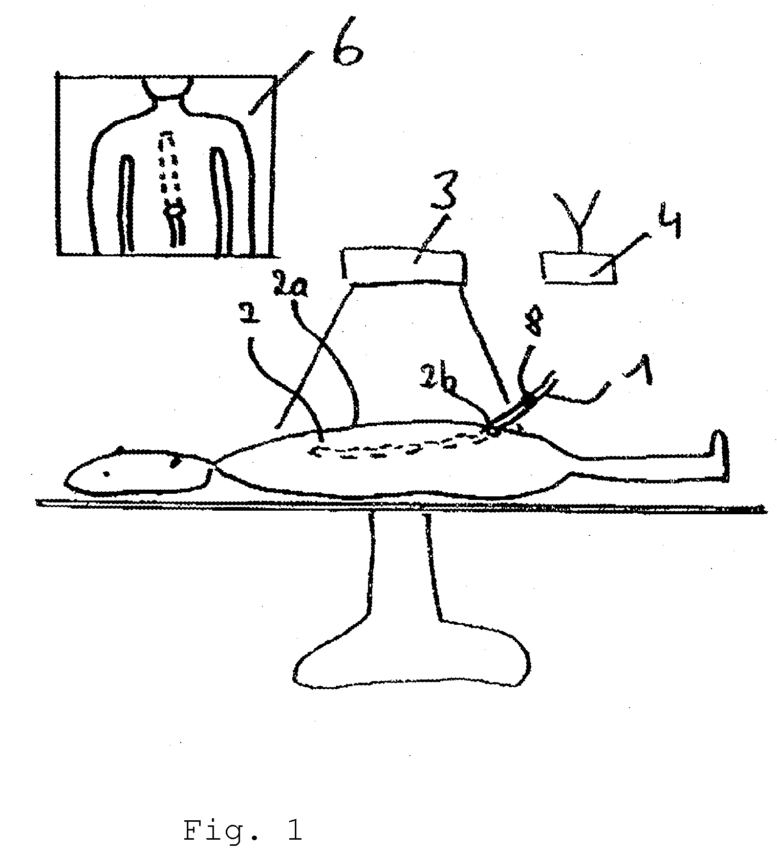
[0038]FIG. 1 shows an execution of the inventive system on the basis of a schematic display of a surgical situation in a body 2 of a patient. According to this embodiment, an endoscope 1 is inserted into a human body 2 in the area of the patient's navel. The endoscope extends in the interior of the body 2 from the navel to the area of the heart, where the distal end of the endoscope 1 comes to rest. It is precisely during the endoscopy process, that is, at the start of the insertion of the endoscope into a body opening (entry channel) and the advancing of the endoscope to the site of the occurrence of the illness, that the patient or the surface of the patient's body is captured by means of an extracorporeal video camera device 3. In the process, the video camera device 3 is positioned directly above the patient and is equipped with a corresponding lens, which makes it possible for the entire duration of the intervention to reliably capture the part of the surface of the patient tha...


PUM


Abstract

A system for orientation assistance and display of an instrument that is inserted or present in the natural or artificially produced hollow cavity (human, animal, object), and that is equipped with one or more sensor units. Multiple measurements of the 3D position of the instrument equipped with one or more sensor units are performed by positioning a measuring system, so that a precise orientation and positioning of the instrument in the body can be computed. The 3D position data are used to compute a virtual image of the instrument synchronously. The virtual images are then either projected directly in exact position onto the body surface of a person or combined in a body surface image (real video camera image of the patient) onto a monitor or superimposed (virtual or augmented reality). The system is especially appropriate for displaying for a user a medical instrument, such as a catheter or a rigid or flexible endoscope, in the body of a person in real time, extracorporeally and in correct position.

Description

CROSS-REFERENCE TO RELATED APPLICATIONS[0001]The present application claims priority of German patent application No. 10 2009 025 077.8 filed on Jun. 10, 2009.FIELD OF THE INVENTION[0002]The present invention relates to a system for orientation assistance and display of an instrument in an object under examination, in particular an endoscope for use in the human or animal body.BACKGROUND OF THE INVENTION[0003]There are already various processes and devices in the art that demonstrate the combination of minimally invasive endoscopic procedure methods with augmented reality and image data supported techniques (R. Mösges, Die Methodik computergestützten Operierens. Habilitationsschrift, 1992, Hochschule Ascher).[0004]Thus the three-dimensional display of the organ systems (individual internal patient anatomy), based first on pre-operatively acquired image data of a 3D-CT or MRT scanner, is ascertained and viewed on a monitor. The position and orientation, for instance, of an endoscope ...

Claims


Application Information

Patent Timeline
Patent Type & Authority Applications(United States)
IPC IPC(8): H04N7/18
CPCA61B19/52A61B19/5212A61B2019/5293A61B2019/5261A61B2019/5289A61B2019/5251A61B90/36A61B2090/366A61B2090/364A61B90/361A61B2034/2051A61B2034/2061
Inventor SOLER, LUCMARESCAUX, JACQUES FRANCOIS BERNARDNICOLAU, STEPHANEIRION, KLAUS-MARTIN
Owner KARL STORZ GMBH & CO KG
Why Eureka
Try Eureka