Cancer Detection Methods and Reagents

a technology of immunological reagents and reagents, applied in the direction of snake antigen ingredients, instruments, viruses, etc., can solve the problems of insufficient detection accuracy of commercial antibodies used in standard insufficient detection accuracy and insufficient sensitivity of commercial antibodies used in standard tests to detect antigens, etc., to achieve the effect of improving the individual sensitivity of each assay

Inactive Publication Date: 2011-04-14
ONCIMMUNE
View PDF50 Cites 25 Cited by
  • Summary
  • Abstract
  • Description
  • Claims
  • Application Information

AI Technical Summary

Benefits of technology

[0013]Known serum assays, are not optimised for the detection of auto-antibodies, particularly for MUC1. Accordingly, in addition to providing more sensitive assays for cancer diagnosis and prognosis as well as identification of suitable therapies, the present invention comprises a novel means of protein

Problems solved by technology

In the past the direct detection of cancer-associated proteins has advantageously been used in routine tests for the diagnosis of cancer but, unfortunately, these assays have many limitations.
In particular, commercial antibodies available for use in standard tests to detect antigen are usually not sensitive enough to detect the low levels of cancer-associated proteins that are found at the very early stages of the disease, for example in asymptomatic patients, when a treatment would be the most effective.
In addition, most commercial antibodies are not specific for modified forms of cancer-associated markers and cross-react with wild-type forms of these proteins.
As a consequence, they are only useful for detecting substantial increases in serum levels of canc

Method used


Image

  • Cancer Detection Methods and Reagents
  • Cancer Detection Methods and Reagents
  • Cancer Detection Methods and Reagents

Examples


example 1

Isolation of ABC MUC1 from Advanced Breast Cancer Patients

Method

[0222]ABC MUC1 was purified from pooled sera taken from 20 patients with advanced breast cancer using immunoaffinity chromatography as follows:

[0223]The mouse monoclonal anti-MUC1 antibody B55 (also known as NCRC 11 and described by Ellis et al. (1984) Histopathology. 8: 501-516 and in International patent application No. WO 89 / 01153) was conjugated to CNBr-sepharose beads. Pooled sera from patients diagnosed with advanced breast cancer was diluted 1 / 10 in phosphate buffered saline (PBS) and then incubated with the antibody conjugated sepharose beads (25 ml diluted sera to 1 ml packed volume of beads) overnight at 4° C. with rolling. The beads were then packed by centrifugation and the supernatant removed. In order to wash away unbound serum components the beads were resuspended in PBS, rolled for 10 minutes, packed by centrifugation and the supernatant removed. This washing sequence was repeated 5 times (or until A28On...

example 2

Immunological Characterisation of ABC MUC1 Isolated from the Serum of Patients with Advanced Breast Cancer

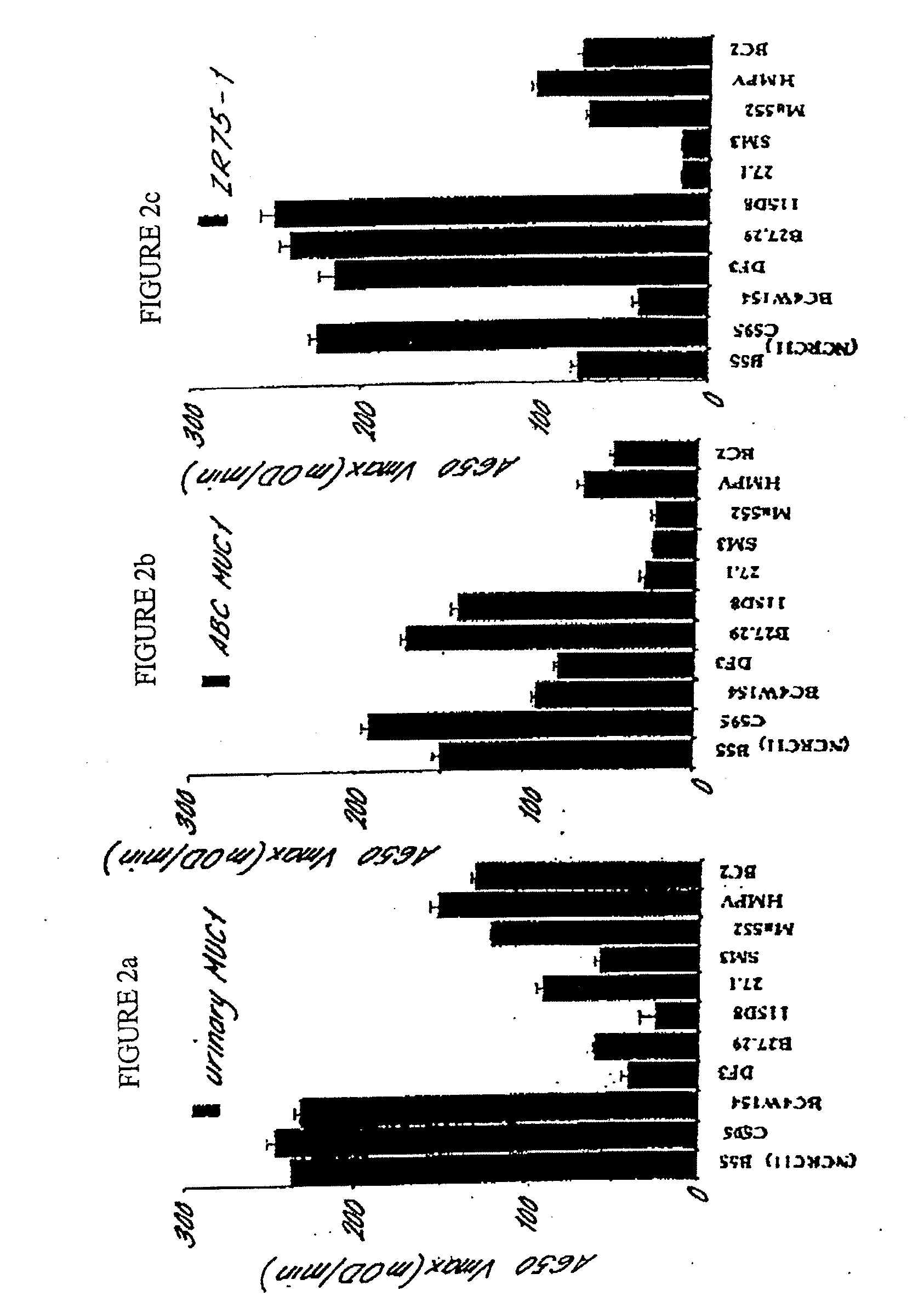
[0224]ABC MUC1 isolated from the serum of at least 20 patients with advanced breast cancer according to the procedure described in Example 1 can be distinguished from MUC1 isolated from the urine of normal human subjects (normal human urinary MUC 1) on the basis of altered affinity for the following mouse monoclonal anti-MUC1 antibodies:

B55 (NCRC 11)C595BC4W154Obtainable from Hybritech, IncDF3Obtainable from CentocorB27.29Obtainable from Biomira, Inc115D8Obtainable from Centocor27.1Obtainable from Austin Research InstituteSM3Obtainable from the Imperial Cancer ResearchFundMa552Obtainable from CanAgHMPVObtainable from Austin Research InstituteBC2Obtainable from Austin Research Institute

[0225]Normal urinary MUC1 is available from Dr M. R. Price, Cancer Research Laboratories, The University of Nottingham, University Park, Nottingham. NG7 2RD, United Kingdom.

[0226]The affinity of ea...

example 3

Cloning of Biotinylated p53

Method

[0227]Commercially available cDNA for p53 (E. coli clone pBH53, deposited in the American Type Culture Collection under accession number 79110) was cloned into the PinPoint™ plasmid vector (Promega Corporation, Madison Wis., USA) using standard molecular biology techniques. The PinPoint™ vector is designed to facilitate the production of fusion proteins comprising a biotinylation domain (consisting of a fragment of a biotin carboxylase carrier protein) fused N-terminally to the target protein of interest. Care was therefore taken during the cloning procedure to ensure that the reading frame of p53 was maintained in the fusion protein. Procedures for cloning in PinPoint™ vectors are described in detail in the Promega Protocols and Applications Guide obtainable from Promega Corporation, Madison Wis., USA.

[0228]Fusion proteins expressed from the PinPoint™ vector in E. coli are biotinylated by an enzyme system of the E. coli host cells and may therefore ...


PUM

PropertyMeasurementUnit
Volumeaaaaaaaaaa
Fractionaaaaaaaaaa
Fractionaaaaaaaaaa

Abstract

The present invention comprises methods and compositions for detecting cancer in an individual comprising autoantibodies to cancer-associated antigens. Specifically, the present invention comprises methods and compositions for detecting autoantibodies to cancer-associated antigen in a bodily fluid as well as use of said autoantibodies as a means to detect the presence of cancer-associated antigens.

Description

RELATED APPLICATIONS[0001]This application is a continuation-in-part of U.S. Patent Application Ser. No. 09 / 881,339, filed Jun. 14, 2001, which claims the benefit of U.S. Provisional Application Ser. No 60 / 211,886 filed Jun. 14, 2000, the disclosure of which is incorporated by reference herein in its entirety.FIELD OF THE INVENTION[0002]This application relates to immunological reagents and methods for detecting and treating cancer in an animal, most preferably a human. In particular, the invention relates to compositions and methods for detecting or quantitatively measuring the immune response of a mammal to circulating tumour markers or tumour markers expressed on the surface of tumour cells, also to tumour marker antigens for use in these methods and to kits for performing the methods. The present invention also relates to highly sensitive and specific compositions and methods for detecting the presence of cancer or tumour marker proteins in the bodily fluids of a mammal, to auto...

Claims


Application Information

Patent Timeline
IPC IPC(8): A61K39/00A61K39/12A61P35/00A61P37/04G01N33/574
CPCG01N33/57484A61P35/00A61P37/04
Inventor ROBERTSON, JOHN FORSYTH RUSSELLGRAVES, CATHERINE ROSAMUND LOUISEPRICE, MICHAEL RAWLINGPRICE, FRANCES MARGARET
Owner ONCIMMUNE
Why Eureka
Try Eureka