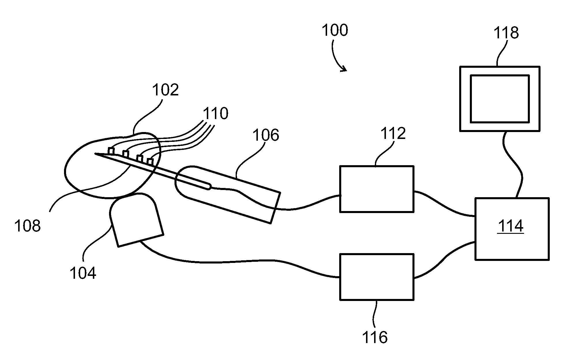
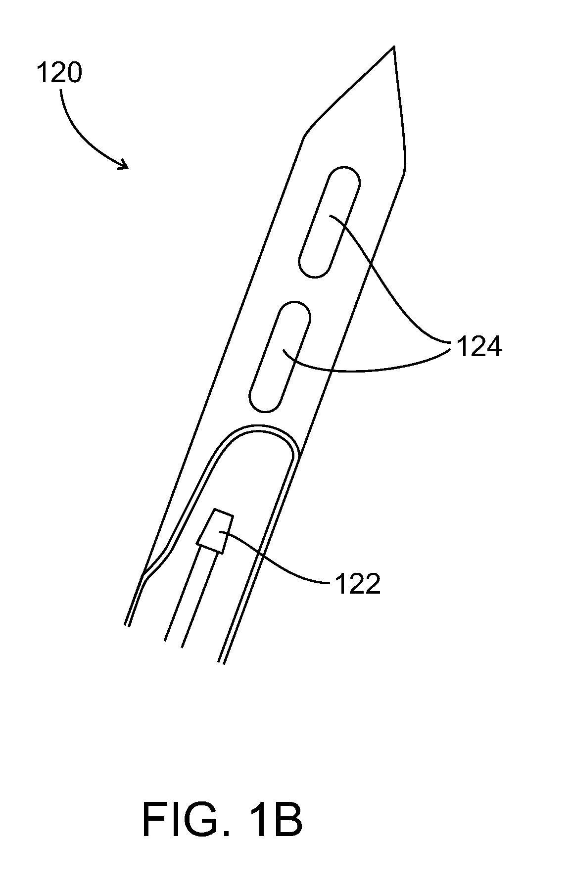
Method and system for tissue recognition
a tissue recognition and tissue technology, applied in the field of tissue recognition, can solve the problems of large mri equipment, limited resolution and noise level of x-ray images, and inability to distinguish soft tissue well between some types of soft tissue,
- Summary
- Abstract
- Description
- Claims
- Application Information
AI Technical Summary
Benefits of technology
Problems solved by technology
Method used
Image


Examples
examples
[0160]Reference is now made to the following examples, which together with the above descriptions, illustrate some embodiments of the invention in a non limiting fashion.
[0161]To demonstrate the feasibility of using different ultrasound heating rates to distinguish between otherwise similar types of tissue, in vitro tests were done in which a small piece of chicken liver was embedded in the surface of a larger piece of beef liver, or a small piece of beef liver was embedded in the surface of a larger piece of chicken liver. The entire sample was then heated uniformly by ultrasound at either 1 MHz or 3 MHz, at a time-averaged intensity lower than 720 mW / cm2, for a long enough duration, which ranged from about 1 minute to about 6 minutes, so that its temperature rose by a few degrees Celsius. An infrared camera was used to image the surface of the sample, and to measure the temperature of the two types of liver. In all cases, it was found that the beef liver heated substantially faste...
PUM
 Abstract
Description
Claims
Application Information
 - R&D
- Intellectual Property
- Life Sciences
- Materials
- Tech Scout
- Unparalleled Data Quality
- Higher Quality Content
- 60% Fewer Hallucinations
Browse by: Latest US Patents, China's latest patents, Technical Efficacy Thesaurus, Application Domain, Technology Topic, Popular Technical Reports.
© 2025 PatSnap. All rights reserved.Legal|Privacy policy|Modern Slavery Act Transparency Statement|Sitemap|About US| Contact US: help@patsnap.com