Method of discovering and analyzing secreted biomarkers of disease from solid tissue

a biomarker and solid tissue technology, applied in the field of biological sample composition and method, can solve the problems of few known biomarkers that are detected in tissue, can not be detected and assayed, and can affect the tissue and cellular structure of the biological sample, and affect the protein cross-linking. , the effect of disrupting the tissue and cellular structur

Inactive Publication Date: 2011-05-05
EXPRESSION PATHOLOGY
View PDF2 Cites 12 Cited by
  • Summary
  • Abstract
  • Description
  • Claims
  • Application Information

AI Technical Summary

Problems solved by technology

These methods can be disruptive and potentially harmful to the patient.
However, very few known biomarkers that are detected in tissue can be detected and assayed in bodily fluids such as serum and urine.

Method used


Image

  • Method of discovering and analyzing secreted biomarkers of disease from solid tissue
  • Method of discovering and analyzing secreted biomarkers of disease from solid tissue

Examples


example 1

[0033]Table 1 is a list of proteins found commonly expressed both in uterine fluid as discovered from a literature survey and in soluble protein lysate from the glandular lumen shown in FIG. 1. Of note is that all of the proteins previously shown to be secreted by endometrial cells through a survey of the published literature between 1986 and 2003 are also identified in the protein profile of the endometrial glandular lumen.

example 2

[0034]Table 2 shows results from an analysis that determined what types of proteins are present in the glandular lumen protein profile and compares that to the types of proteins from a protein profile determined by mass spectrometry of protein from epithelial cells. Note that the highest percentage of protein in the glandular lumen sample represents proteins that are extracellular associated and membrane bound, whereas a smaller percentage of the total protein is of intracellular origin. The highest percentage of protein in the epithelial cell sample is of intracellular origin. These results indicate that the lumen does in fact contain predominantly secreted extracellular protein and that this invention is advantageous for specifically identifying secreted protein by analysis of protein present within organ and glandular lumens.

example 3

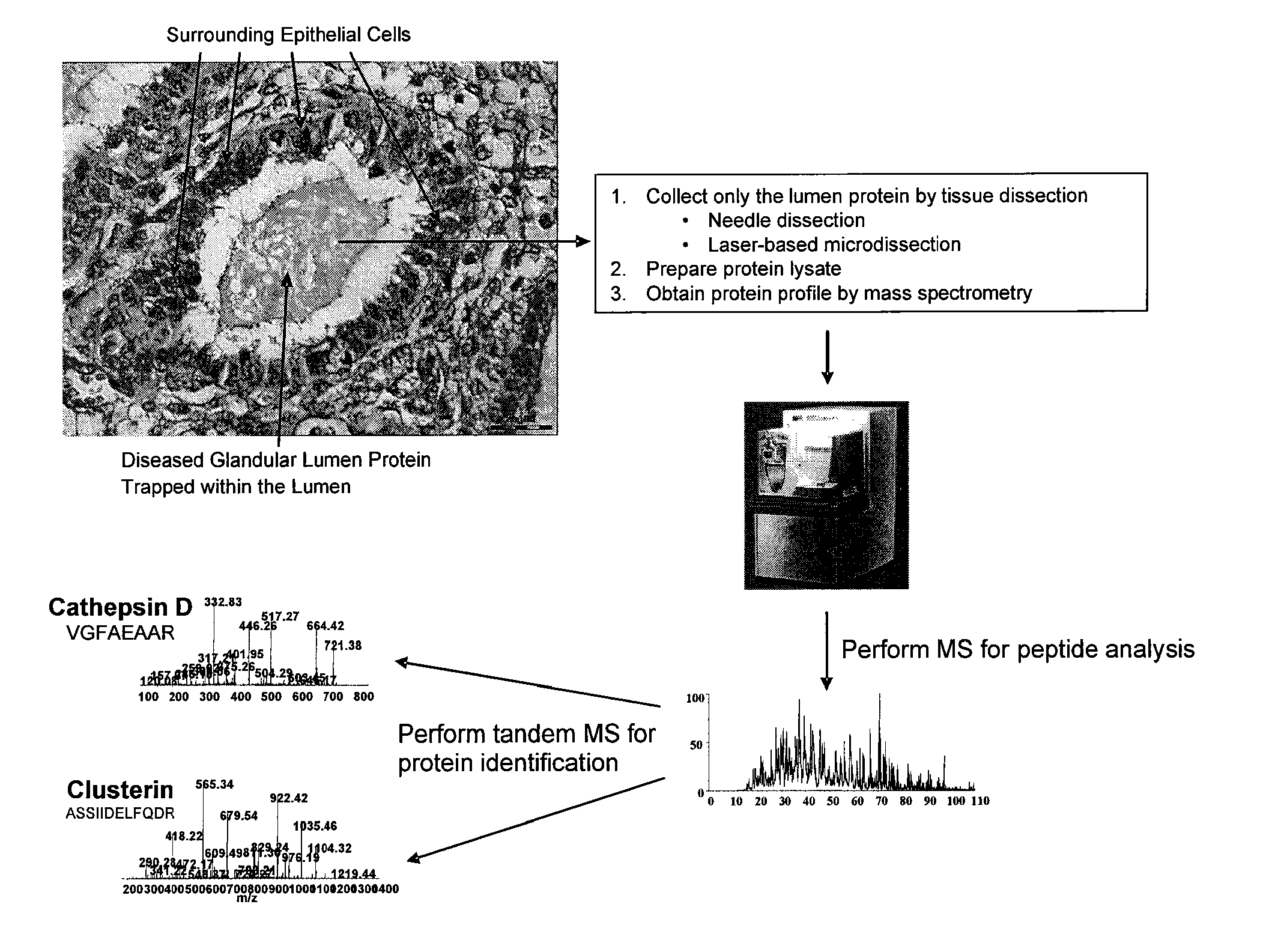
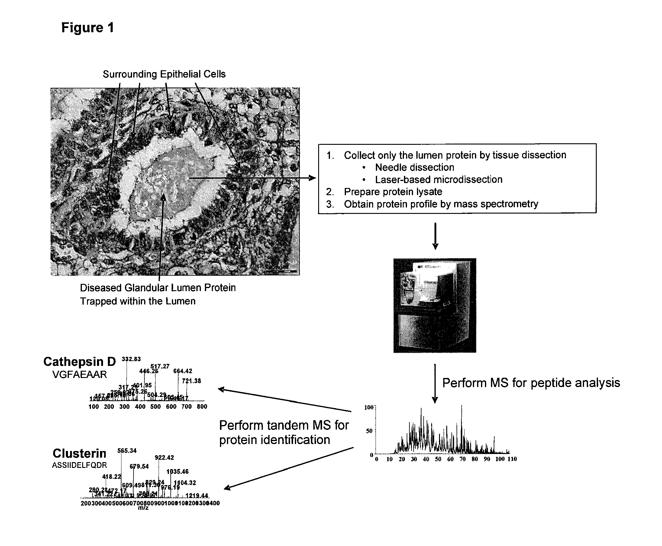
[0035]FIG. 4 shows the complete list of proteins identified by mass spectrometry analysis of a soluble protein lysate derived from the organ / glandular lumen from a single histopathologically processed solid tumor tissue sample of endometrial origin shown in FIG. 1.

TABLE 1Galectin-3 binding protein precursor (Lectin galactoside-binding soluble 3 binding protein)Gelsolin precursor (Actin-depolymerizing factor)(ADF)(Brevin)(AGEL)Glycodelin precursor (GD)(Pregnancy-associated endometrial alpha-2 globulin)Heat shock cognate 71 kDa protein (Heat shock 70 kDa protein 8)Heat shock protein HSP 90-beta (HSP 84)(HSP 90)Heat shock-related 70 kDa protein 2 (Heat shock 70 kDa protein 2)Insulin-like growth factor binding protein 7 precursor (IGFBP-7)(IBP-7)Lipocalin-1 precursor (Von Ebner gland protein)(VEG protein) (Tear prealbumin)Serotransferrin precursor (Transferrin)(Siderophilin)(Beta-1-metal binding globulin)

TABLE 2Cancer CellLumen ProteinExtracellular Proteins9.31%37.88%Intracellular Prote...


PUM

PropertyMeasurementUnit
mass spectrometryaaaaaaaaaa
freezingaaaaaaaaaa
temperatureaaaaaaaaaa

Abstract

The current invention provides a method for discovering protein biomarkers of disease for use in diagnostic assays of bodily fluids by determining differential expression patterns of proteins secreted or released directly by normal and diseased epithelial cells into glandular lumens. Determining those secreted or released proteins directly from the glandular lumen of diseased and normal solid tissue would lead to a catalogue of proteins that have a high degree of probability to be present in various bodily fluids in persons with specific diseases. This would prove useful as a means to diagnose specific conditions and diseases by simply assaying easily acquired bodily fluids. Past efforts to discover such diagnostic / screening markers in bodily fluids have proven difficult at best due to overriding complexity of the proteins within bodily fluids. This invention is a method of discovering those biomarkers in a more focused and less complex protein subpopulation, namely the secreted or released proteins present in glandular lumens.

Description

FIELD OF THE INVENTION[0001]The invention provides compositions and methods for detecting of disease biomarkers in biological samples, including samples prepared from formalin-fixed tissue specimens.BACKGROUND OF THE INVENTION[0002]Trained pathologists and histotechnologists perform molecular analyses on cancer tissue to determine the expression of specific molecular biomarkers. Information derived from such analyses is valuable prognostic tool. For instance, a number of different protein biomarkers are associated with specific stages or grades of cancer and are relied upon to guide therapies. The current method of immunohistochemistry (IHC) is very capable of analyzing biomarkers that are highly predictive of a specific stage or grade of cancers. However, in order to perform such a clinical analysis on a tissue sample, it is necessary to procure tissue from a cancer patient either by invasive surgical or by biopsy procedures. These methods can be disruptive and potentially harmful ...

Claims


Application Information

Patent Timeline
Patent Type & Authority Applications(United States)
IPC IPC(8): C12Q1/00C40B20/00
CPCG01N33/6842G01N33/57488
Inventor KRIZMAN, DAVID B.
Owner EXPRESSION PATHOLOGY
Why Eureka
Try Eureka