Use of PEDF in an Encapsulated Cell-Based Delivery System

a cell-based delivery and encapsulation technology, applied in the direction of genetically modified cells, prosthesis, peptide/protein ingredients, etc., can solve the problems of ineffective protein or peptide-based therapeutics in particular, difficult to administer to the eye, and ineffective oral administration of protein or peptide-based therapeutics, etc., to achieve inhibition of neural or retinal degradation

Inactive Publication Date: 2011-05-12
NEUROTECH USA
View PDF49 Cites 9 Cited by
  • Summary
  • Abstract
  • Description
  • Claims
  • Application Information

AI Technical Summary

Benefits of technology

[0020]The invention also provides methods for inhibiting neural or retinal degradation or degeneration in a host comprising implanting (e.g., intraocularly or periocularly) any of the cell culture devices

Problems solved by technology

Protein or peptide-based therapeutics in particular have proven difficult to administer to the eye.
Oral administration is typically not effective to provide the desired dosage to the eye.
Topical administration of liquids, gels, or ointments tends to be ineffective for protein or peptide-based therapeutics which are not easily formulated for topical delivery and which may be unable to cross the cornea.
In addition, topical formulations tend to be ineffective for delivery to the sclera, vitreous

Method used


Image

  • Use of PEDF in an Encapsulated Cell-Based Delivery System
  • Use of PEDF in an Encapsulated Cell-Based Delivery System
  • Use of PEDF in an Encapsulated Cell-Based Delivery System

Examples


example 1

Sub-Cloning

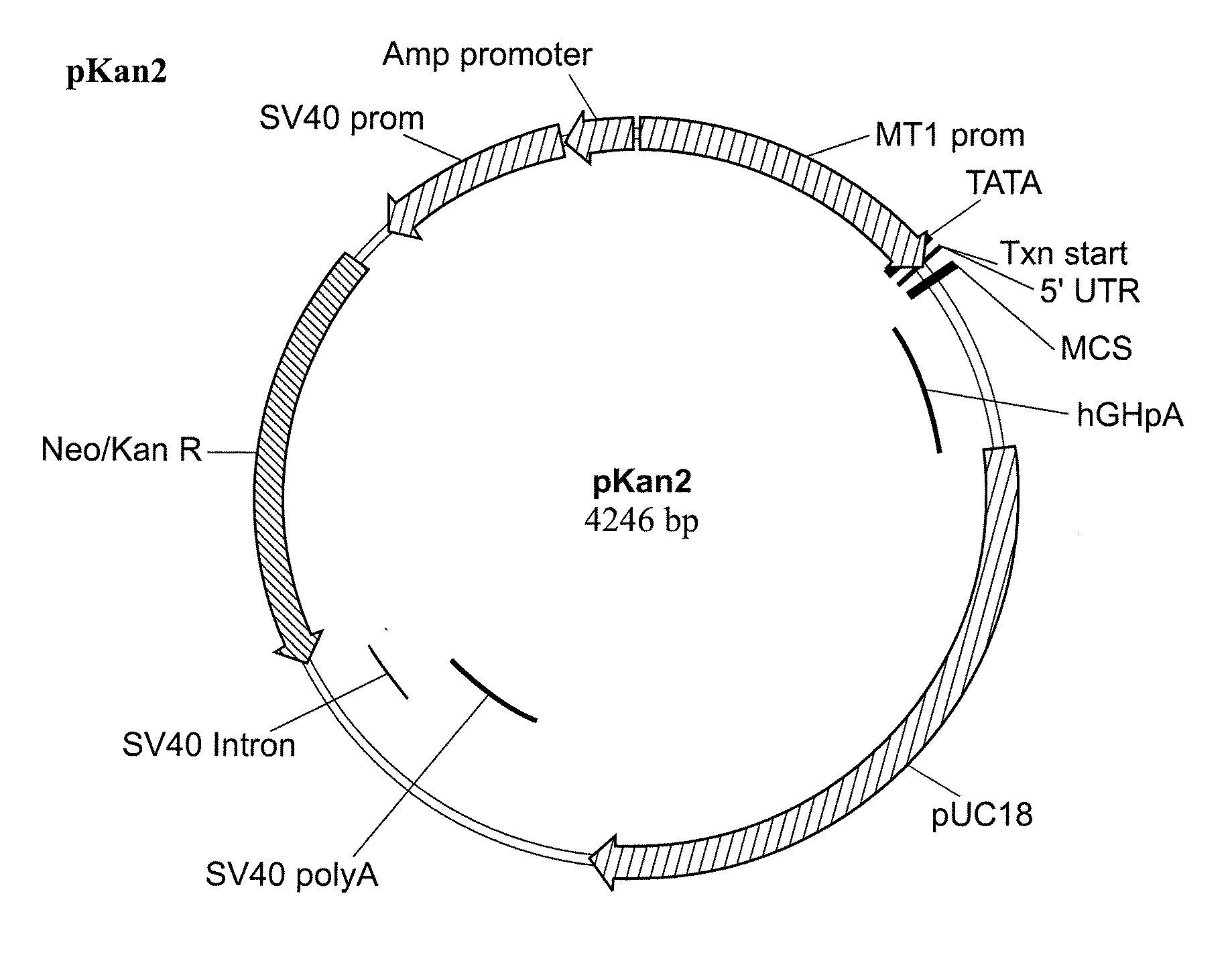
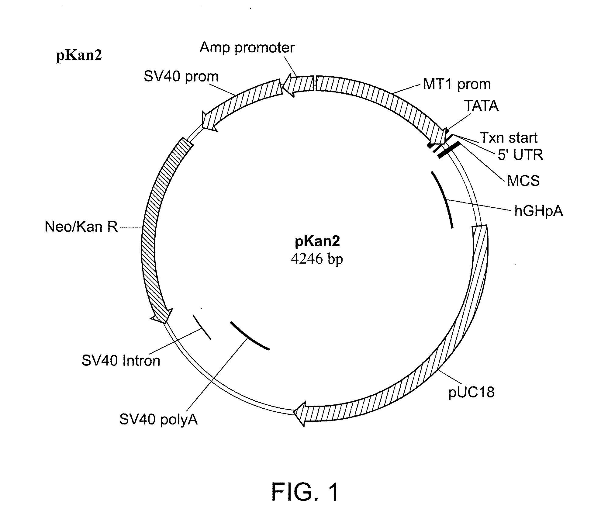
[0209]cDNA encoding human PEDF (GenBank Accession No. NM—002615) was subcloned into Neurotech mammalian expression vector pKAN2, a schematic of which is shown in FIG. 1. The pKAN2 backbone is based on the pNUT-IgSP-hCNTF expression plasmid used to create the ARPE-19-hCNTF cell lines.

[0210]The nucleotide sequence of pKAN2 is shown below in SEQ ID NO: 3:

cttggtttttaaaaccagcctggagtagagcagatgggttaaggtgagtgacccctcagccctggacattcttagatgagccccctcaggagtagagaataatgttgagatgagttctgttggctaaaataatcaaggctagtctttataaaactgtctcctcttctcctagcttcgatccagagagagacctgggcggagctggtcgctgctcaggaactccaggaaaggagaagctgaggttaccacgctgcgaatgggtttacggagatagctggctttccggggtgagttctcgtaaactccagagcagcgataggccgtaatatcggggaaagcactatagggacatgatgttccacacgtcacatgggtcgtcctatccgagccagtcgtgccaaaggggcggtcccgctgtgcacactggcgctccagggagctctgcactccgcccgaaaagtgcgctcggctctgccaaggacgcggggcgcgtgactatgcgtgggctggagcaaccgcctgctgggtgcaaaccctttgcgcccggactcgtccaacgactataaagagggcaggctgtcctctaagcgtcacccctagagtcgagctgtgacggtccttacactcgagac...

example 2

Cell Line and Device Construction

[0212]Verified plasmid clones were used to transfect NTC-200 cells to obtain stable polyclonal cell lines. Briefly, 200-300K cells, plated 18 hours previously, were transfected with 3.0 ug of plasmid DNA using 6.0 ul of Fugene 6 transfection reagent (Roche Applied Science, Indianapolis Ind.) according to the manufacturer's recommendations. Transfections were performed in 3.0 ml of DMEM / F12 with 10% FBS, Endothelial SFM or Optimem media (Invitrogen Corp, Carlsbad, Calif.). Twenty four to 48 hours later cells were either fed with fresh media containing 1.0 ug / ul of G418 or passaged to a T-25 tissue culture flask containing G418. Cell lines were passaged under selection for 14-21 days until normal growth resumed, after which time drug was removed and cells were allowed to recover (˜1 week) prior to characterization.

[0213]Expression stability of the recombinant protein from these cell lines was measure over the course of several weeks using the Human PED...

example 3

Protein Characterization

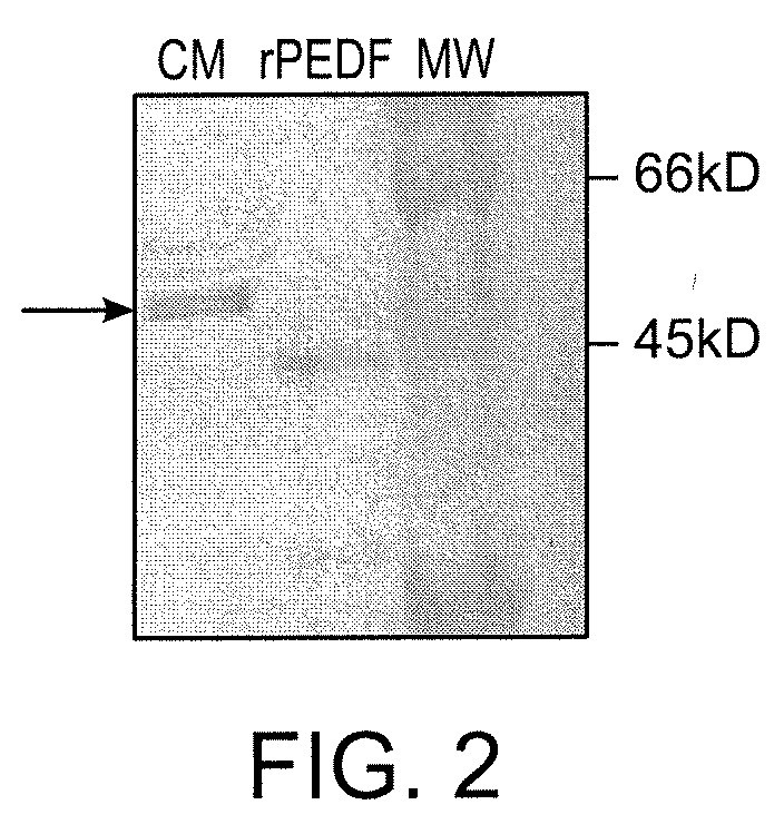
[0217]Expression of PEDF from stably transfected cell lines was quantified by analyzing conditioned media from cell monolayers using a commercially available ELISA kit (BioProducts Maryland, Middletown, Md.). Conditioned media from stably transfected cells was analyzed by a colorimetric Western blot assay to determine protein integrity.


PUM

PropertyMeasurementUnit
Massaaaaaaaaaa
Diameteraaaaaaaaaa
Diameteraaaaaaaaaa

Abstract

The invention relates to a device for delivery of pigment epithelium derived factor (PEDF) to the eye utilizing encapsulated PEDF-secreting cells and related methods for the treatment and prevention of ophthalmic diseases and disorders.

Description

RELATED APPLICATIONS[0001]This application claims priority to U.S. Ser. No. 61 / 249,787, filed Oct. 8, 2009, the contents of which are herein incorporated by reference in its entirety.FIELD OF THE INVENTION[0002]The present invention relates to the use of pigment epithelium derived factor (PEDF), and biologically active variants thereof, in a delivery system utilizing encapsulated cells engineered to secrete PEDF, and related methods for the treatment of ophthalmic diseases and disorders using encapsulated PEDF-secreting cells.INCORPORATION BY REFERENCE OF SEQUENCE LISTING[0003]The contents of the text file named “19141-556001USSeqList.txt”, which was created on Dec. 13, 2010 and is 13 KB in size, are hereby incorporated by reference in their entirety.BACKGROUND OF THE INVENTION[0004]Pigment epithelium derived factor (PEDF) was first identified in the conditioned medium of cultured fetal human retinal pigment epithelial cells as a 50-kDa protein having neurotrophic activity (Tombran-...

Claims


Application Information

Patent Timeline
IPC IPC(8): A61F2/00A61K35/12C12N11/02A61F9/007
CPCA61K9/0051A61K2035/128A61K38/00C12N2510/02C12N5/0621A61F2220/0008A61F2240/001A61F2250/0068A61P27/02Y02A50/30A61F9/0017
Inventor TAO, WENGKAUPER, KONRADSTABILA, PAULLING, VINCENT
Owner NEUROTECH USA
Why Eureka
Try Eureka