Image processing apparatus and image processing method

a technology of image processing apparatus and image processing method, which is applied in the field of image processing technique for processing a tomogram, can solve the problems of inability to obtain effective diagnosis information data, and inability to appropriately obtain all layer position information

Inactive Publication Date: 2011-06-09
CANON KK
View PDF5 Cites 85 Cited by
  • Summary
  • Abstract
  • Description
  • Claims
  • Application Information

AI Technical Summary

Benefits of technology

[0012]According to the present invention, it is possible to obtain diagnosis information data effective for eye disease diagnosis independently of an eye state.

Problems solved by technology

For this reason, it may be impossible to appropriately obtain all layer position information depending on the eye state (the presence / absence or type of a disease).
If the same image analysis algorithm is executed for the region, it may be impossible to obtain effective diagnosis information data.

Method used


Image

  • Image processing apparatus and image processing method
  • Image processing apparatus and image processing method
  • Image processing apparatus and image processing method

Examples


first embodiment

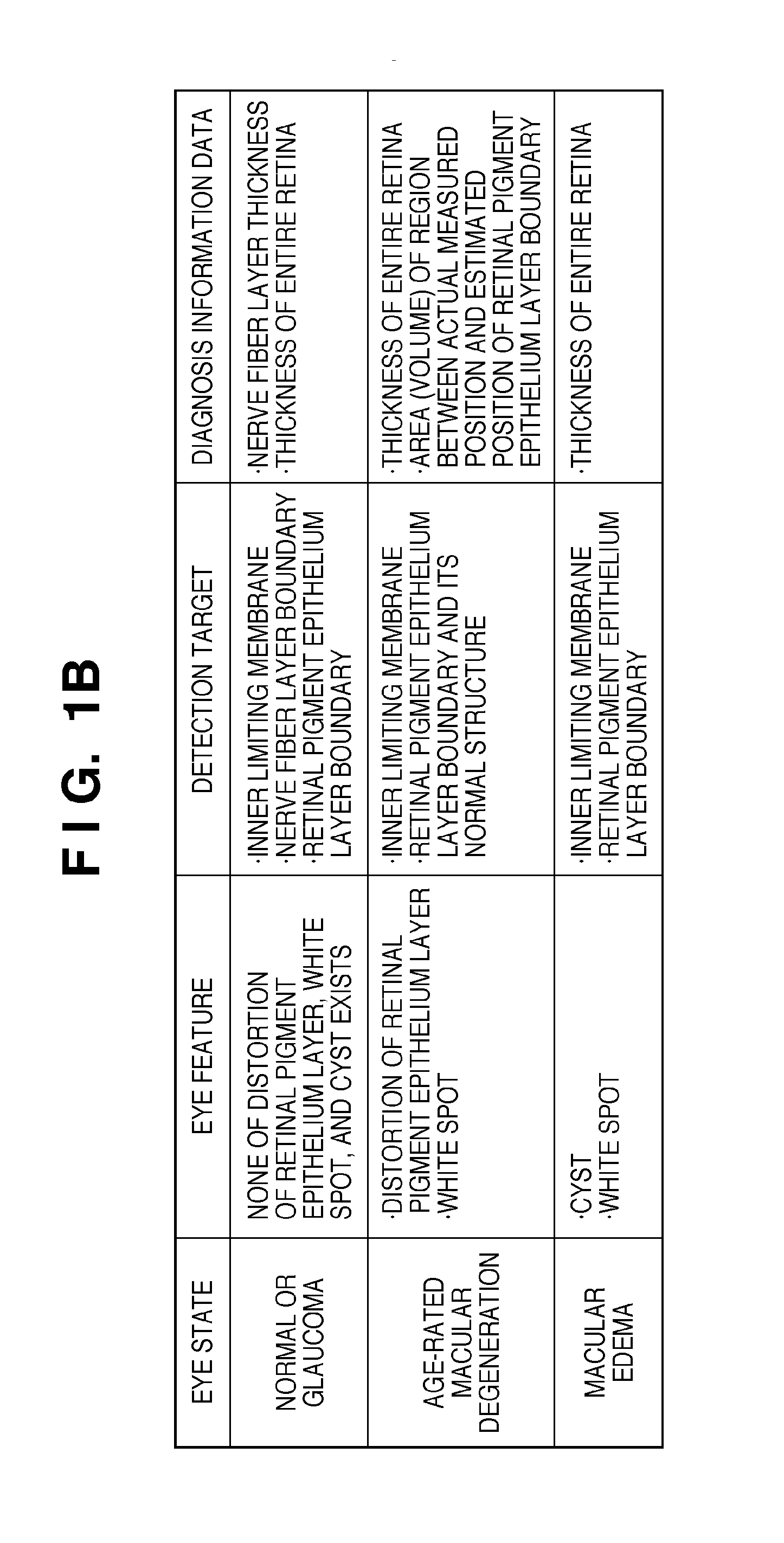
[0035]An image processing apparatus according to this embodiment is characterized by diagnosing an eye (disease) state in advance based on information (to be referred to as an “eye feature”) about the shape or presence / absence of a predetermined tissue such as the presence / absence of distortion of the retinal pigment epithelium layer boundary or the presence / absence of a white spot or cyst. The apparatus is also characterized by acquiring diagnosis information data corresponding to an eye state by applying an image analysis algorithm capable of acquiring diagnosis information data corresponding to the diagnosed eye state. The image processing apparatus according to the embodiment will now be described below in detail.

[0036]

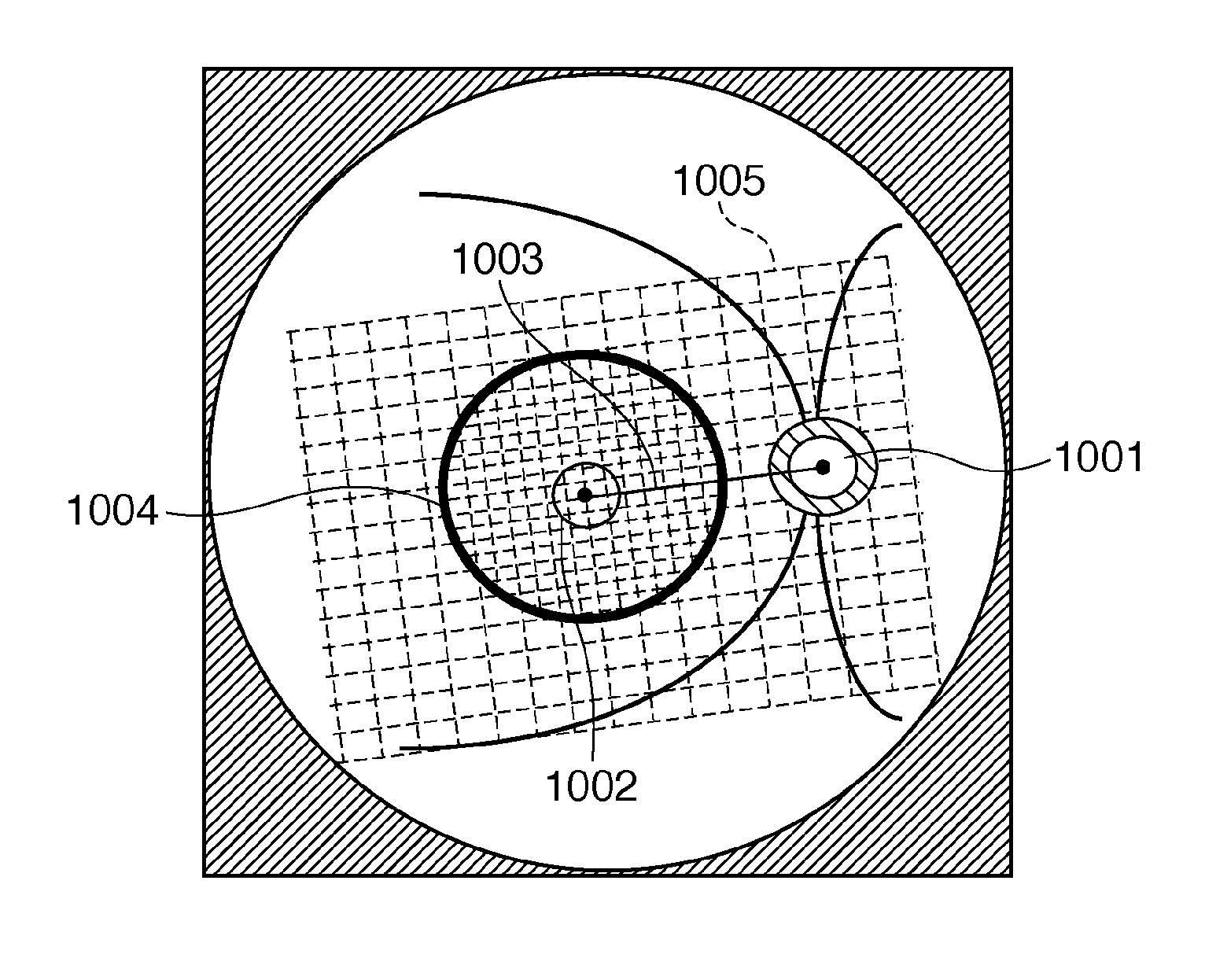
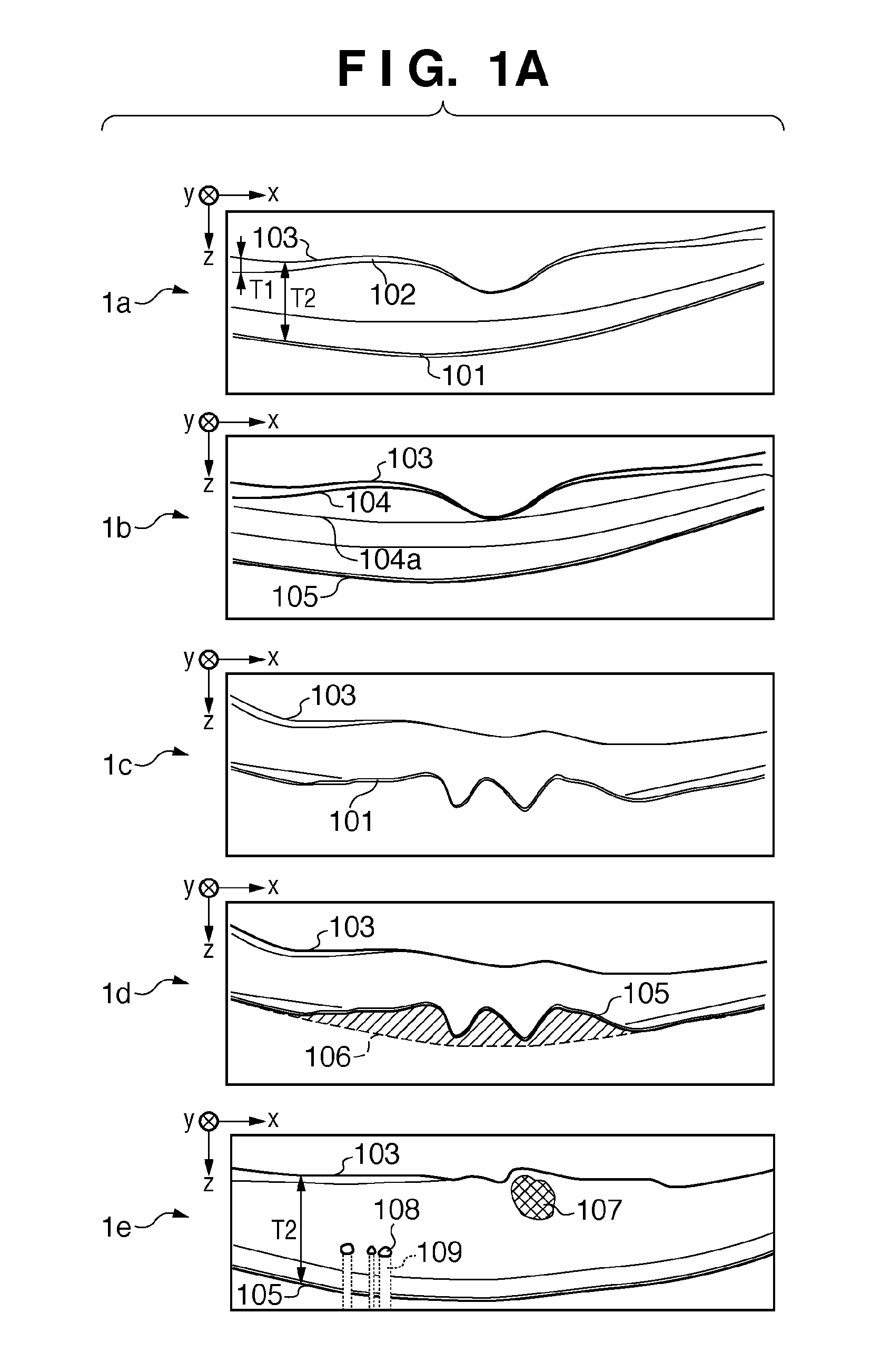
[0037]The relationship between eye states, eye features, detection targets, and diagnosis information data will be explained first. In FIG. 1A, 1a to 1e are schematic views showing the tomograms of a macular portion of retina captured by an OCT. In FIG. 1B is a ta...

second embodiment

[0151]In the above-described first embodiment, assuming that the image analysis target is a tomogram of a macular portion, eye features are extracted, and the eye state is determined based on the extracted eye features. However, the tomogram of image analysis target is not limited to the tomogram of a macular portion. It may be, for example, a wide-angle tomogram including not only a macular portion but also an optic disc portion. In the second embodiment, an image processing apparatus will be described which, when the tomogram of image analysis target is a wide-angle tomogram including a macular portion and an optic disc portion, specifies each part and executes an image analysis algorithm for each part.

[0152]Note that the overall arrangement of the diagnostic imaging system and the hardware configuration of the image processing apparatus are the same as in the first embodiment, and a description thereof will not be repeated here.

[0153]

[0154]A wide-angle tomogram including a macula...

third embodiment

[0195]In the above-described first and second embodiments, the apparatus is configured to calculate, as diagnosis information data, the nerve fiber layer thickness, the thickness of entire retinal layer, the area (volume) of a region between the actual measured position and the estimated position of retinal pigment epithelium layer boundary, and the like. However, the present invention is not limited to this. For example, the apparatus may be configured to obtain diagnosis information data from tomograms of different imaging dates / times (imaging timings), compare them with each other to quantify the time-rate change, and output new diagnosis information data (follow-up diagnosis information data). More specifically, two tomograms of different imaging dates / times are aligned based on a predetermined alignment target included in each tomogram, and the difference between corresponding diagnosis information data is obtained, thereby quantifying the time-rate change between the two tomog...


PUM


Abstract

This invention concerns the acquisition of diagnosis information data effective for diagnosing eye disease independently of the eye state. An image processing apparatus for processing a tomogram of an eye includes a unit configured to determine an eye feature based on the tomogram and thus determine the eye state, a unit configured to detect, from the tomogram, a detection target to be used to calculate diagnosis information data quantitatively representing the determined eye state, and a unit configured to calculate the diagnosis information data using position information of the detection target. In accordance with the eye state, the detection unit changes the detection target or an algorithm to be used to detect the detection target.

Description

BACKGROUND OF THE INVENTION[0001]1. Field of the Invention[0002]The present invention relates to an image processing technique for processing a tomogram.[0003]2. Description of the Related Art[0004]Ophthalmic examinations are conventionally made aimed at providing an early diagnosis of lifestyle diseases or high-ranking diseases that lead to blindness. An ophthalmic tomography imaging apparatus such as an OCT (Optical Coherence Tomography) is generally used for the ophthalmic examinations. This is because using the tomographic ophthalmic imaging apparatus such as an OCT makes it possible to observe the internal state of a retinal layer three-dimensionally, and thus render a more reliable diagnosis.[0005]On the other hand, when diagnosing an eye disease (for example, glaucoma, age-rated macular degeneration, or macular edema) using obtained tomograms, it is important to analyze the tomograms and quantitatively extract information effective for diagnosis.[0006]To do this, an image pro...

Claims


Application Information

Patent Timeline
Patent Type & Authority Applications(United States)
IPC IPC(8): A61B5/05
CPCG06T7/0012G06T2207/30041G06T2207/10081
Inventor IMAMURA, HIROSHINAKANO, YUTAIWASE, YOSHIHIKOSATOH, KIYOHIDEKATAYAMA, AKIHIRO
Owner CANON KK
Why Eureka
Try Eureka