Miniaturized multi-spectral imager for real-time tissue oxygenation measurement

a multi-spectral imager, real-time technology, applied in the field of multi-spectral imaging, can solve the problems of few techniques that can demarcate, few techniques that can be used, and the raw spectral or imaging measurement seldom reveals directly the property of clinical interest, so as to achieve the convergence of correct alignment more quickly and more reliably

Inactive Publication Date: 2011-06-16
HYPERMED IMAGING
View PDF11 Cites 19 Cited by
  • Summary
  • Abstract
  • Description
  • Claims
  • Application Information

AI Technical Summary

Benefits of technology

[0024]Building a joint-intensity model does require having access to a registered pair of images of the same modality and approximately the same coverage as the novel pair to be registered. Mutual information approaches do not need to draw upon previously registered ...

Problems solved by technology

However, the volume of information contained in spectroscopic images can make standard data processing techniques cumbersome.
Furthermore, there are few techniques that can demarcate which regions of a spectroscopic image contain similar spectra without a priori knowledge of either the spectral data or the sample's composition.
There are few techniques that can demarcate which regions of a sample contain similar substances without a priori knowledge of the sample's composition.
While it is now clear that both spectroscopy and spectroscopic imaging can play roles in providing medically relevant information, the raw spectral or imaging measurement seldom reveals directly the property of clinical interest.
For example using spectroscopy, one cannot easily determine whether the tissue is cancerous, or determine blood glucose concentrations and the adequacy of tissue perfusion.
The very reason why multiple scans are useful is what makes the registration process difficult.
As each modality images tissue differently and has its own artifacts and noise characteristics, accurately modeling the intensity relationship between the scans, and subsequently aligning them, is difficult.
The main drawback of this method is that the markers must remain attached to the patient at...

Method used


Image

  • Miniaturized multi-spectral imager for real-time tissue oxygenation measurement
  • Miniaturized multi-spectral imager for real-time tissue oxygenation measurement
  • Miniaturized multi-spectral imager for real-time tissue oxygenation measurement

Examples


Embodiment Construction

[0038]As embodied and broadly described herein, the disclosures herein provide detailed embodiments of the invention. However, the disclosed embodiments are merely exemplary of the invention that may be embodied in various and alternative forms. Therefore, there is no intent that specific structural and functional details should be limiting, but rather the intention is that they provide a basis for the claims and as a representative basis for teaching one skilled in the art to variously employ the present invention.

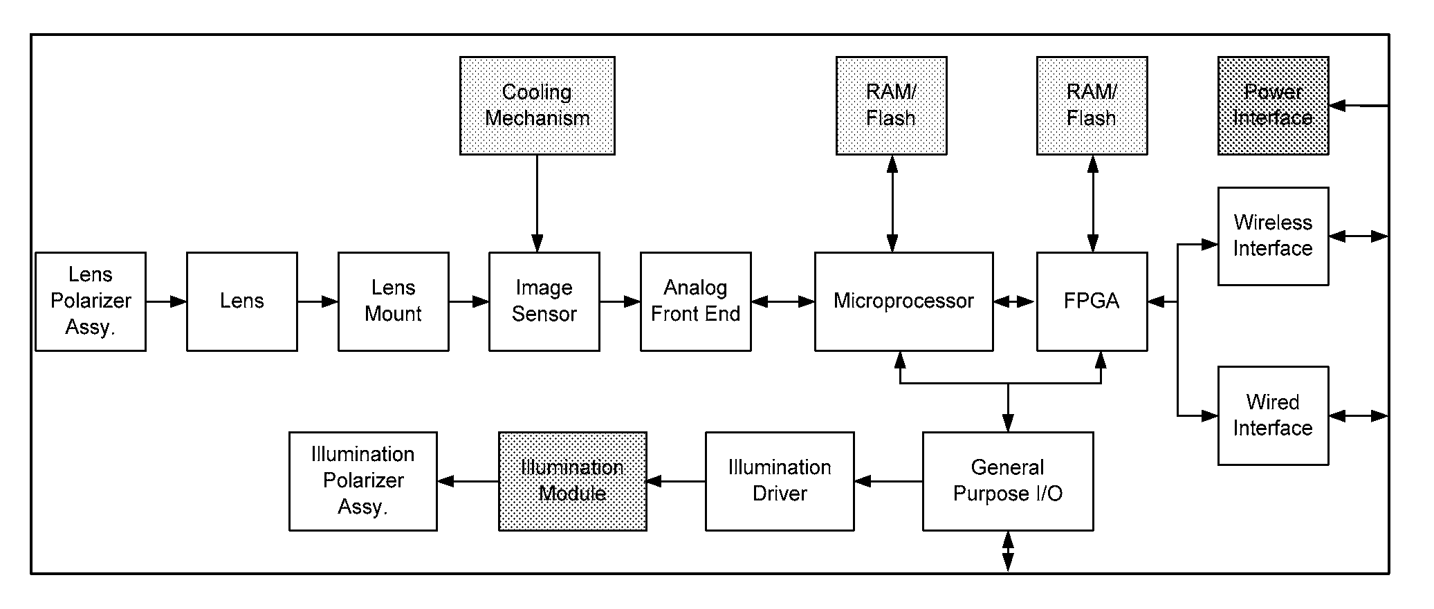
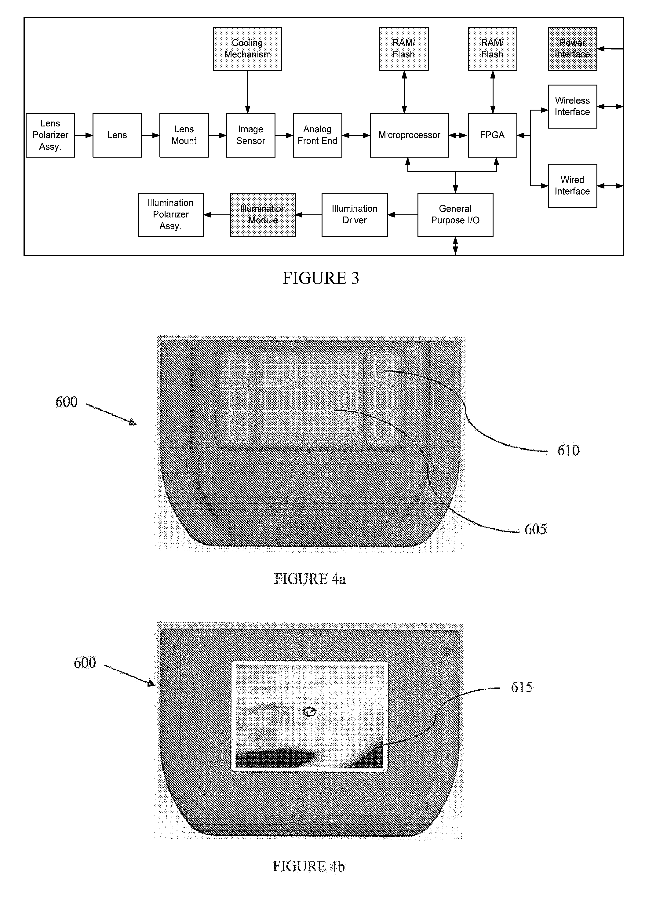
[0039]A problem in the art capable of being solved by the embodiments of the present invention is producing a miniaturized medical multi-spectral imaging (MSI) sensor capable of providing real-time measurements of oxygen saturation StO2t in skin, serving as an excellent indicator of oxygenation status in patients with multiple medical conditions, including but not limited to diabetes, wound care, vascular disease and pressure ulcers as well as providing an early warning f...


PUM


Abstract

A portable multi-spectral imaging system and device is disclosed. The system includes at least one image acquisition device for acquiring an image from a subject, a filtering device to filter the light received by the image acquisition device, a processor for processing the image acquired by the image acquisition device, and a display. There is software running on the processor that determines oxygenation values of the subject based on the processed image.

Description

REFERENCE TO RELATED APPLICATIONS[0001]This application claims priority to U.S. Provisional Application No. 61 / 037,780 entitled “MINIATURIZED MULTI-SPECTRAL IMAGER FOR REAL-TIME TISSUE OXYGENATION MEASUREMENT” filed Mar. 19, 2008, the entirety of which is hereby specifically incorporated by reference.BACKGROUND[0002]1. Field of the Invention[0003]The invention is directed to multi-spectral imaging. Specifically, the invention is directed to a portable multi-spectral imager.[0004]2. Background of the Invention[0005]Spectroscopy, whether it is visible, near infrared, infrared or Raman, is an enormously powerful tool for the analysis of biomedical samples. The medical community, however, has a definite preference for imaging methods, as exemplified by methods such as MRI and CT scanning as well as standard X-ray photography and ultrasound imaging. This is entirely understandable as many factors need to be taken into account for a physician to make a clinical diagnosis. Imaging methods ...

Claims


Application Information

Patent Timeline
IPC IPC(8): A61B5/1455
CPCA61B5/0059A61B5/14552G01J3/02G01J3/0272G01N2021/3155G01J3/40G01N21/31G01N2021/3144G01J3/2823
Inventor LIFSITZ, RICKJIANG, CHUNSHENGGUSYATIN, OLEGSHUBENSTOV, ILYA
Owner HYPERMED IMAGING
Why Eureka
Try Eureka