Fluorescent microscope slide

a fluorescent microscope and slide technology, applied in the field of fluorescent microscope slides, can solve the problems of low sensitivities of bright field microscopy, the risk of active tb infection of persons with hiv, and the global health problem of tuberculosis, etc., to achieve accurate identification of the field of view

Inactive Publication Date: 2011-07-21
QBC DIAGNOSTICS
View PDF8 Cites 23 Cited by
  • Summary
  • Abstract
  • Description
  • Claims
  • Application Information

AI Technical Summary

Benefits of technology

[0017]The present teachings provide a useful device comprising a microscopy slide imprinted with strategically placed ink that fluoresces such that when the slide is read with fluorescent microscopy technology, the pattern deposited on the slide enables a microscopist to find a field of focus and to accurately identify a field of view.

Problems solved by technology

Tuberculosis (TB) disease presents a global health problem.
Persons with HIV who become infected with Mycobacterium tuberculosis also suffer from an increased risk of developing active TB.
Despite its role in TB healthcare, however, bright field microscopy suffers from low sensitivities of about 60%.
Moreover, in laboratories where assay volumes are high, examination time may be attenuated, resulting in further loss of sensitivity.
With this technology, and with use of other bright field methods, it can be difficult to identify and distinguish the bacilli from the background material, and proper examination requires significant time from a skilled technician.
Time and skill resources are often limited in many TB lab settings.
However, because the morphology of the bacteria are unique and have a variety of distinguishing characteristics, and because of the difficulties with ZN staining referenced above, it takes a fairly sophisticated technician and / or lab expertise to detect tuberculosis.
This procedure is extremely time consuming and limits the number of slides a microscopist or technician can accurately read in a given day.
Despite its recognized benefits, FM (both conventional and LED based) introduces significant problems that affect its utility and indication for widespread use.
These problems center around the fact that FM is a dark field technique, and, as such, requires the sample to provide a light signal.
Therefore, it becomes difficult to ensure the quality of assays where a negative result is found.
Furthermore, the signal from fluorescently stained AFB is relatively low (compared to bright field methods) and can only be seen within a small number of focal planes.
Difficulty in focusing a fluorescence microscope is not only a problem for the inexperienced but is also a problem for the seasoned technician.
As a result, it is very difficult for a microscopist to ascertain whether what he or she is viewing is in focus, and thus, the microscopist often does not have great confidence in reporting that there is no tuberculosis.
Finally, it remains very difficult for FM microscopists to ascertain whether they are looking at the same lateral field of view or new fields of view, especially when there is little to no background material to provide internal reference points when moving from field of view to field of view, and as a result, there is a lack of predictability in results, and a lower than desired level of accuracy in reading slides, even with the fluorescence microscopy technology.
In addition, FM has been unfeasible (and in some instances unsuitable) for most healthcare facilities around the world.
This lack of feasibility is due to significant problems introduced by the technology, including: expensive capital costs; complex, short-lived and expensive light sources; technician fears of UV induced health hazards; notoriously delicate instrumentation; the need for stable power requirements; and significant training period requirements.

Method used


Image

  • Fluorescent microscope slide
  • Fluorescent microscope slide
  • Fluorescent microscope slide

Examples


examples

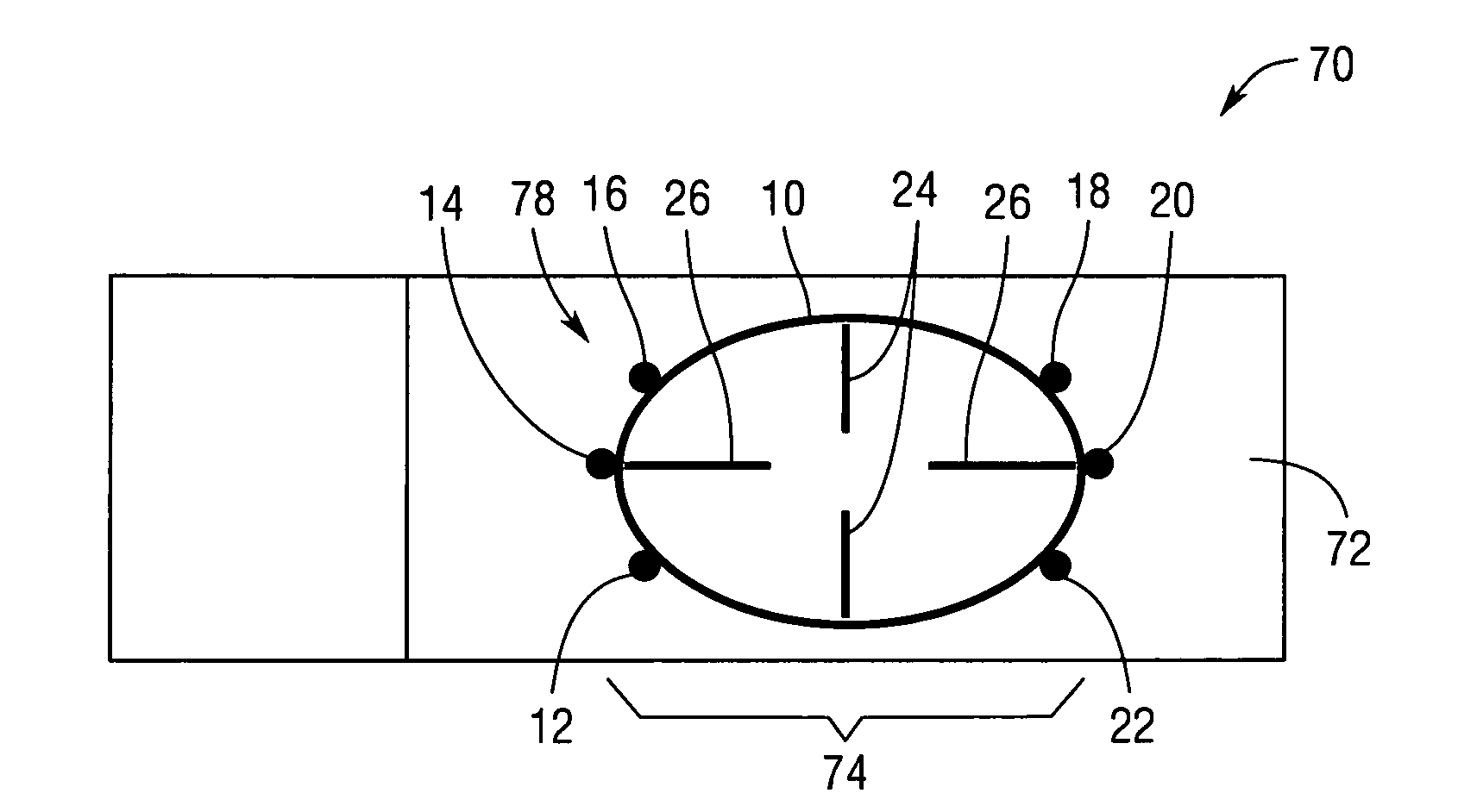
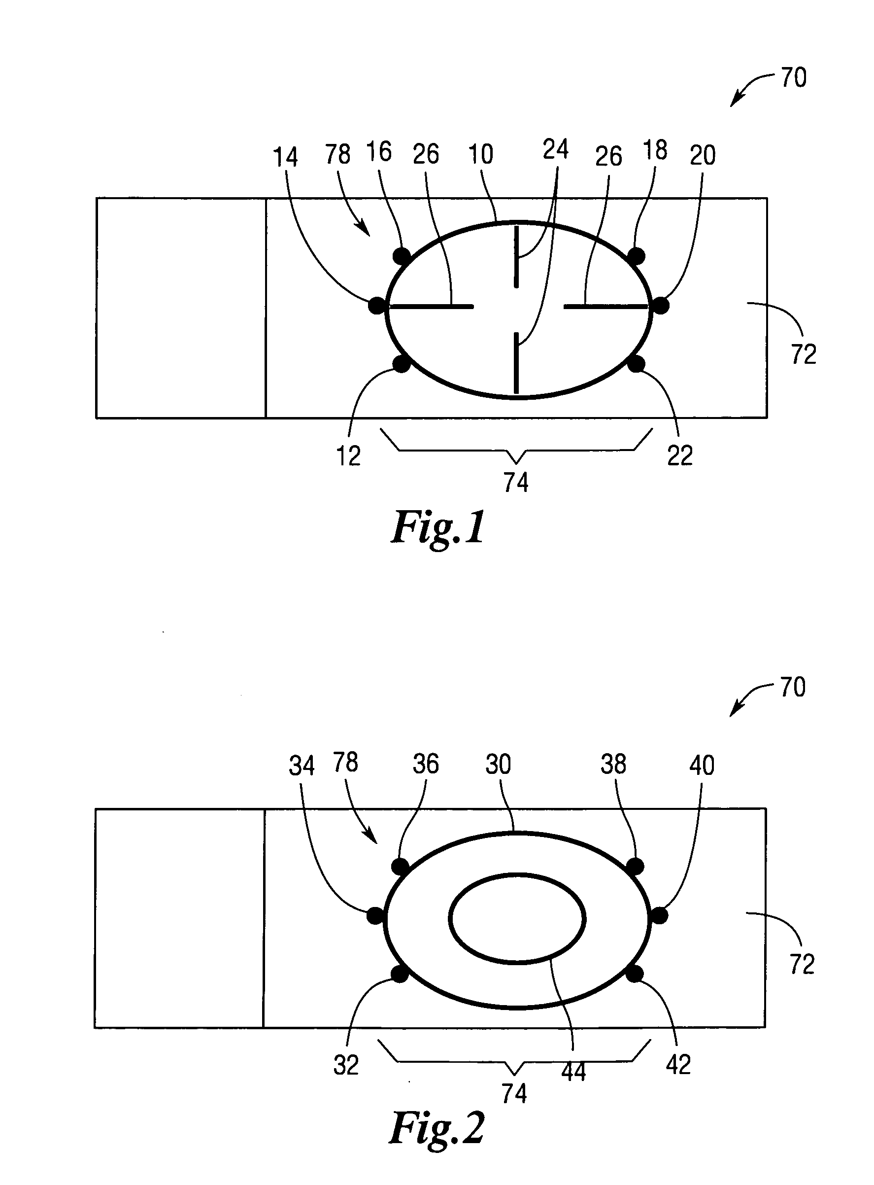
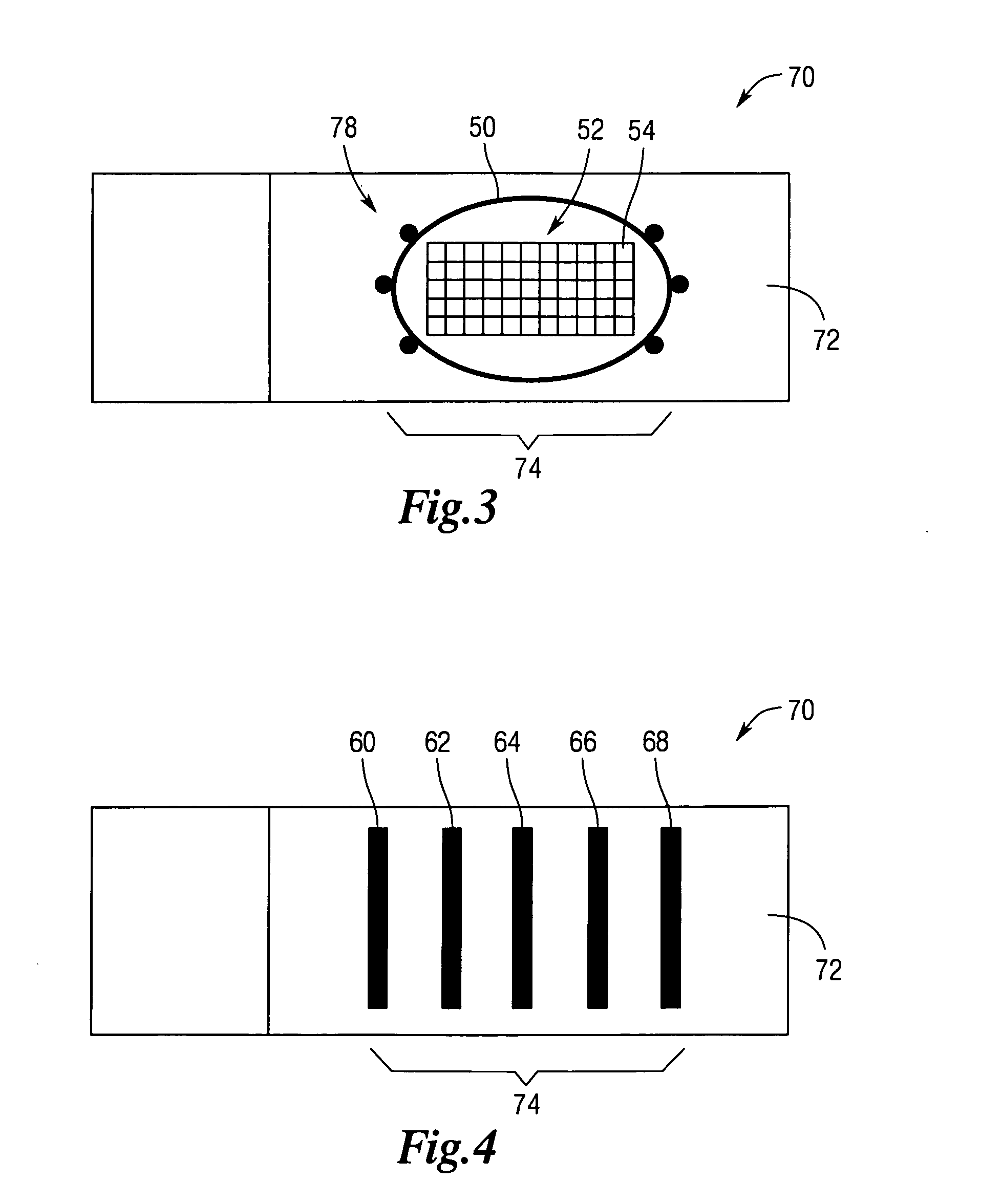
[0038]The device of the present teachings enables rapid and accurate detection of the presence of components of interest (e.g. acid fast bacilli) in a sample using fluoroscopic microscopy. The fluorescent microscope substrate 70 features a specimen receptacle (e.g. a microscope slide or cover) 72 comprising markings 74 on a surface of the receptacle that: fluoresce, demarcate a sample specimen area 78, identify examination start areas, and demarcate examination distances.

[0039]Stated otherwise, the fluorescent markings 74 define a target area 12 for a sample. In addition, fluorescent markings 74 about (i.e. proximate, adjacent to, or within) the target area 12 enable a microscopist or other user to ascertain a field of focus. Fluorescent markings 74 about the target area 12 also allow a user to identify a field of view.

[0040]Markings 74 of the fluorescent microscope slide 70 can assume any desired form including, but not limited to: lines, etchings, symbols, numbers, text, geometric...


PUM

PropertyMeasurementUnit
thicknessaaaaaaaaaa
heightaaaaaaaaaa
heightaaaaaaaaaa

Abstract

An apparatus and method for increasing the efficiency of finding a field of focus, and for increasing the accuracy of field of view in reading slides with fluorescent microscopy technology, including tuberculosis slides.

Description

REFERENCE TO RELATED APPLICATIONS[0001]This application claims priority to U.S. Provisional Patent Application No. 61 / 336,115, filed Jan. 15, 2010.INTRODUCTION[0002]The present teachings relate to a device and methodology for increasing the efficiency of finding a field of focus, and for increasing the accuracy of a field of view in reading slides with fluorescent microscopy technology.[0003]Tuberculosis (TB) disease presents a global health problem. An estimated one-third of the world's population is infected with the causative agent of TB, Mycobacterium tuberculosis. While in most cases the disease remains latent, about 5 to 10% of persons infected will go on to develop active disease sometime in their life. Left undetected and / or untreated, patient mortality is about 50%. Furthermore, statistical studies show that an untreated infected individual will go on to infect 10 to 15 other individuals per annum.[0004]The worldwide incidence of active TB has been on an increasing trend si...

Claims


Application Information

Patent Timeline
Patent Type & Authority Applications(United States)
IPC IPC(8): C12Q1/06C12Q1/02C12M1/34
CPCG01N21/01G01N21/6458G02B21/34G02B21/241G02B21/16G02B27/32
Inventor GRAHAM, CHRISTOPHER J.STOUT, CRAIG
Owner QBC DIAGNOSTICS
Why Eureka
Try Eureka