Method and apparatus for magnetic resonance imaging to create t1 maps

a magnetic resonance imaging and t1 map technology, applied in the field of magnetic resonance imaging, can solve the problems of inability to use conventional methods for t1/b> quantification to depict cardiac muscle, inability to quantify cardiac muscle, etc., and achieve the effect of improving the accuracy of t1 maps and improving the accuracy of t1 results

Active Publication Date: 2011-07-28
SIEMENS HEALTHCARE GMBH
View PDF5 Cites 52 Cited by
  • Summary
  • Abstract
  • Description
  • Claims
  • Application Information

AI Technical Summary

Benefits of technology

[0008]An object of the present invention is to provide an improved method for spatially resolved quantification of the T1 relaxation time that in particular is suitable for volumetric scanning and generation of a T1 map, for example of a beating heart.

Problems solved by technology

However, these methods are not quantitative, meaning that they merely enable a delimitation between healthy and destroyed tissue but not a differentiation in the transition range or in ischemic but not infracted tissue regions.
However, conventional methods for T1 quantification can not be used to depict the cardiac muscle because heart beat and breathing movement prevent an artifact-free depiction.
Due to the properties of the single shot readout steps that are used, the method is subject to limitations with regard to the possible temporal and spatial resolution.
In particular, the method enables the acquisition of only one slice so that a volumetric coverage of the heart during a breath hold phase is not possible with the MOLLI acquisition scheme.

Method used


Image

  • Method and apparatus for magnetic resonance imaging to create t1 maps
  • Method and apparatus for magnetic resonance imaging to create t1 maps
  • Method and apparatus for magnetic resonance imaging to create t1 maps

Examples


Embodiment Construction

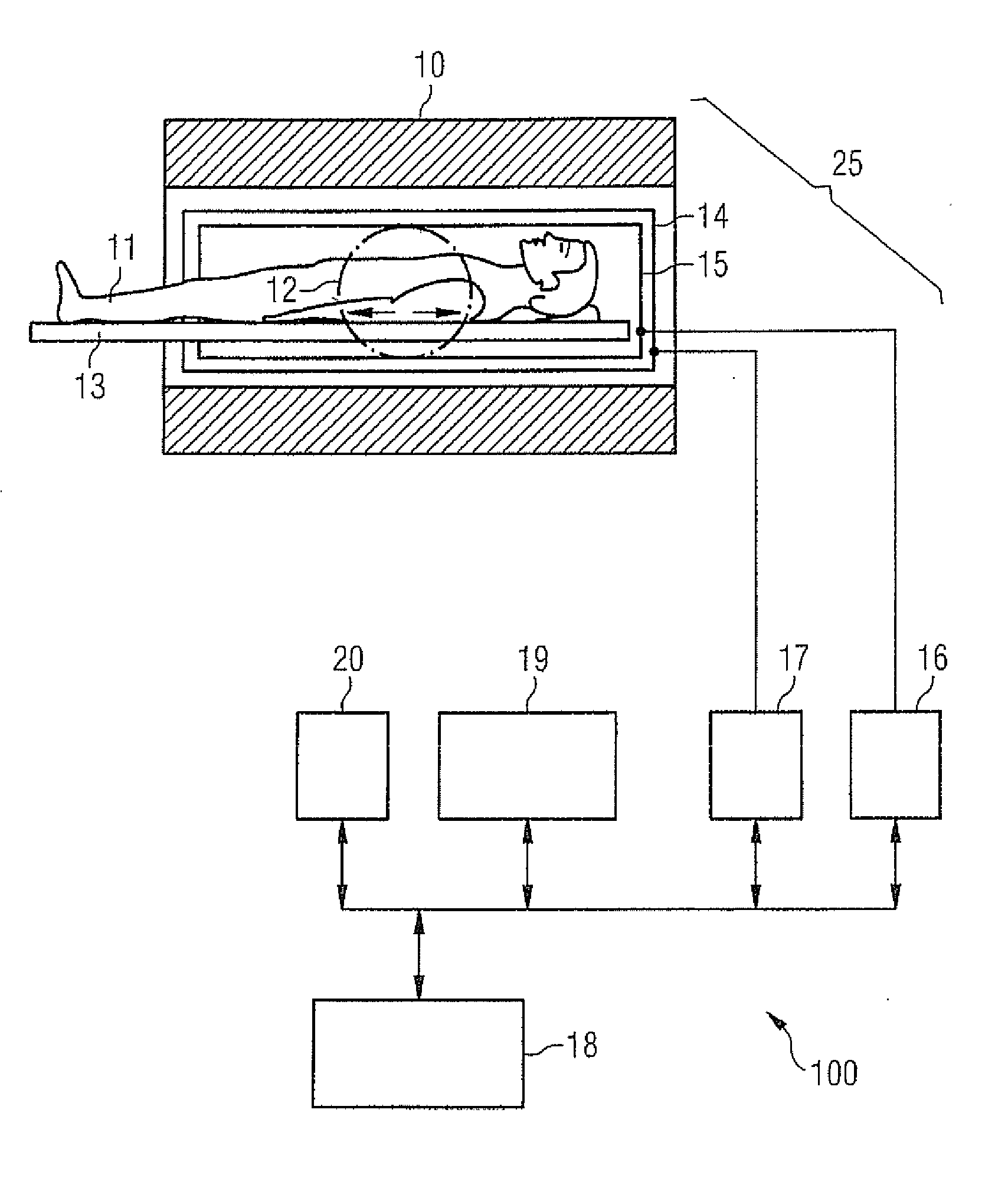
[0040]FIG. 1 schematically shows a magnetic resonance system 100 which is designed to implement acquisition sequences for spatially resolved quantification of T1 relaxation times in parallel. Such a magnetic resonance system possesses a magnet 10 to generate a polarization field B0. To increase the signal-to-noise ratio, field strengths of at least 2.5 T (advantageously 3.0 T) can thereby be used. An examination subject (here an examined person 11) on a bed table 13 can be slid into the magnet, as is schematically depicted by the arrows. The MR system furthermore possesses a gradient system 14 to generate magnetic field gradients that are used for the imaging and spatial coding. To excite the polarization resulting in the basic magnetic field, a radio-frequency coil arrangement 15 is provided that radiates a radio-frequency (RF) field into the examined person 11 in order to deflect the magnetization out of the steady state. A gradient unit 17 is provided to control the magnetic fiel...


PUM


Abstract

In a method and apparatus for MR imaging, a data acquisition sequence is executed wherein at least two slices of an examination subject are imaged in parallel with a gradient echo method for spatially resolved quantification of the T1 relaxation time.
At least one first acquisition sequence is implemented to acquire MR data from a first slice of the examination subject and at least one second acquisition sequence is implemented to acquire MR data from a second slice of the examination subject. The acquisition sequences each include an inversion pulse and at least two successive readout steps. The first and second acquisition sequences are temporally offset from one another such that they at least partially overlap.

Description

BACKGROUND OF THE INVENTION[0001]1. Field of the Invention[0002]The present invention concerns a method for magnetic resonance (MR) imaging in which a spatially resolved quantification of the T1 relaxation time is created with a gradient echo method. The invention furthermore concerns a magnetic resonance system to implement such a method.[0003]2. Description of the Prior Art[0004]Magnetic resonance tomography (MRT) is an examination modality with which regions inside an examination subject can be shown with high resolution and good contrast, in particular even soft tissues such as muscles and organs. A dependency of the measured MR signal on the T1 and T2 relaxation times can be achieved by appropriate acquisition sequences. The relaxation times T1 and T2 are the time constants of the decay of the macroscopic magnetization excited in the tissue, which vary for different tissues. The spin lattice relaxation time T1 is characteristic of a process that produces the reestablishment of ...

Claims


Application Information

Patent Timeline
Patent Type & Authority Applications(United States)
IPC IPC(8): G01R33/48
CPCA61B5/7207A61B5/055G01R33/4835G01R33/50
Inventor GREISER, ANDREAS
Owner SIEMENS HEALTHCARE GMBH
Why Eureka
Try Eureka