
Diagnosis assisting apparatus, coronary artery analyzing method and recording medium having a coronary artery analyzing program stored therein

a technology of coronary artery and recording medium, which is applied in the field of diagnosis assisting apparatus, coronary artery analyzing method and recording medium having a coronary artery analyzing program stored therein, can solve the problems of difficult to determine a single optimal phase for analysis, difficult for a computer to automatically select an optimal phase, and difficult to accurately extract coronary artery regions

Active Publication Date: 2011-09-08
FUJIFILM CORP
View PDF7 Cites 14 Cited by
  • Summary
  • Abstract
  • Description
  • Claims
  • Application Information

AI Technical Summary

Benefits of technology

[0015]The index value integrating means evaluates the character of plaque at positions within the coronary artery regions, by integrating the index values which are calculated at the plurality of analysis points corresponding to each of the positions. For example, the index value integrating means may evaluate the character of plaque based on a total sum of a plurality of index values. Alternatively, the index value integrating means may calculate weighted averages of the plurality of index values by multiplying the index values by weighting coefficients which are set for each of the phases, and evaluate the character of plaque based on the values of the weighted averages. The index value integrating means evaluates the character of plaque based on a plurality of index values. Therefore, the influence imparted by inaccurate values to the evaluation results is lessened, even in cases that some of the plurality of the index values are inaccurate.
[0016]In the case that the weighted averages of the index values are calculated by the index value integrating means, it is preferable for the weighting coefficients that the index values calculated for analysis points corresponding to positions within a right coronary artery region are multiplied by to be set higher for the telesystolic phase than for other phases. Meanwhile, it is preferable for the weighting coefficients that the index values calculated for analysis points corresponding to positions within a left coronary artery region are multiplied by to be set higher for the middiastolic phase than for other phases. By setting the weighting coefficients in this manner, the influence imparted on the evaluation results by index values calculated from sets of volume data obtained during periods when the movement of the cardiac muscles is small can be relatively increased. Thereby, the accuracy of evaluations can be improved.
[0018]A configuration may be adopted, in which the diagnosis assisting apparatus further comprises alert required region detecting means, for detecting alert required regions based on the evaluation results regarding the character of plaque in addition to the aforementioned means. In this case, the output control means displays or prints the detected alert required regions in a discernable manner during output of the evaluation results. Here, the expression “alert required regions” refers to regions at which careful observation is thought to be required during diagnosis. In other words, the alert required regions are regions which have possibilities of being factors that may cause serious disorders. The alert required region detecting means may detect regions having index values that indicate a stenosis rate greater than a predetermined threshold value as alert required regions. Alternatively, the alert required region detecting means may detect regions having index values that indicate instability of plaque greater than a predetermined threshold value as alert required regions. Further, the alert required region detecting means may detect regions having index values that indicate a stenosis rate greater than a predetermined threshold value and index values that indicate instability of plaque greater than a predetermined threshold value as alert required regions. If the alert required region detecting means is provided, regions of interest can be focused on in advance, to reduce the burden of observations placed on physicians, thereby improving efficiency of diagnosis.
[0019]According to the present invention, it is not necessary to select specific phases to perform analysis. Accordingly, the burden of selecting phases is not placed on physicians or technicians. In addition, a plurality of sets of volume data that represent states in different phases are utilized to perform analysis. Therefore, the influence of sets of volume data having poor image quality can be reduced. As a result, great errors do not occur in the analysis results, and constantly uniform accuracy in analysis can be guaranteed.

Problems solved by technology

Sets of volume data which are obtained when the movement of cardiac muscles is great often include problems, such as motion artifacts and faulty contrast caused by shifts in the injection timing of contrast agents, resulting in accurate extraction of coronary artery regions becoming difficult.
Therefore, it is difficult to determine a single optimal phase for analysis.
It is difficult for a computer to automatically select an optimal phase, and it had been necessary to rely on the visual evaluations of physicians and technicians.
There is a problem that accurate evaluation results cannot be obtained if the selection of an optimal phase is erroneous.

Method used


Image

  • Diagnosis assisting apparatus, coronary artery analyzing method and recording medium having a coronary artery analyzing program stored therein
  • Diagnosis assisting apparatus, coronary artery analyzing method and recording medium having a coronary artery analyzing program stored therein
  • Diagnosis assisting apparatus, coronary artery analyzing method and recording medium having a coronary artery analyzing program stored therein

Examples


Embodiment Construction

[0043]Hereinafter, embodiments of a diagnosis assisting apparatus, a coronary artery analyzing method, and a recording medium in which a coronary artery analyzing program is recorded of the present invention will be described with reference to the attached drawings.

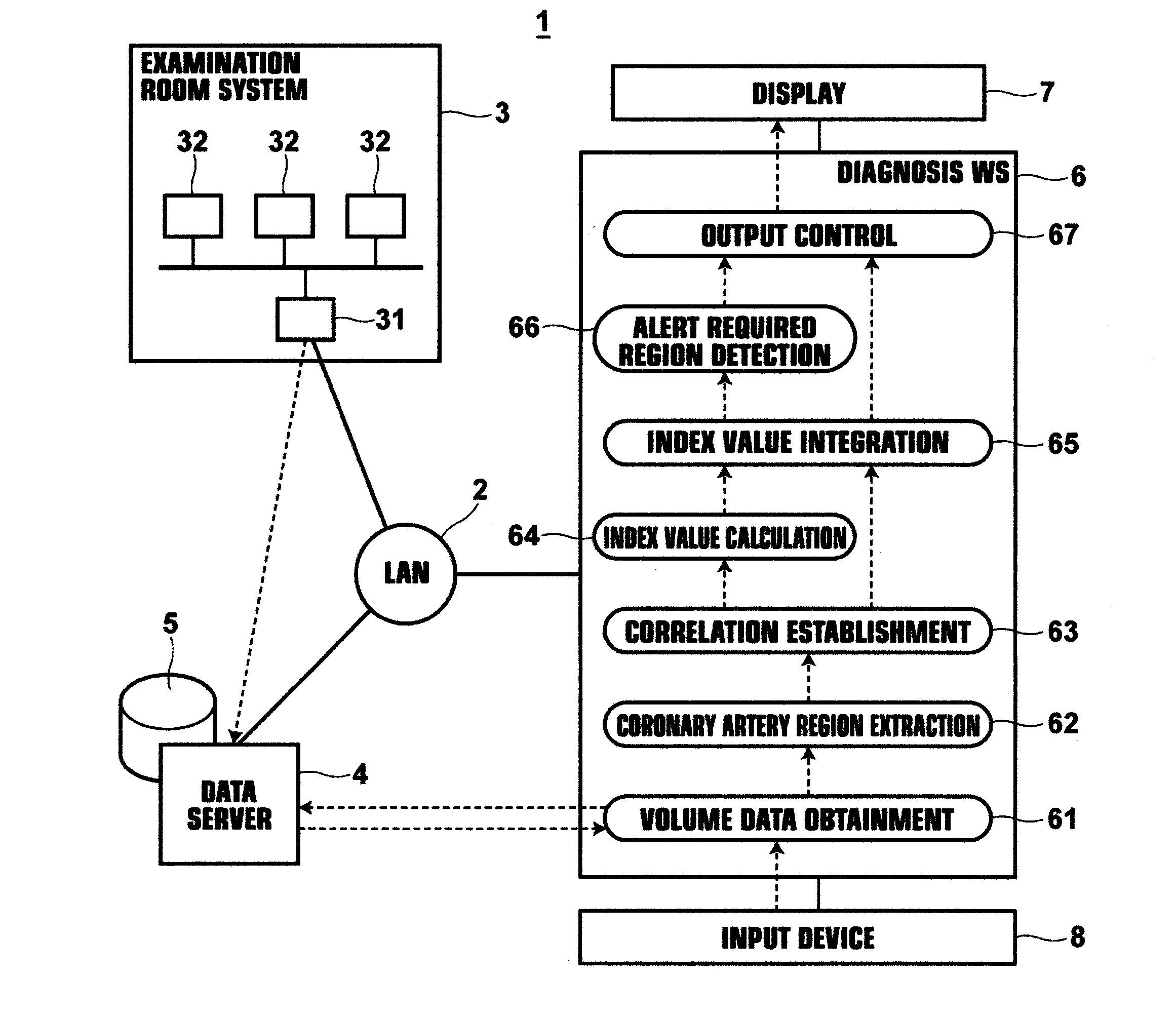
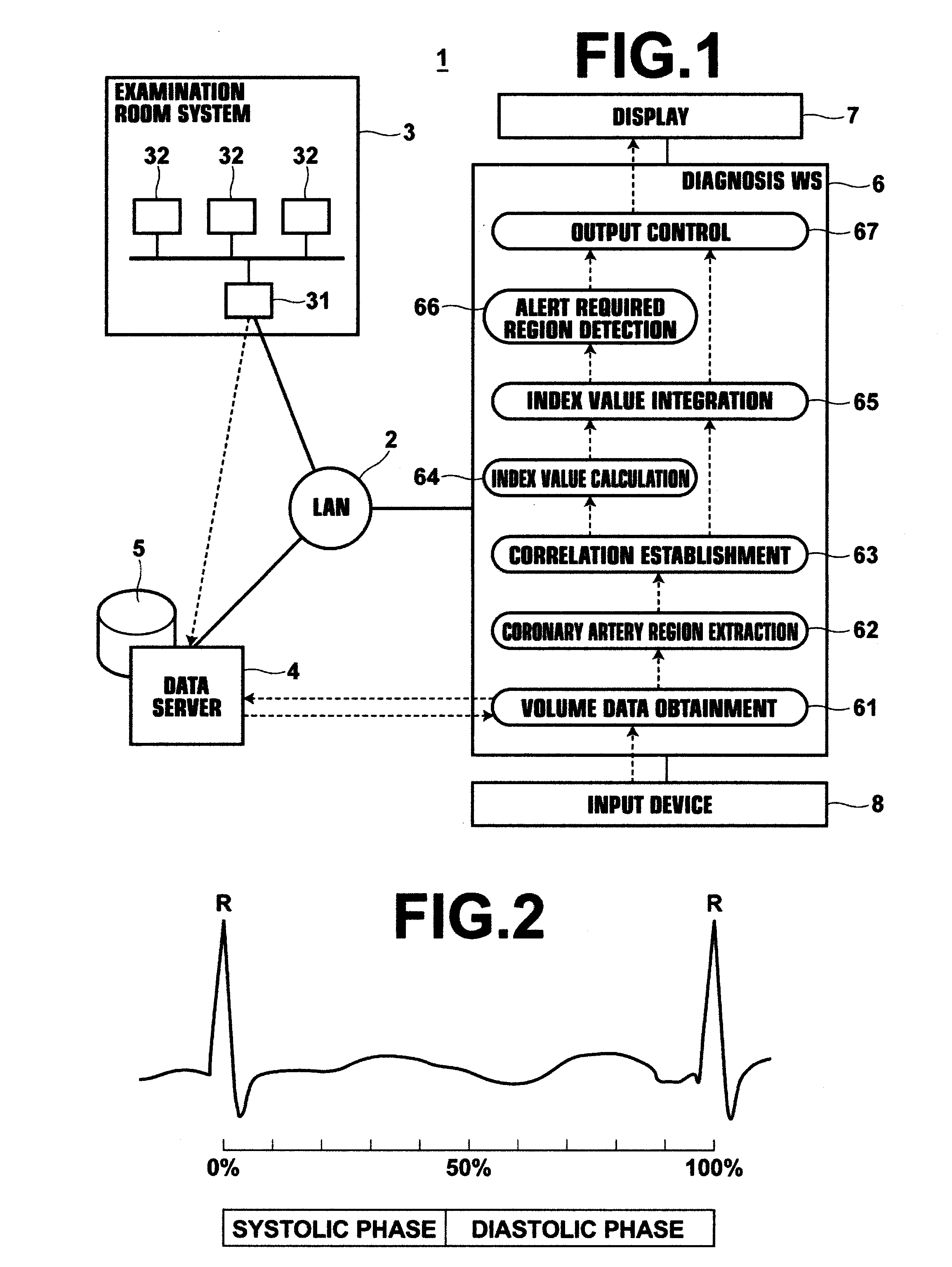
[0044]FIG. 1 illustrates the schematic structure of a hospital system 1 that includes a diagnosis assisting apparatus according to an embodiment of the present invention. The hospital system 1 is constituted by: an examination room system 3; a data server 4; and a diagnosis workstation 6 (WS 6); which are connected to each other via a local area network 2 (LAN 2).

[0045]The examination room system 3 is constituted by: various modalities 32 for imaging subjects; and an examination room workstation 31 (WS 31) for confirming and adjusting images output from each modality. Examples of the modalities 32 include: an X ray imaging apparatus; an MSCT (Multi Slice Computed Tomography) apparatus; a DSCT (Dual Source Computed Tomogra...


PUM


Abstract

A plurality of sets of volume data, each of which represent the state of a beating heart in different phases, are obtained. Coronary artery regions are extracted from at least two sets of volume data from among the obtained sets of volume data. A plurality of analysis points are set in each extracted coronary artery region. Correlations are established among analysis points set at the same anatomical positions within the coronary artery regions. Index values that indicate the character of plaque are calculated at each analysis point within all of the coronary artery regions. The character of plaque is evaluated at positions within the coronary artery regions, by integrating the index values calculated at the analysis points corresponding to each of the positions. The evaluation results regarding the character of plaque at each of the positions within the coronary artery regions are output, correlated with information regarding the positions.

Description

BACKGROUND OF THE INVENTION[0001]1. Field of the Invention[0002]The present invention is related to a method and an apparatus for assisting diagnosis by physicians by analyzing the states of coronary arteries based on there dimensional data. The present invention is also related to a recording medium in which a program that causes at least one computer to execute the diagnosis assisting method is recorded.[0003]2. Description of the Related Art[0004]Apparatuses and software programs that analyze the states of organs and blood vessels based on three dimensional image data (volume data) obtained by CT (Computed Tomography) examinations are being provided as tools for assisting image diagnosis by physicians. Cardiac function analyzing functions and coronary artery analyzing functions are widely utilized as functions for assisting diagnosis of the heart. With respect to the cardiac artery analyzing functions, Japanese Unexamined Patent Publication No. 2009-195561 discloses an apparatus ...

Claims


Application Information

Patent Timeline
Patent Type & Authority Applications(United States)
IPC IPC(8): A61B5/05
CPCG06T7/0016G06T2207/10076G06T2207/30172G06T2207/30048G06T2207/30101G06T2207/20072A61B6/5217A61B6/5211A61B6/5205A61B5/02007A61B5/02
Inventor KITAMURA, YOSHIRO
Owner FUJIFILM CORP
Learn More