Gene expression signature of genomic instability in breast cancer

Inactive Publication Date: 2011-09-15
US DEPT OF HEALTH & HUMAN SERVICES +1
View PDF0 Cites 1 Cited by
  • Summary
  • Abstract
  • Description
  • Claims
  • Application Information

AI Technical Summary

Benefits of technology

[0004]The invention provides a method of assessing genomic instability in breast cancer tissue comprising (a) measuring the expression level of (i) CDKN2A, SCYA18, STK15, NXF1, cDNA Dkfzp762M127, p28, KIAA0882, MYB, Human clone 23948, RERG, HNF3A, and (ii) ACADSB

Problems solved by technology

Genomic instability is the tendency of the cells of a tissue to exhibit diversity in the amount of nuclear DNA present, principally due to chromosomal rearrangement and duplication.

Method used


Image

  • Gene expression signature of genomic instability in breast cancer
  • Gene expression signature of genomic instability in breast cancer
  • Gene expression signature of genomic instability in breast cancer

Examples


example 1

[0053]The following example demonstrates the correlation of gene expression with genomic instability.

[0054]Tumor samples were collected from 48 patients diagnosed with breast cancer. Clinical material was collected from surgically removed tumors, diagnosed on H&E-stained tissue sections, and graded according to Elston et al., Histopathology, 19(5), 403-10 (1991). The clinical data are summarized in Table 1. After surgery, clinical tissue was used for touch preparation slides for quantitative measurement of the nuclear DNA content. The tissue samples were then snap frozen until further processing with TRIzol reagent (Invitrogen, Carlsbad, Calif.) for DNA and RNA extraction. In addition, paraffin-embedded specimens of the same tumors were used for histopathology and immunohistochemistry.

[0055]Image cytometry was performed on Feulgen-stained touch preparation slides. The staining procedure, internal standardization, and tumor cell selection were based on methods described previously (A...

example 2

[0061]The following example demonstrates the correlation between genomic instability and the expression of a 12-gene signature.

[0062]A supervised machine learning approach was applied in two sample settings to identify a genomic instability signature from the expression profiles of 7,657 genes in 44 of the primary breast cancer samples described in Example 1. Using random forests, the gene expression data from the three groups of tumors (dGS, aGS, and aGU) was compared. This comparison and classification generated a list of 70 genes. The top 10 genes of this set were selected with Relief, and then used to individually classify the three groups. This classification achieved an accuracy of 80%.

[0063]Random forests was then used to establish a gene list that discerns the genomically stable (dGS and aGS) tumors from the genomically unstable ones (aGU). Seven genes were identified from the analysis. In leave-one-out cross-validation, this 7-gene signature allowed classification of genomi...

example 3

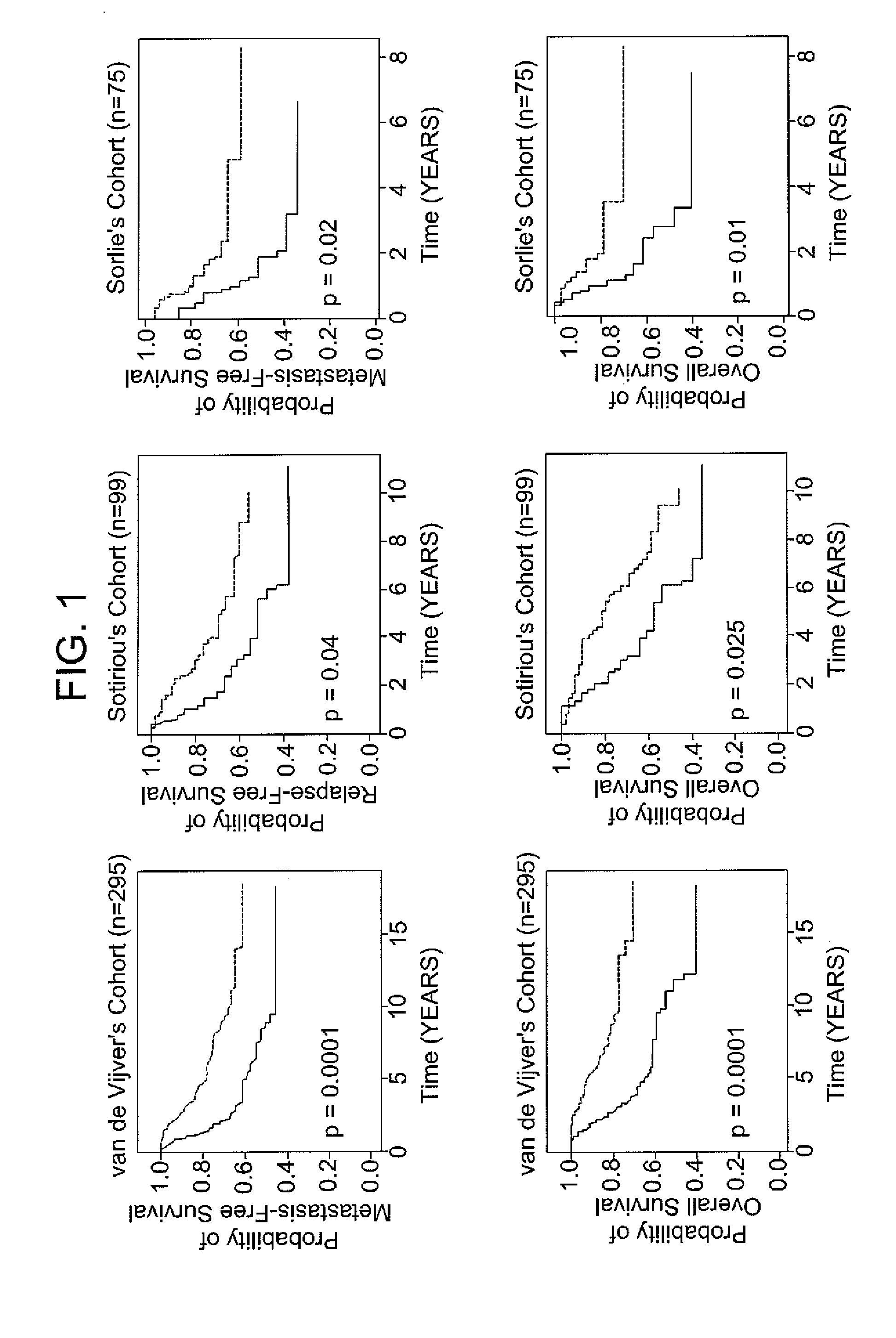
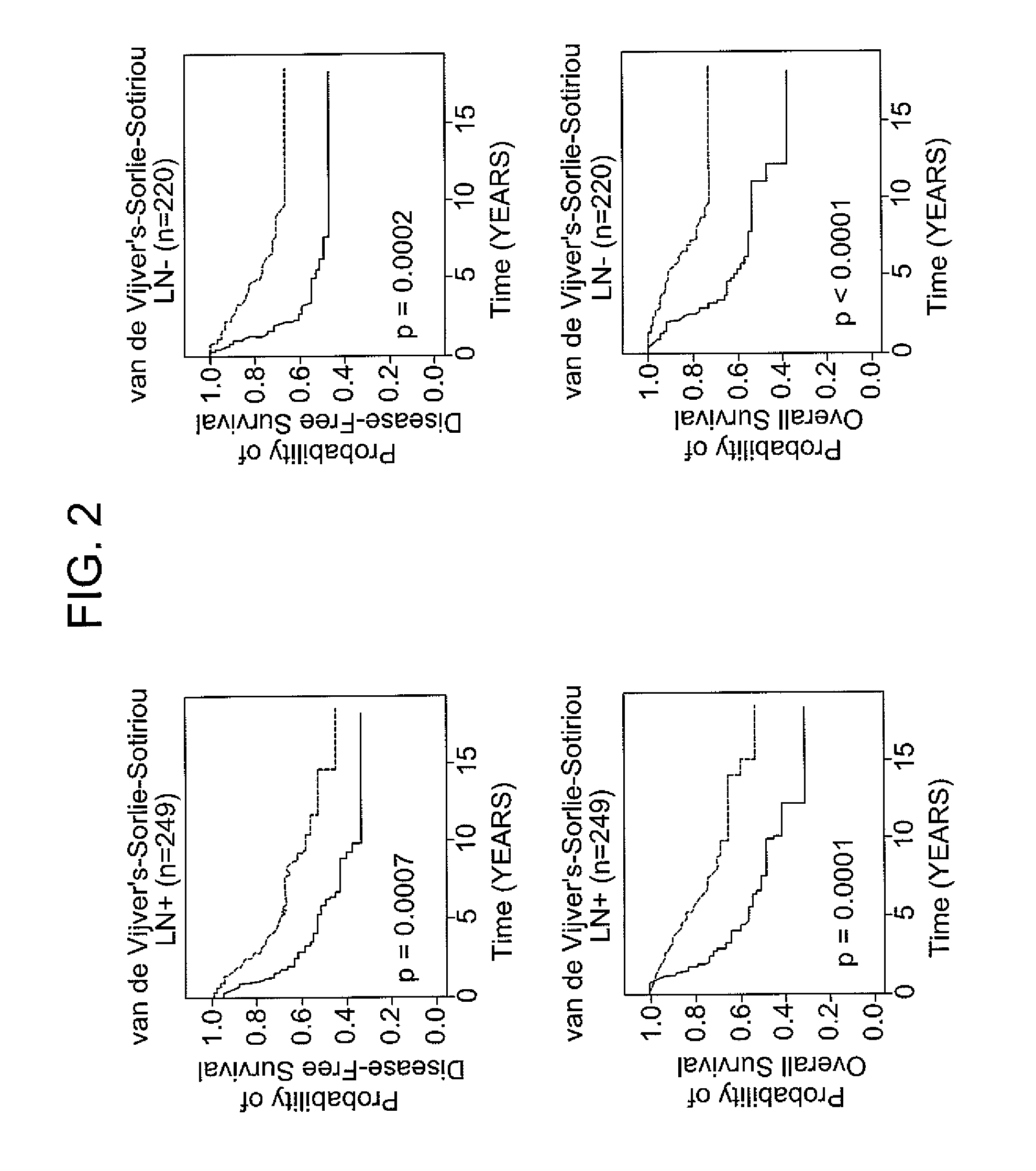
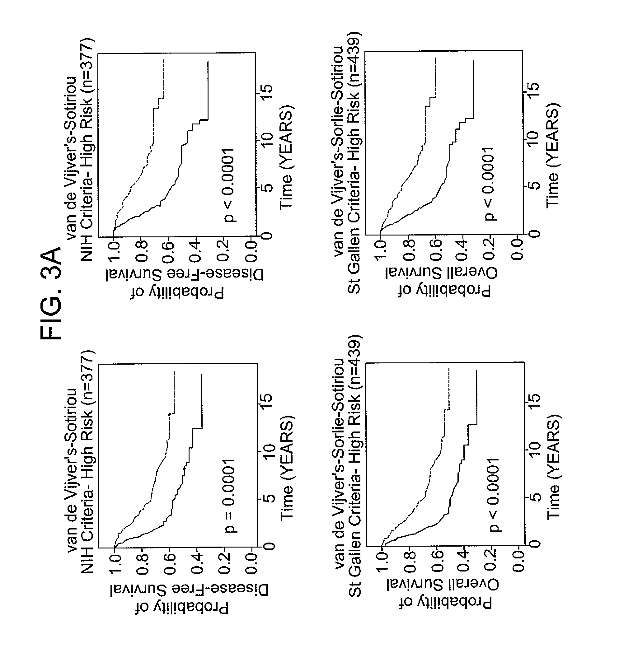
[0066]The following example demonstrates the use of the 12-gene signature to classify tumors in an independent validation set.

[0067]Patients with breast cancer from three data sets (n =469), for which different clinical endpoints were available, were used to validate the 12-gene signature. The three data sets are described in van de Vijver et al., N Engl J Med, 347 (25), 1999-2009 (2002), Sotiriou et al., Proc Natl Acad Sci USA, 100 (18), 10393-8 (2003), and Sorlie et al., Proc Natl Acad Sci USA, 100 (14), 8418-23 (2003). These datasets comprise gene expression profiles from 469 patients with heterogeneous histology and different disease stages. The clinical parameters included tumor grade, tumor size, lymph node status, estrogen receptor status, progesterone receptor status, age, and histology. The clinical endpoints used for the validation of the classifiers included relapse-free survival, metastasis-free survival, disease-free survival (here a disease event refers to either breas...


PUM

PropertyMeasurementUnit
Fractionaaaaaaaaaa
Dynamic viscosityaaaaaaaaaa

Abstract

Methods of assessing genomic instability in breast cancer tissue by measuring the expression level of genes CDKN2A, SCYA18, STK15, NXF1, cDNA Dkfzp762M127, p28 KIAA0882, MYB, Human clone 23948, RERG, HNF3A, and ACADSB or a nucleic acid sequence comprising about 90% or greater sequence identity to SEQ ID NO: 21 in breast cancer tissue, an array suitable for use in such methods, and related methods and compositions.

Description

CROSS-REFERENCE TO RELATED APPLICATIONS[0001]This patent application claims the benefit of U.S. Provisional Patent Application No. 61 / 097,101, filed Sep. 15, 2008, which is incorporated by reference.SEQUENCE LISTING[0002]Incorporated by reference in its entirety herein is a nucleotide / amino acid sequence listing submitted concurrently herewith.BACKGROUND OF THE INVENTION[0003]Breast cancers are among the most frequently occurring tumors in women worldwide, and the clinical course and disease free survival times for patients with breast cancers are extremely heterogeneous. Accordingly, intensive efforts have been made over the past decades to develop biological markers that allow for precise disease staging and prognostication. Such markers include tumor size, grade and stage, lymph node and hormone receptor status, expression of growth factor receptors, DNA content, S-phase fraction and proliferative activity, and, more recently, specific gene expression signatures that suggest a po...

Claims


Application Information

Patent Timeline
IPC IPC(8): C40B30/00C40B40/06C12Q1/68
CPCC12Q1/6886C12Q2600/112C12Q2600/118G01N33/57415C12Q2600/158G01N33/5011C12Q2600/136
Inventor RIED, KARL THOMASHABERMANN, JENSGUO, NANCY LANAUER, GERT
Owner US DEPT OF HEALTH & HUMAN SERVICES
Why Eureka
Try Eureka