
Therapeutic agent for mll leukemia and moz leukemia of which molecular target is m-csf receptor, and use thereof
- Summary
- Abstract
- Description
- Claims
- Application Information
AI Technical Summary
Benefits of technology
Problems solved by technology
Method used
Image

Examples
example 1
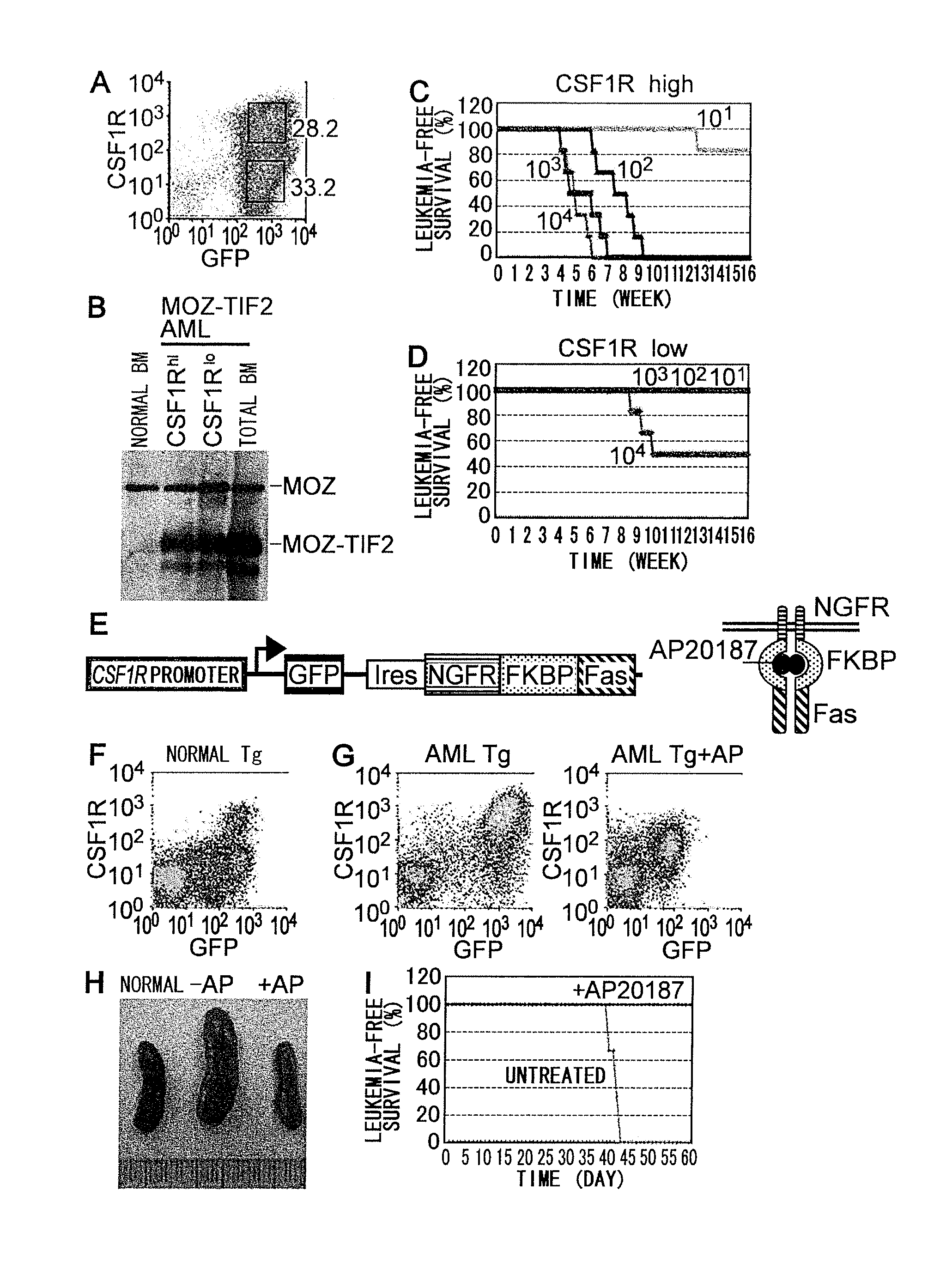
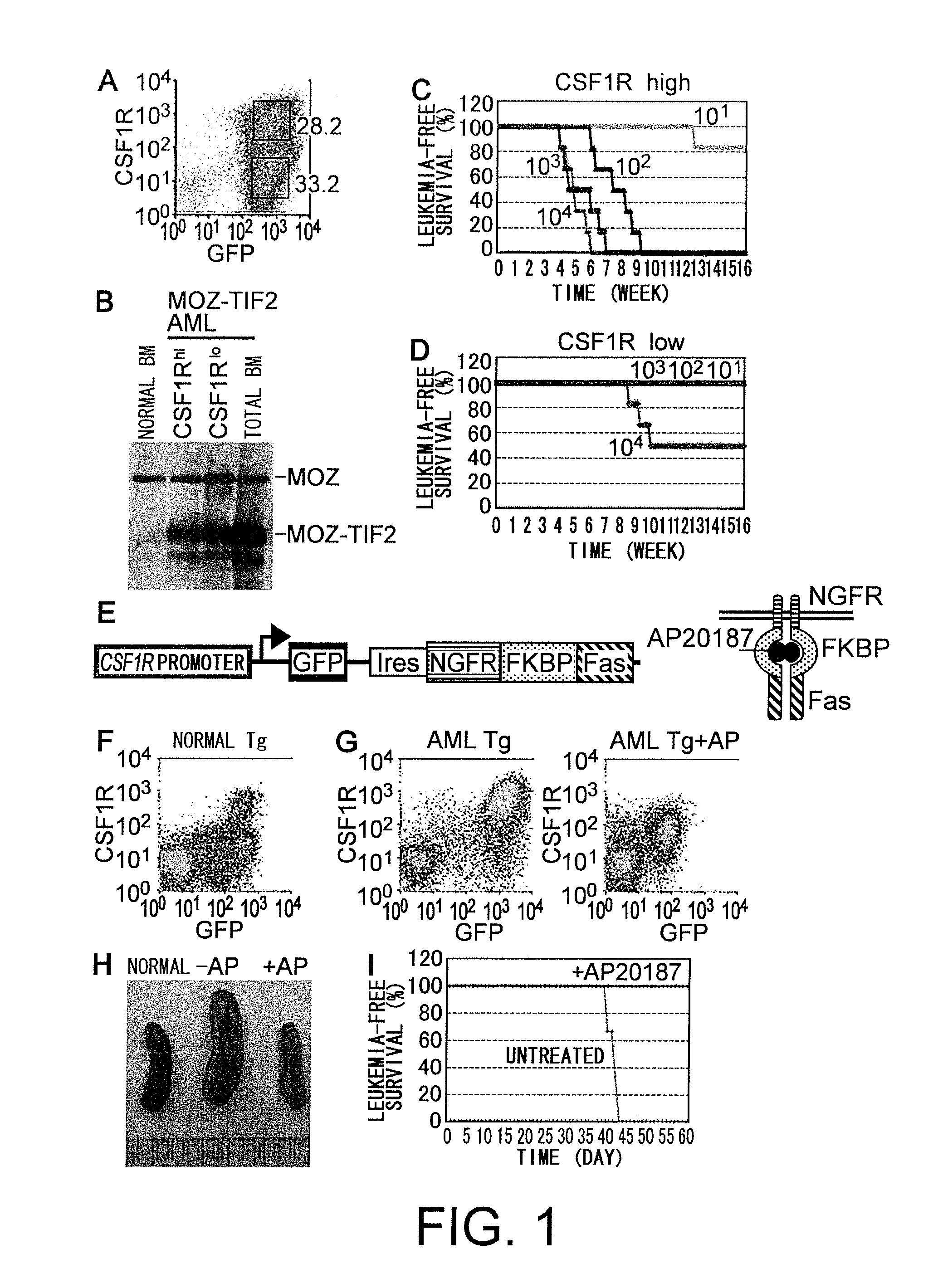
[0189]To generate a mouse model for acute myelogenous leukemia (herein below, abbreviated as AML), c-KIT+ bone marrow (BM) cells were infected with MSCV-MOZ-TIF2-ires-EGFP retrovirus and transplanted into lethally-irradiated mice. AML mice models are generated as described (Yokoyama, A. et al., Cell (2005) 123: 207-18). The mice developed AML about two months after transplantation as reported by Deguchi et al. (Deguchi, K. et al., Cancer Cell (2003) 3: 259-271). When limited numbers of BM cells derived from the AML mice were transplanted into irradiated mice, these mice developed AML three to six weeks after transplantation, suggesting that transplantable leukemia-initiating cells (herein after, abbreviated as LICs) were present in the BMs of AML mice (Huntly, B. J. et al., Cancer Cell (2004) 6: 587-96).
[0190]In order to identify LICs, the present inventors investigated the BM cells of the AML mice for various cell surface markers by fluorescence-activated cell sorting (FACS) analys...
example 2
[0192]To determine if leukemogenesis can be suppressed by removing M-CSFR-expressing cells that exhibit stronger LIC activity, the present inventors used transgenic mice expressing a drug-inducible FKBP-Fas suicide gene and EGFP under the control of the M-CSFR promoter (Burnett, S. H. et al., J Leukoc Biol (2004) 75: 612-23) (FIG. 1E). In the transgenic mice, expression levels of endogenous M-CSFR were proportional to those of EGFP and FKBP-Fas (FIG. 1F), and conditional ablation of M-CSFR-expressing cells can be induced by injection of the AP20187 dimerizer (Burnett, S. H. et al., J Leukoc Biol (2004) 75: 612-23). c-KIT+ BM cells of the transgenic mice were infected with the MOZ-TIF2 retrovirus, and transplanted into lethally-irradiated wild-type mice. These mice developed AML about two months after transplantation, and their BM cells (105 cells / mouse) were transplanted into secondary recipient mice. Seven days after transplantation, the mice were injected with AP20187 as described...
example 3
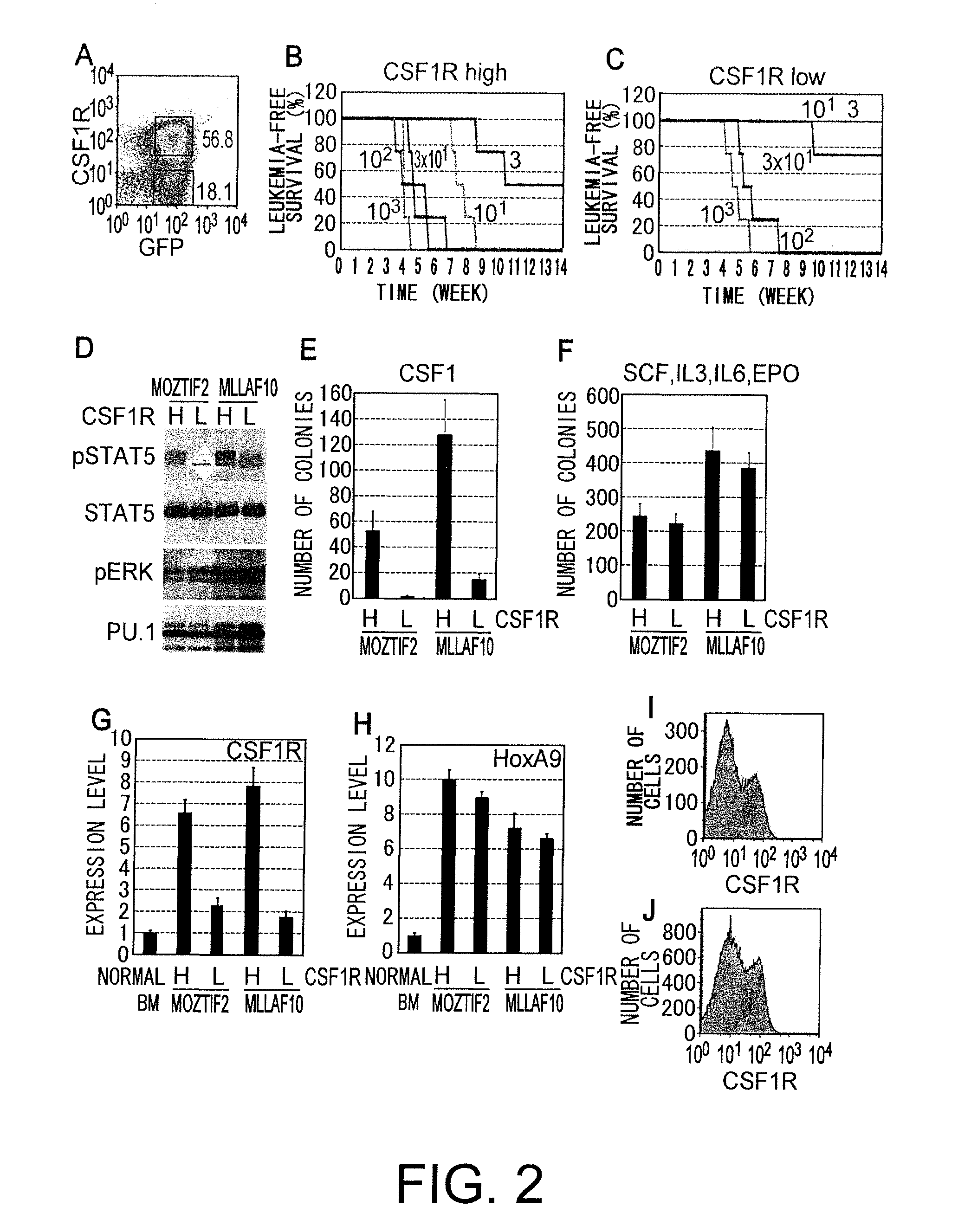
[0193]Then, the present inventors investigated the M-CSFR expression in AML mice with MLL-AF10 (DiMartino, J. F. et al., Blood (2002) 99: 3780-59), and found that M-CSFRhigh and M-CSFRlow / - cells were also present in BM of MLL-AF10-induced AML mice (FIG. 2A). When transplanted, cell sorter-sorted M-CSFRhigh cells exhibited more than 10 times stronger leukemia-initiating activity than M-CSFRlow / - cells (FIGS. 2B and 2C). STATS and ERIC, which are downstream effectors for M-CSFR, are activated in a variety of leukemia and myeloproliferative disorders. The present inventors investigated the phosphorylation status of these proteins in M-CSFRhigh and M-CSFRlow / - cells from MOZ-TIF2- and MLL-AF10-induced AML mice using immunoblot analysis with phospho-specific anti-STATS and anti-ERK antibodies. STATS was highly phosphorylated in M-CSFRhigh cells but not in M-CSFRlow / - cells (FIG. 2D), while ERK1 / 2 were phosphorylated in both M-CSFRhigh and M-CSFRlow / - cells. These results suggest that ST...
PUM
| Property | Measurement | Unit |
|---|---|---|
| Length | aaaaa | aaaaa |
| Level | aaaaa | aaaaa |
Abstract
Description
Claims
Application Information