[0010]The object underlying the present invention is therefore to disclose a method for recording and reconstructing a three-dimensional image dataset which permits improved imaging in relation to low-contrast structures in particular by means of a C-arm X-ray apparatus.
[0012]The basic concept of the present invention is to acquire, not one, but a plurality of projection images for realizing an improved
image quality in the low-contrast range in the determination of three-dimensional image datasets by means of C-arm X-ray apparatuses for at least one, in particular, however, for every recording geometry of a three-dimensional scan. This is because in this way different sets of information that can be processed collectively are acquired in the corresponding recording geometries, permitting
noise and / or
artifact reduction in the final reconstruction result by means of correct, in particular adaptive combination into an overall set of information. By means of such a combination mechanism it is possible in particular to take into account the plurality of projection images and to perform their fusion in order to produce a
single image dataset in such a way that what is involved is a pre-
processing or post-
processing, yet the actual
reconstruction algorithm—a multiplicity of such being known in the prior art—can remain unchanged. Then the innovative approach proposed here can be combined with any
reconstruction method, filtered back-projection for example.
[0014]In an alternative embodiment it can be provided that the number of images to be recorded for a recording geometry is determined adaptively during the recording session, in particular taking into account a
dose measurement and / or an
image analysis of a first recorded
projection image, and / or taking into account information concerning the object to be recorded, in particular the
path length through the object in the recording geometry. It is therefore possible, for each recording geometry, to adaptively adjust the number of projection images to be recorded there, wherein firstly preliminary information can be used, for example such information as concerns the object that is to be recorded, from which it emerges how long the
path length of the X-
ray radiation in the object is likely to be, so that for example in regions in which the
path length is very long, and consequently the attenuation very great, the number of projection images to be produced can be increased. In addition or alternatively, however, it is also possible to specify the number of projection images still to follow during the recording itself, for example by considering the
dose received in the specific recording geometry. A
dose measuring device is already provided in many X-ray apparatuses in order to enable automatic dose regulation to be performed. From the dose it can also be derived, for example, how strong is the attenuation due to the object and whether a particularly low
signal-to-
noise ratio is to be expected so that then a plurality of projection images can be recorded. Such information can also be deduced from an
image analysis which can relate, for example, also to the
total dose received by the
detector. In this way it is therefore possible to bring about improvements in the most recently obtained
image quality at the points at which a particularly poor contrast or a particularly high
signal-to-noise ratio is to be expected by recording an especially large number of projection images adapted to the current situation.
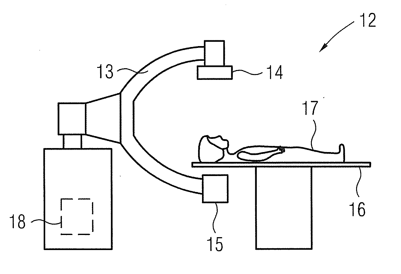
[0016]The different projection images of a recording geometry can be recorded during a single period in which a recording arrangement of the X-ray apparatus remains in the recording geometry and / or during a plurality of passes of the recording arrangement through the recording geometry. In the example of the C-arm
system this means that for example all images can be recorded during a single revolution of the C-arm if a single assumption of the recording geometry is to be sufficient for recording all the images. Using a single recording movement can make sense if the number of recorded projection images varies with the recording geometry. The presence of
motion artifacts can be also minimized with only a single recording movement. Nonetheless, a multiple recording movement can also be useful, for example when a constant number of projection images are to be acquired and at least the target volume in the object exhibits little or no movement, in particular is well fixed. For example, it can advantageously be provided that the C-arm performs the standard recording movement around the object multiple times, e.g. as a sequence of forward and backward passes.
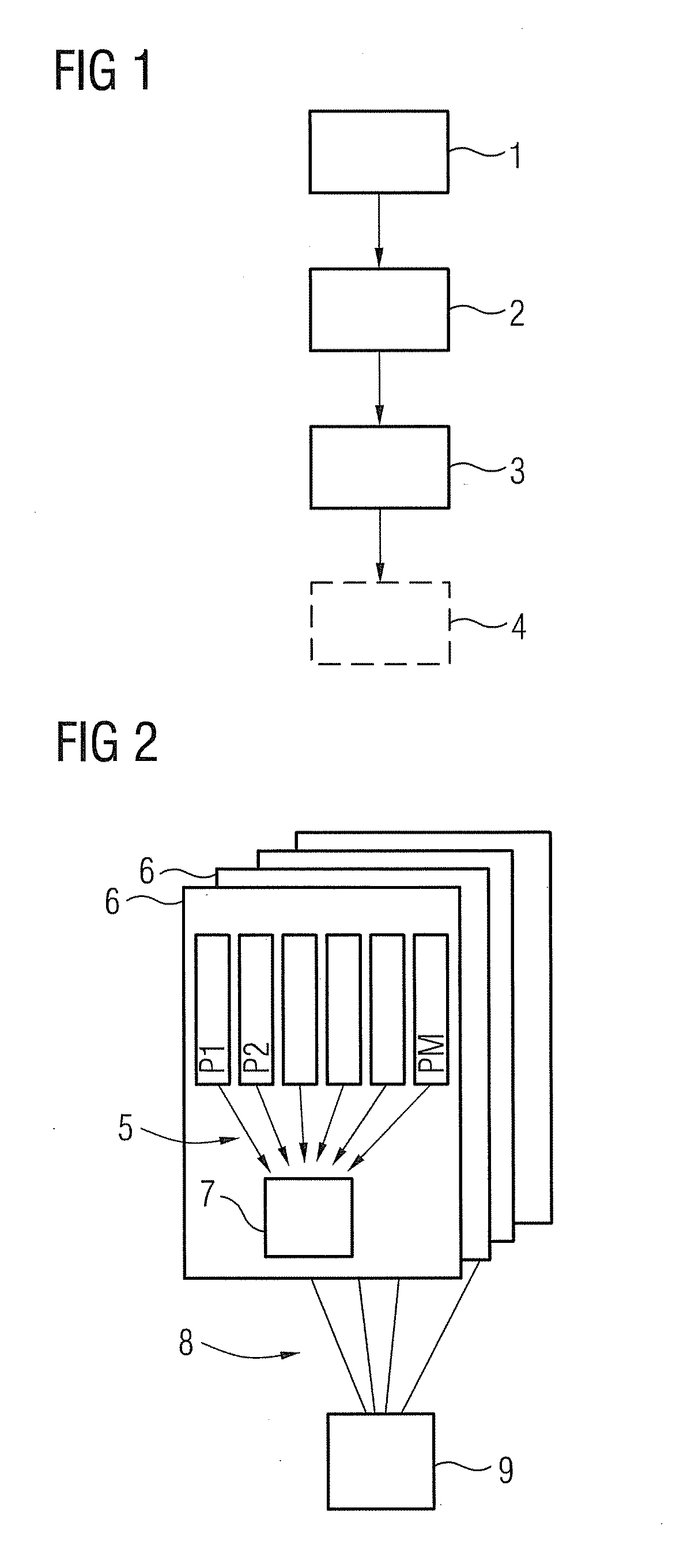
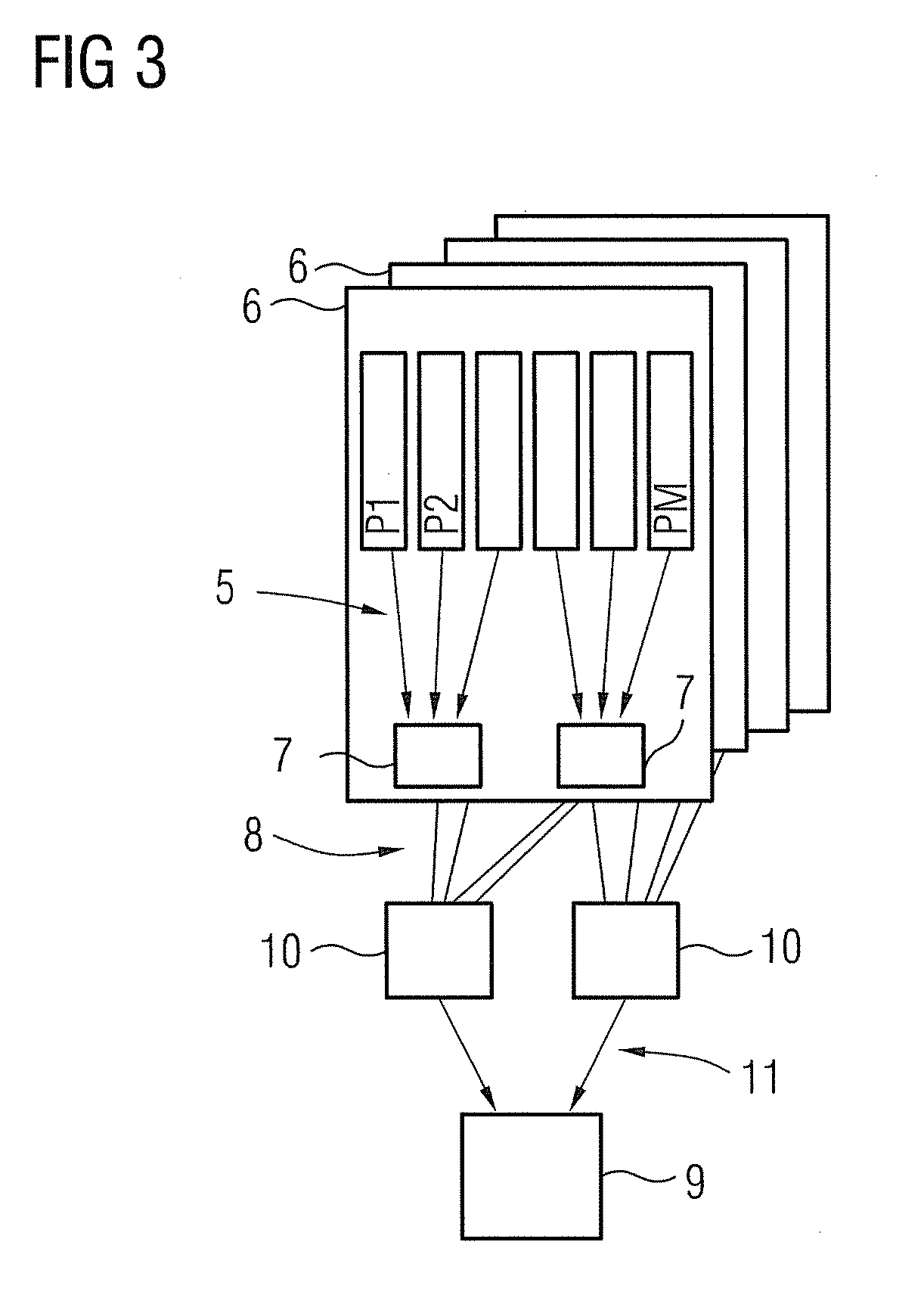
[0017]In a particularly advantageous embodiment of the method according to the invention it can be provided that the plurality of projection images of one recording geometry are fused in at least one combination step to form at least one combination image and / or at least two reconstruction datasets are determined from different projection images and / or combination images and the image dataset is consolidated herefrom. According to the invention the described combination or fusion of the different projection images can therefore be carried out already on the basis of the projection images themselves, which is preferred according to the invention, and / or also through reconstruction of different reconstruction datasets and their combination. In order to keep the reconstruction overhead to a minimum and to be able to work already at the level of the projection images where certain occurring effects impairing the
image quality in the low-contrast range can be handled better, it is advantageous to provide in every case a combination step in which combination images are fused. It is particularly advantageous in this case if at least two different combination steps are performed. This means that different methods for combining or fusing the projection images / combination images can be applied in order to improve the image quality in different ways, as will be discussed in greater depth below. This can additionally, if it is beneficial, be supplemented in that in actual fact more than one reconstruction dataset is produced, these reconstruction datasets then being combined to form the image dataset in a further method for improving the image quality, particularly in relation to the low-contrast range.


