Methods of Producing RPE Cells and Compositions of RPE Cells

a technology of rpe cells and compositions, applied in the direction of drug compositions, biocide, cardiovascular disorders, etc., can solve the problems of macular degeneration, serious consequences of thinning of the retina, etc., and achieve the effect of maximizing the likelihood of cell survival and reducing the risk of cell damag

Inactive Publication Date: 2011-11-10
ADVANCED CELL TECH INC
View PDF3 Cites 43 Cited by
  • Summary
  • Abstract
  • Description
  • Claims
  • Application Information

AI Technical Summary

Benefits of technology

[0066]In certain embodiments of any of the foregoing, said substantially purified culture of RPE cells (with or without mature RPE cells) are frozen for storage. The cells may be stored by any appropriate method known in the art, e.g., cryogenically frozen and may be frozen at any temperature appropriate for storage of the cells. For example, the cells may be frozen at approximately −20° C., −80° C., −120° C., or at any other temperature appropriate for storage of cells. Cryogenically frozen cells are stored in appropriate containers and prepared for storage to reduce risk of cell damage and maximize the likelihood that the cells will survive thawing. In other embodiments, RPE cells can be maintained at room temperature, or refrigerated at, for example, approximately 4° C.
[0067]In certain embodiments of any of the foregoing, human RPE cells are produced in accordance with Good Manufacturing Practices (GMP). In certain embodiments of any of the foregoing, the human embryonic stem cells from which the RPE cells are differentiated were derived in accordance with Good Manufacturing Practices (GMP). In certain embodiments of any of the foregoing, the human embryonic stem cells from which the RPE cells are differentiated were derived from one or more blastomeres removed from an early stage embryo without destroying the remaining embryo. As such, the invention provides GMP compliant preparations of RPE cells, including substantially purified preparations of RPE cells. Such preparations are substantially free of viral, bacterial, and / or fungal contamination or infection.

Problems solved by technology

Breakdown of the RPE interferes with the metabolism of the retina, causing thinning of the retina.
Thinning of the retina can have serious consequences.
For example, thinning of the retina may cause “dry” macular degeneration and may also lead to the inappropriate blood vessel formation that can cause “wet” macular degeneration).

Method used


Image

  • Methods of Producing RPE Cells and Compositions of RPE Cells
  • Methods of Producing RPE Cells and Compositions of RPE Cells
  • Methods of Producing RPE Cells and Compositions of RPE Cells

Examples


example 1

RPE Differentiation and Culture

[0213]Cryopreserved hES cells were thawed and placed into suspension culture on Lo-bind Nunclon Petri dishes in MDBK-Growth Medium (Sigma—SAFC Biosciences) or OptimPro SFM (Invitrogen) supplemented with L-Glutamine, Penicillin / Streptomycin, and B-27 supplement. The hES cells had been previously derived from single blastomeres biopsied from early cleavage stage human embryos. The remainder of the human embryo was not destroyed. Two hES cell line derived from single blastomeres were used—MAO1 and MAO9. The cells were cultured for 7-14 days as embryoid bodies (EBs).

[0214]After 7-14 days, the EBs were plated onto tissue culture plates coated with gelatin from porcine skin. The EBs were grown as adherent cultures for an additional 14-28 days in MDBK-Growth Medium or OptimPro SFM supplemented with L-Glutamine, and Penicillin / Streptomycin, without B-27 supplement.

[0215]From amongst the cells in the adherent culture of EBs, RPE cells become visible and are rec...

example 2

RPE Isolation and Propagation

[0216]As differentiated RPE cells continue to appear in the adherent cultures, clusters of differentiated RPEs become visibly noticeable based on cell shape. Frozen collagenase IV (20 mg / ml) was thawed and diluted to 7 mg / ml. The collagenase IV was applied to the adherent culture containing RPE clusters (1.0 ml to each well in a 6-well plate). Over approximately 1-3 hours, the collagenase IV dissociated the cell clusters. By dissociating the RPE clusters from other cells in the culture, an enriched suspension of RPE cells was obtained. The enriched RPE cell suspension was removed from the culture plate and transferred to a 100 mm tissue culture dish with 10 ml of MEF medium. Pigmented clumps are transferred with a stem cell cutting tool (Swemed-Vitrolife) to a well of a 6-well plate containing 3 ml of MEF media. After all clumps have been picked up, the suspension of pigmented cells is transferred to a 15 ml conical tube containing 7 ml of MEF medium and...

example 3

RPE-Specific mRNA Expression Measured by Quantitative, Real-Time, Reverse Transcription PCR (qPCR)

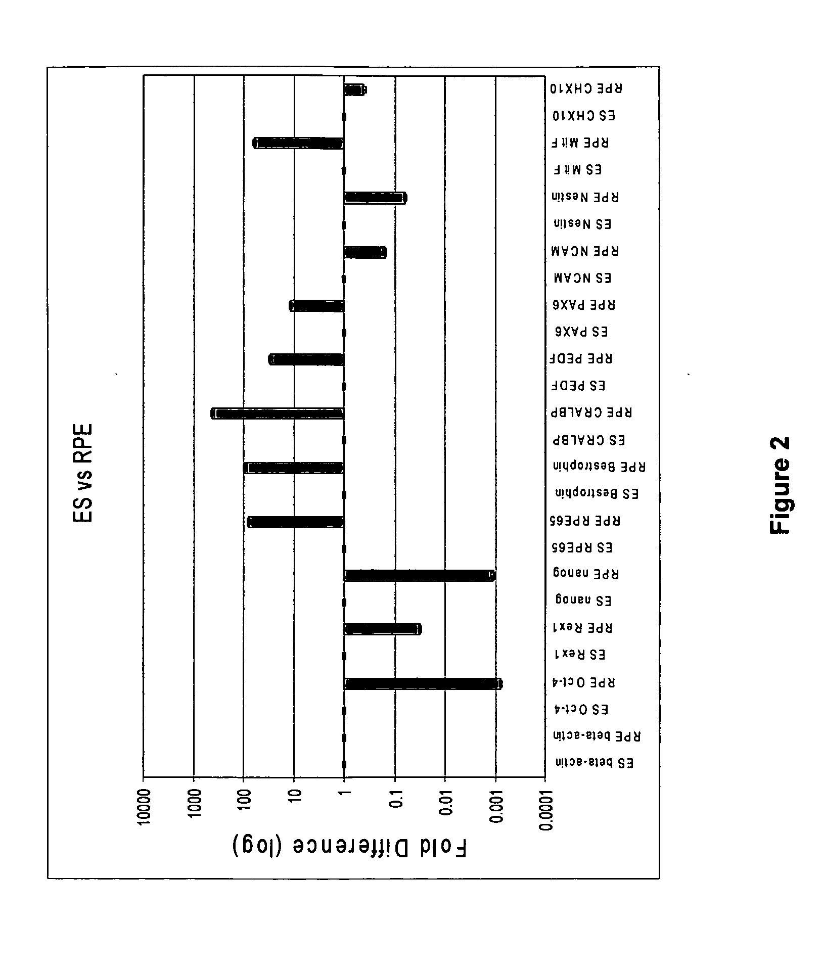
[0219]In order to characterize developmental stages during the human embryonic stem cell (hES) differentiation process into retinal pigmented epithelium (RPE) several assays have been employed to identify the expression levels of genes key to each representative stage of development. qPCR was developed to provide a quantitative and relative measurement of the abundance of cell type-specific mRNA transcripts of interest in the RPE differentiation process. qPCR was used to determine genes that are uniquely expressed in human embryonic stem cells, in neuroretinal cells during eye development, and in RPE cells differentiated from human embryonic stem cells. The genes for each cell type are listed below in Table 1.

TABLE 1Genes specific to hES, neuroretina / eye, and hRPE cellshESc-SpecificNeuroectoderm / NeuroretinaRPE-Specific GenesOct-4 (POU5F1)CHX10PAX-6NanogNCAMPAX-2Rex-1NestinRPE-65TDGF-1Be...


PUM

PropertyMeasurementUnit
temperatureaaaaaaaaaa

Abstract

The present invention provides improved methods for producing RPE cells from human embryonic stem cells or from other human pluripotent stem cells. The invention also relates to human retinal pigmented epithelial cells derived from human embryonic stem cells or other human multipotent or pluripotent stem cells. hRPE cells derived from embryonic stem cells are molecularly distinct from adult and fetal-derived RPE cells, and are also distinct from embryonic stem cells. The hRPE cells described herein are useful for treating retinal degenerative diseases.

Description

RELATED APPLICATIONS[0001]This application claims the benefit of priority to U.S. provisional application Nos. 60 / 998,766, filed Oct. 12, 2007, 60 / 998,668, filed Oct. 12, 2007, 61 / 009,908, filed Jan. 2, 2008, and 61 / 009,911, filed Jan. 2, 2008. The disclosures of each of the foregoing applications are hereby incorporated by reference in their entirety.BACKGROUND OF THE INVENTION[0002]The retinal pigment epithelium (RPE) is the pigmented cell layer just outside the neurosensory retina. This layer of cells nourishes retinal visual cells, and is attached to the underlying choroid (the layer of blood vessels behind the retina) and overlying retinal visual cells. The RPE acts as a filter to determine what nutrients reach the retina from the choroid. Additionally, the RPE provides insulation between the retina and the choroid. Breakdown of the RPE interferes with the metabolism of the retina, causing thinning of the retina. Thinning of the retina can have serious consequences. For example...

Claims


Application Information

Patent Timeline
Patent Type & Authority Applications(United States)
IPC IPC(8): A61K35/12C12N5/071A61P27/02C12Q1/02
CPCA61K35/12C12N5/0621C12N2506/02C12N2509/00C12N2501/734A61K35/30C12N2500/32C12N2501/06C12N2501/10C12N2533/54A61P25/16A61P27/02A61P27/06A61P43/00A61P9/00A61P9/10
Inventor MALCUIT, CHRISTOPHERLEMIEUX, LINDAHOLMES, WILLIAMHUERTAS, PEDROVILNER, LUCY
Owner ADVANCED CELL TECH INC
Why Eureka
Try Eureka