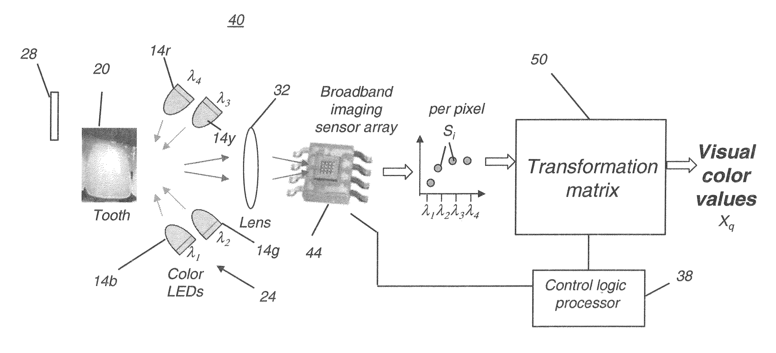
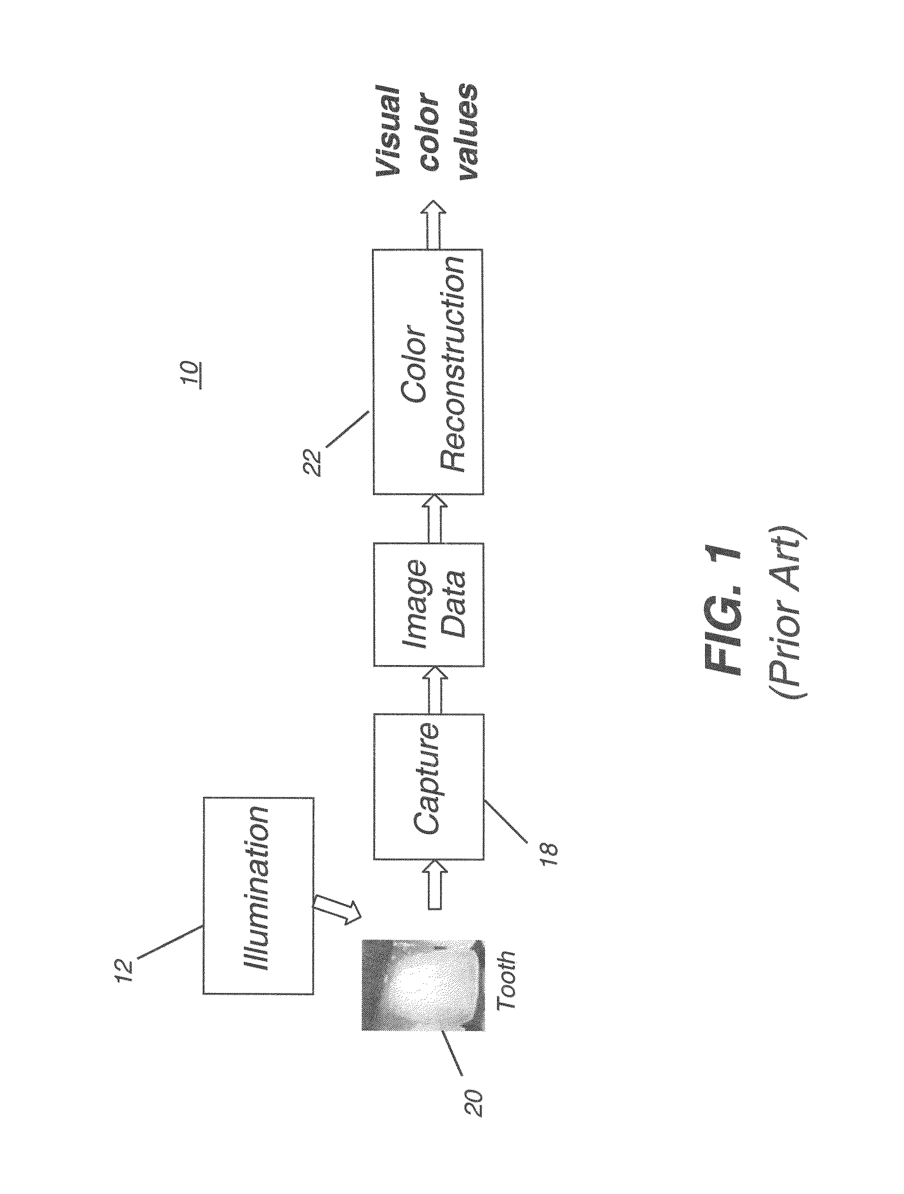
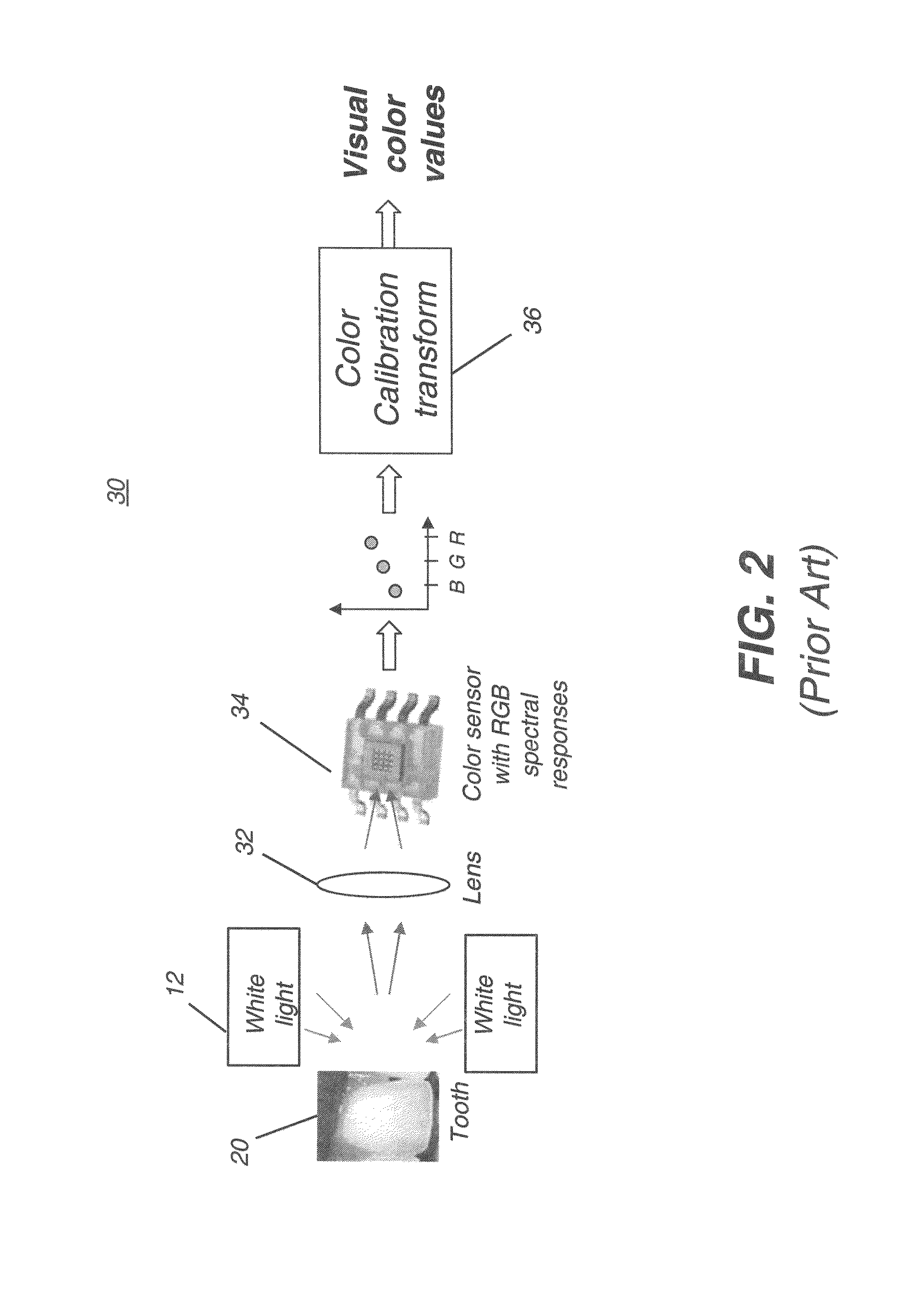
The visual shade matching process can be highly subjective and subject to a number of problems.
The initial matching procedure is often difficult and tedious, and it is not unusual for the process to take twenty minutes or longer.
The problem of accurately modeling the color of a tooth is more complex than obtaining a close color match using shade tabs.
One result of this complexity is that color appearance and
color measurement are greatly influenced by lighting geometry, spectrum, surrounding colors, and other environmental factors.
As a further complication, color within a
single tooth is generally not uniform.
Color non-uniformities can result from spatial variations in composition, structure, thickness, internal and external stains, surface texture, fissures, cracks, and degree of wetness.
As a result, measurements taken over relatively large areas produce averaged values that may not be representative of a tooth's dominant color.
In addition, natural color variations and non-uniformities make it unlikely that a given tooth can be matched exactly by any single shade tab.
Further,
tooth color is seldom uniform from tooth to tooth.
Therefore, the ideal color of a restoration may not be in visual harmony with that of an adjacent tooth or of any other
single tooth in a patient's mouth.
Understandably, they are quite intolerant of restorations that appear inappropriate in color.
It is often difficult to decide which tab matches most closely (or, conversely, which has the least mismatch) and to provide accurate information on color variation over the
tooth surface.
Frequently, the practitioner determines that the patient's teeth are particularly difficult to match, requiring that the patient go in person to the laboratory that will be fabricating the restoration.
Considering the relative difficulty of the
color matching task, and the further complexity of color mapping, a
high rate of failure is not at all surprising.
Visual color evaluation of relatively small color differences is always difficult, and the conditions under which dental color evaluations must be made are likely to give rise to a number of complicating psychophysical effects such as local chromatic
adaptation, local brightness
adaptation, and lateral-brightness
adaptation.
Moreover, shade tabs provide at best a metameric (that is, non-spectral) match to real teeth; thus, the matching is illuminant-sensitive and subject to variability due to normal variations in human
color vision, such as observer metamerism, for example.
Maintaining accuracy tends to be difficult due to metamerism, in which the color measured is highly dependent upon the illuminant.
This is particularly troublesome since dental measurement and imaging are generally carried out under conditions that differ significantly from natural lighting conditions.
As with RGB-based devices described in (i), measurements from this type of device also suffer from metamerism.
It is significant to note, however, that the spectrophotometer is not an imaging device.
Although the data obtained using the spectrophotometric approach provides advantages for more accurate
color matching over colorimetric and RGB approaches, including
elimination of metamerism, this approach has been found difficult to implement in practice.
The use of a
light scanning component for measuring different spectral components, generally employing a
grating or
filter wheel, tends to make the spectrophotometric
system fairly bulky and complex.
This makes it difficult to measure teeth toward the back of the mouth, for example.
Attempts to alleviate this problem have not shown great success.
However, as is true of spectrophotometric devices in general (iii, above), this device measures only a small area of the
tooth surface at a time.
The
image capture process is time-consuming and does not provide consistent results.
Color mappings can be inaccurate using such an approach, since there can be considerable sensitivity to illumination and
image capture angles and probe orientation during the imaging process.
In general, conventional methods that employ color filters, either at the illuminant end or at the sensor end, can be less desirable because they are subject to the limitations of the filter itself.


