
Method and apparatus for closing an opening in thick, moving tissue

a tissue opening and moving technology, applied in the field of moving tissue opening methods and apparatuses, can solve the problems of difficult establishment and closure of access wounds in tissue structures that are thicker walls, and are not fixed in position, and achieve the effect of avoiding overly compressing the tissu

Active Publication Date: 2012-01-19
LSI SOLUTIONS
View PDF7 Cites 61 Cited by
  • Summary
  • Abstract
  • Description
  • Claims
  • Application Information

AI Technical Summary

Benefits of technology

[0020]The present invention reliably places two concentric horizontal mattress sutures around a targeted tissue site, which can be thicker walled and potentially moving. To protect a suture closure site, an additional element called a pledget is sometimes used. The pledget, also called a bolster, can be made from sturdy but soft and compliant pad-like material, such as Teflon cloth or rolled cotton linen. The pledget is placed between the narrow suture and the delicate tissue to avoid overly compressing the tissue with the suture during tightening and healing. The outside segment of both sutures at the flat bottom of the box passes over a central portion of a single four holed pledget held in the distal end of the device. The two ends of each suture pass through four corresponding holes in the pledget so that after taking the four tissue bites, the pledget is pulled down onto the tissue. To close the top of the box, a second single four holed pledget or alternatively two, two holed pledgets can be used to secure the free ends of the suture that have exited the proximal side of the tissue suturing site. The free ends of the suture pass through holes in the pledgets and provide a cushion buffer between the knotted suture connection and the underlying tissue. Knotting or otherwise securing the suture together essentially permanently closes the top of box and compresses together the tissue between the sutures and pledgets.
[0021]This is a novel suturing instrument for the remote placement of multiple pledgeted sutures centered circumferentially surrounding a targeted tissue wound site in relatively thick and less compliant tissue, such as the beating heart and aortic wall structures. Its sutures are precisely delivered to predetermined tissue depths and for pre-set distances. It does not require the distal end of the device to enter the wound site and can place the secure sutures around an existing guide wire before the access wound is fully opened. The suturing instrument includes a pistol grip style handle and a hand actuated lever for the precise placement of multiple pledgeted horizontal mattress sutures at the targeted access site, such as the anterior surface of the heart near its apex or the ascending aorta. The shaft of the instrument connects at its proximal end to the handle, which remains outside of the patient and in direct control of the surgeon. For example, in creating and closing a transapical access site, the shaft enters and traverses the patient's body at the left chest wall to deliver the device distal end where suture placement occurs to the targeted anatomic location (e.g., the apex of the beating heart).
[0022]This suturing instrument incorporates a low profile rotating, lockable, mechanical tissue alignment guide to enable more automatic controlled positioning of its distal end at its desired tissue location. The unlocked alignment mechanism rotates to allow the device end and its indwelling guide wire to pass more freely through narrow openings. When locked, the alignment guide provides a stable guide mechanism orienting the indwelling guide wire in a favorable position perpendicular to the tissue receiving jaw or welting trough of the suturing instrument for placing the sutures.
[0023]Further, this instrument provides a mechanical assembly to accurately and securely engage the distal end of this suture delivery instrument with the targeted tissue site. One tissue engagement assembly provides two linked balloons. When both balloons are filled and expanded, a tissue ridge or welt is compressed and sandwiched in the space between the balloons and the receiving gap or welting trough in the distal end of the instrument, thereby moving and re-conforming the targeted tissue into the most appropriate suturing position. An alternative tissue engagement assembly provides an expandable, mechanical assembly such as an internal hinged mechanical anchor that pulls tissue up into the suturing position against the welting trough in the undersurface of the suturing jaw, while an external compression spring exerts force on the top of the rotating alignment guide mechanism to further press the welting trough onto the tissue suturing site.
[0025]Transcatheter therapies are rapidly becoming mainstream for the treatment of structural heart disease. A transthoracic non-rib spreading single port option providing short distance, non-torturous direct access and including a secure transapical wound closure could further advance the benefits of this antegrade procedure.
[0028]Advanced customized tools are needed to assure cardiothoracic surgeons continue to lead in the critically important arena of minimally invasive therapeutic heart procedures. This automated transapical access wound closure technology and technique developed for endoscopic use was demonstrated to be ergonomic, fast, effective and highly reliable. The fresh porcine heart bursting pressure study showed these remote suture mediated wound closures remained hermetic beyond the supra physiologic infusion pressures intolerable to other structural elements of the hearts tested in this model. The early successful results illustrating hemostatic closures in porcine in vivo beating hearts and transthoracic totally endoscopic apical closures through a single port in the human cadaver model encourage further evaluation.

Problems solved by technology

Access wounds made in tissue structures that are thicker walled, not fixed in position (e.g., mobile or actively moving) or acting as barriers to hold pressurized fluids or gases can be more challenging to establish and close.
While transcatheter transapical heart valve replacement products are already helping many patients, especially in Europe, until now an excellent means to remotely close transapical access wounds has remained elusive.
Hand sewing a traditional double purse string suture into the ascending aorta through a minimally invasive small remote port site using standard needle drivers is so challenging that for most surgeons it would not be worth the additional risks.
These balloons tend to migrate to less appropriate locations and frequently require repositioning.
Any catheter traversing the arch of the aorta risks displacing embolic material and inducing stroke and other complications.
The use of non-specialized laparoscopic or thoracoscopic needle drivers presents significant limitations to ergonomic and accurate remote suture placement.
None of these products readily permits the accurate and simultaneous placement of concentric sutures at the tissue locations required in the applications.
Typically these devices are suture mediated and their device distal ends enter the hole they are intended to close, which may be problematic in the above mentioned examples.
While many variations of trocar wound closure devices have come into use over the past two decades (U.S. Pat. Nos. 5,368,611 and 5,620,456), none are known to enable this transapical wound or aortic cannulation site preparation and closure.
Since these devices have also been available for some time, they appear unacceptable for the proposed related applications, including transapical access and aortic cannulation site closure.
Thicker walled structures may not be held reliably enough by vacuum alone.
This previous instrument is not intended for use on tissue which is acting as a barrier to hold back pressurized fluids.
The needles generally penetrate the full thickness of the tissue, which could cause immediate leakage of the pressurized fluid.
Also the use of integrated cutting blade could cause an immediate hemorrhage, for example, in a beating heart.
The previous technology does not teach a mechanical instrument-to-tissue alignment mechanism or an instrument-to-tissue secure engagement means based on compression between external and internal anchors.
The previous technology was not intended for use with thick walled moving structures.
Despite a long recognized critical clinical need, no technology is known to exist that provides for the safe and effective minimally invasive closure of certain access wounds required for many therapeutic interventions, especially several related to cardiac procedures involving thicker tissue.

Method used


Image

  • Method and apparatus for closing an opening in thick, moving tissue
  • Method and apparatus for closing an opening in thick, moving tissue
  • Method and apparatus for closing an opening in thick, moving tissue

Examples


Embodiment Construction

[0080]In accordance with a presently preferred embodiment of the invention, a remote suturing instrument is provided for the precise placement of multiple pledgeted horizontal mattress sutures at a guide wire targeted location in a predetermined orientation, depth and length for securing thicker, less compliant tissue structures. An embodiment of the innovation disclosed here includes a multiple needle drive feature along with a pledgeted suture storage and release concept, a rotating tissue alignment guide mechanism, which includes an extended tube, and two alternative mechanical tissue engagement assemblies (one incorporating balloons and the other a hinged anchor and compression spring) to better hold and position the tissue for suturing.

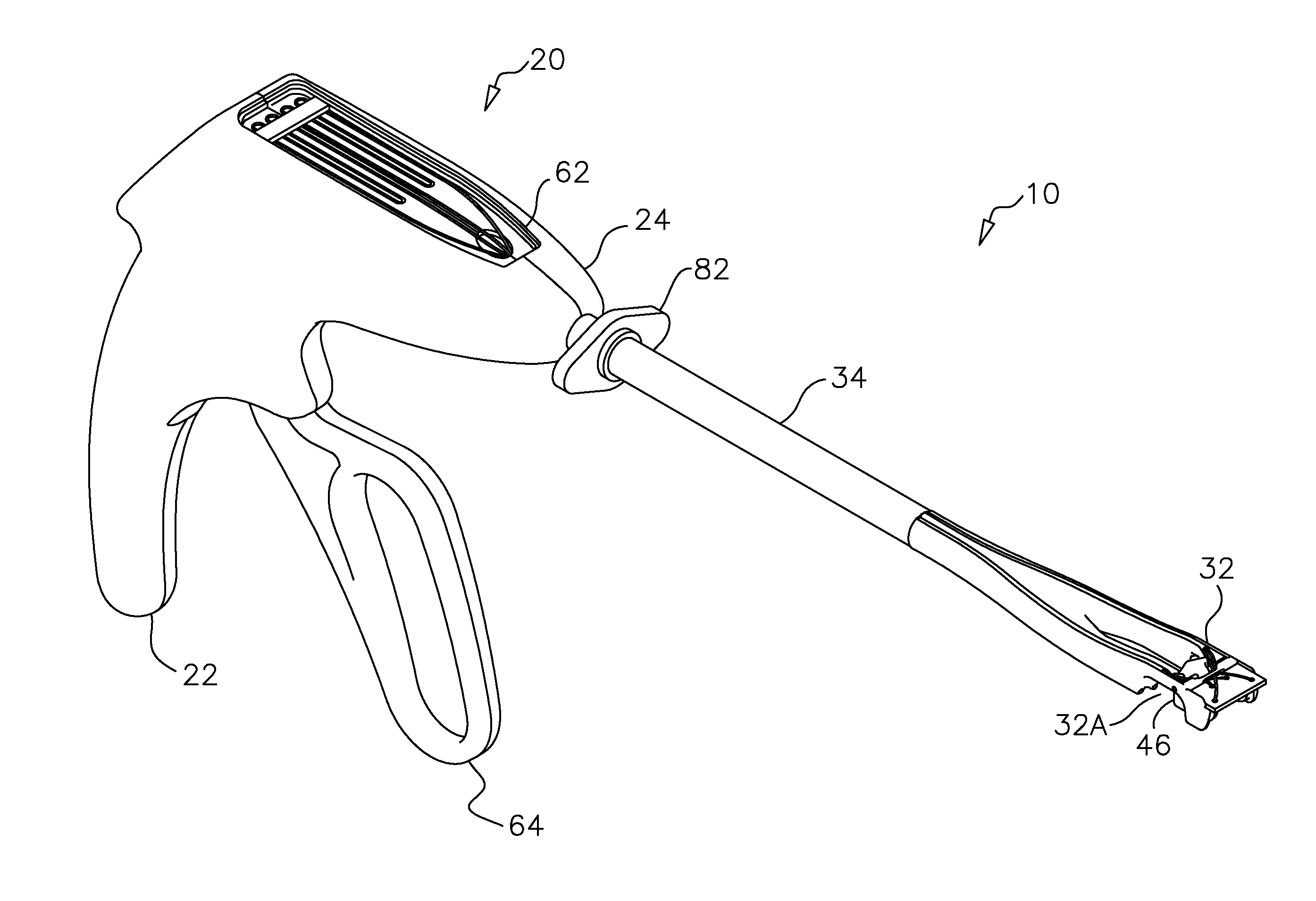
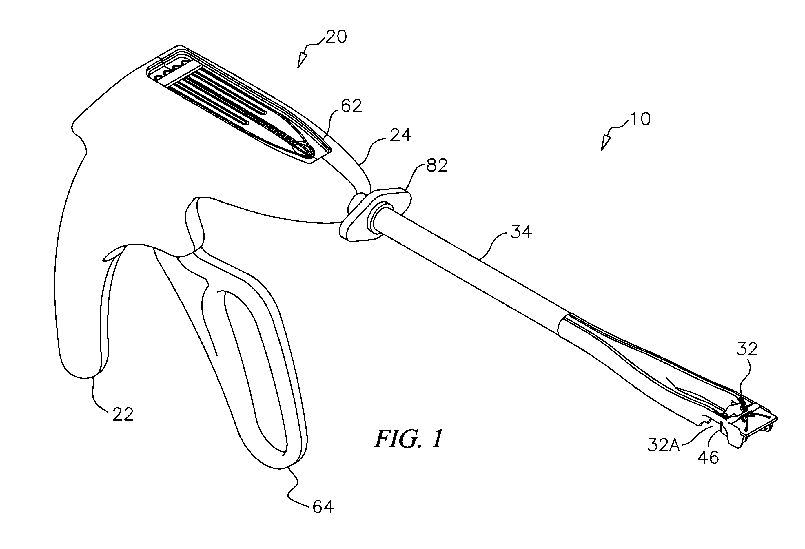
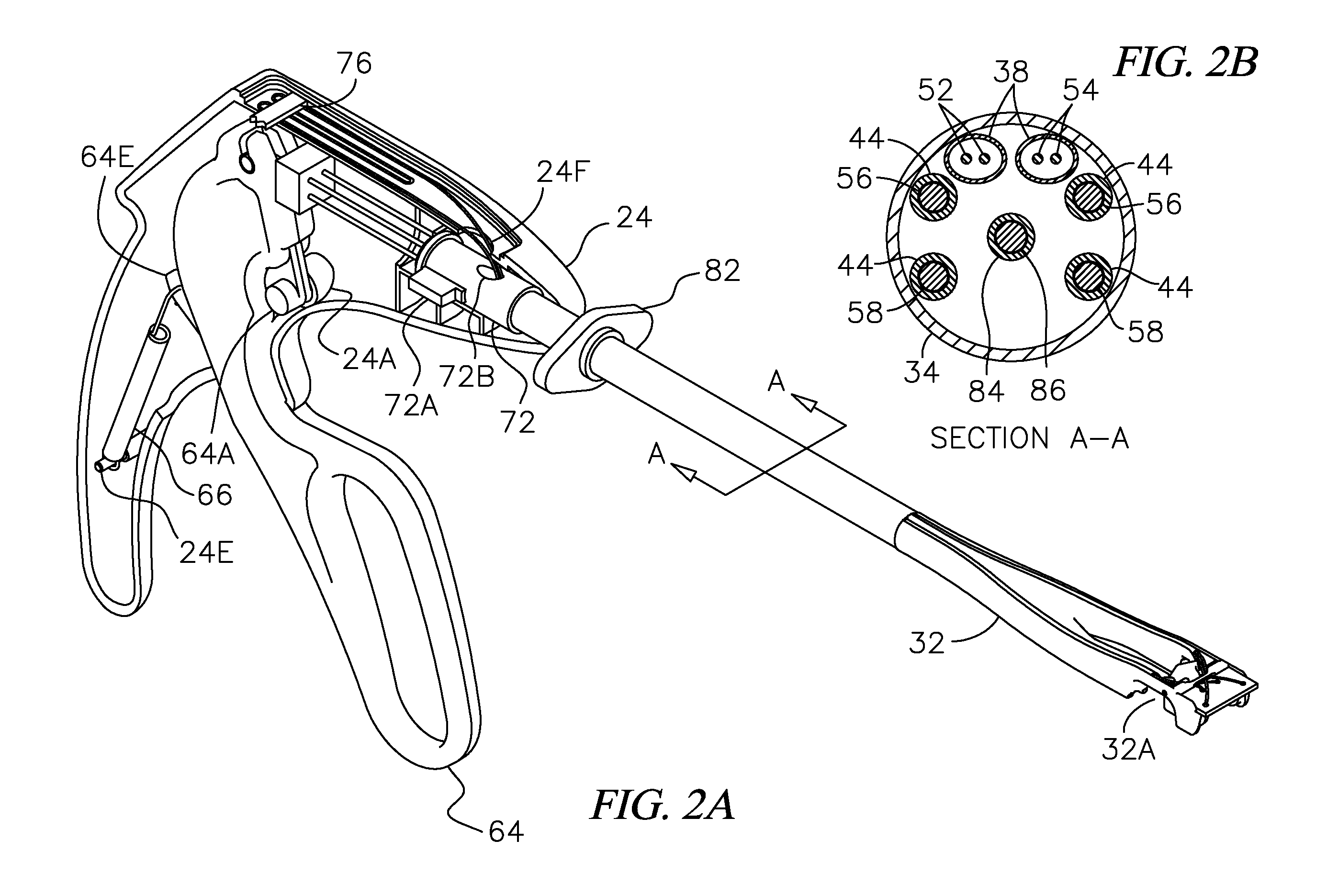
[0081]FIG. 1 is a perspective view of a tissue suturing instrument 10 in accordance with a first embodiment of the present invention. A pistol grip style handle assembly 20 is constructed from a right handle portion 22 and a left handle portion 2...


PUM


Abstract

A device for placing one or more sutures through a section of tissue, especially a section of thick and / or moving tissue such as the wall of a beating heart. The device includes a tissue welting tip having a trough for forming a welt in a tissue section, an alignment guide pivotally mounted in the distal end adjacent to the trough, and having an opening receiving a guide wire and an elongated sleeve slidably engagable with the guide wire there through. The device also includes one or more expandable tissue engaging member(s) on the sleeve selectively expandable from a collapsed configuration having a diameter small enough to pass through the opening in the tip to an expanded configuration having a diameter large enough to engage a tissue section and urge it into the trough to form a welt in the tissue section and a retractable needle extendable through at least two portions of a tissue section while the tissue section is engaged with the trough.

Description

BACKGROUND OF THE INVENTION[0001]1. Field of the Invention[0002]This invention relates generally to new methods and devices for placing stitches in relatively thick, moving tissue so that when the ends of the suture are drawn tightly together, the tissue within the suture is constricted and more particularly to methods and devices for the minimally invasive remote simultaneous and hemostatic placement of multiple horizontal mattress sutures centered circumferentially around a placed guide wire traversing through a tissue wound site that will subsequently be enlarged (to provide a passageway to facilitate a medical intervention) and then hermetically closed. An embodiment of the invention includes a hand actuated suturing instrument with multiple needles that drive through relatively thick engaged tissue each picking up an end of a strand of suture for the precisely oriented suture placement at predetermined depths of multiple concentric horizontal mattress sutures with pre-loaded pl...

Claims


Application Information

Patent Timeline
IPC IPC(8): A61B17/04
CPCA61B2017/0472A61B2017/00986A61B17/0469A61B2017/0243A61B2017/0404A61B17/0057A61B2017/00663A61B17/0483A61B2017/00557
Inventor SAUER, JUDE S.BOVARD, MARK A.HAMMOND, JOHN F.
Owner LSI SOLUTIONS
Learn More