System for diagnosing pathological change of lipid in blood vessels using non-linear optical microscopy

a non-linear optical microscopy and pathological technology, applied in the field of diagnostics, can solve problems such as difficult to read pathological, difficult to indicate plaque vulnerability, and damage to tissu

Inactive Publication Date: 2012-03-01
KOREA RES INST OF STANDARDS & SCI
View PDF4 Cites 5 Cited by
  • Summary
  • Abstract
  • Description
  • Claims
  • Application Information

AI Technical Summary

Benefits of technology

[0021]The system and the method in accordance with the present invention can selectively image lipids without damag...

Problems solved by technology

Recently, Virmani et al. reported that ruptured plaques contain greater amounts of cholesterol clefts or crystals in necrotic cores than erosion or stable plaques from cross-sectioned coronary arteries, potentially indicating plaque vulnerability.
For the micropathological reading of vessel walls, current imaging techniques require tissue staining for micropathological reading, but this brings about damage to tissue.
Further, the only images obtained are cross-sectional images from which it is very difficult to read pathological causes as existing in the tissue.
In addition, there are no staining techniques which allow individual lipid compon...

Method used


Image

  • System for diagnosing pathological change of lipid in blood vessels using non-linear optical microscopy
  • System for diagnosing pathological change of lipid in blood vessels using non-linear optical microscopy
  • System for diagnosing pathological change of lipid in blood vessels using non-linear optical microscopy

Examples


experimental example 1

En Face CARS 3D Imaging of Atherosclerotic Lesion

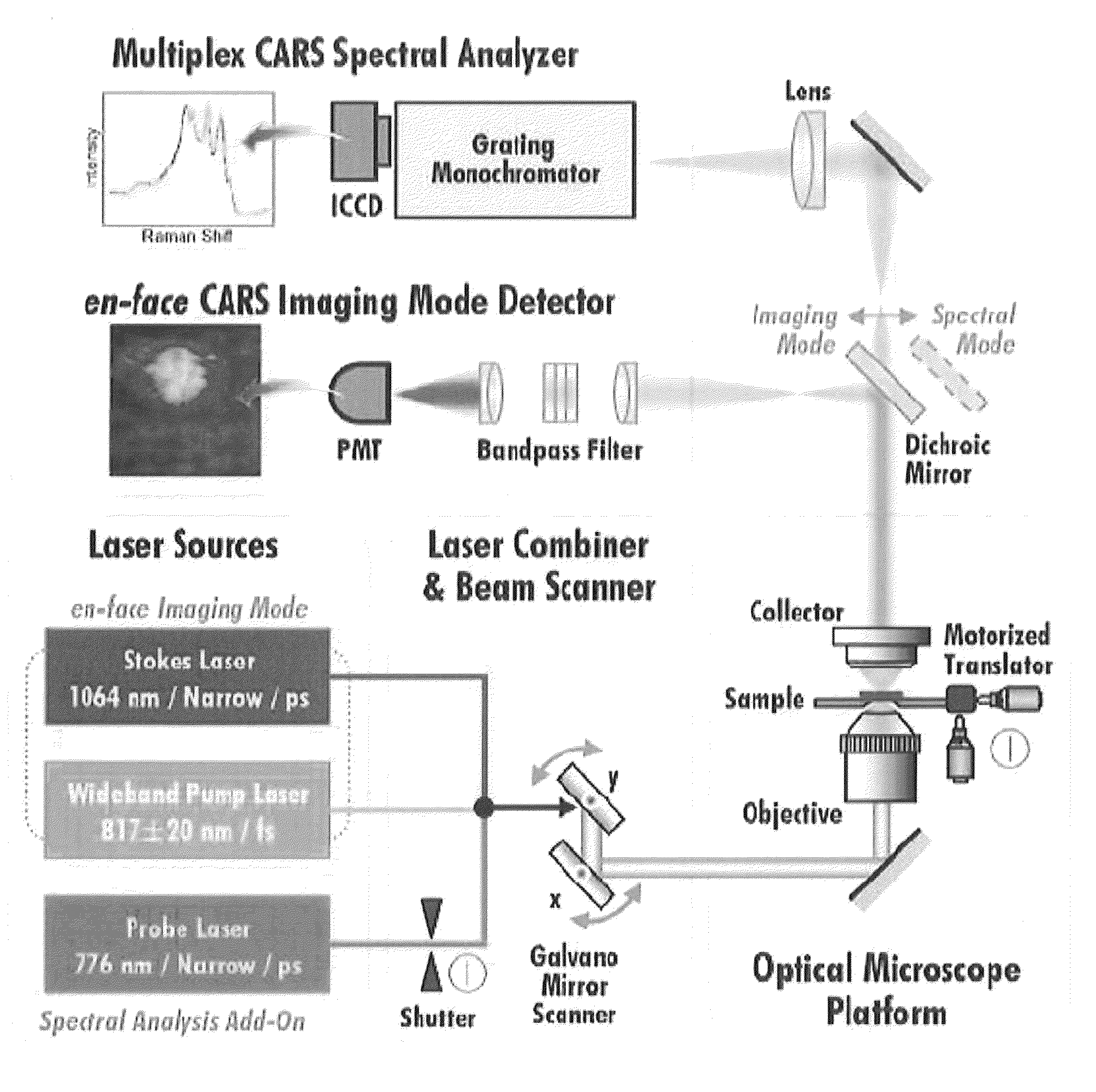
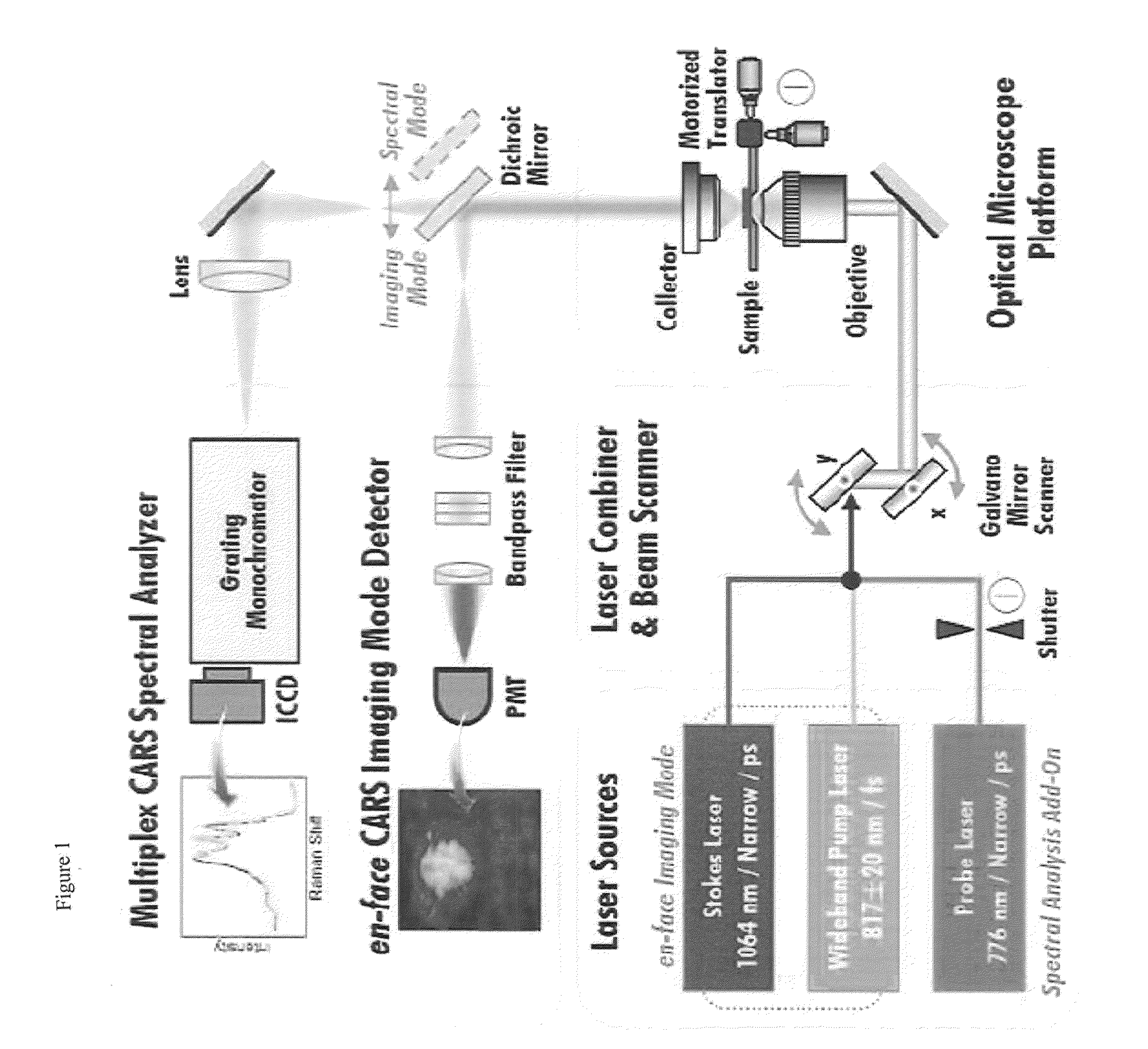
[0078]En face chemical imaging of mouse and human atherosclerotic plaques was performed using the CARS microscope of Example 1. After whole aortas were harvested from atherosclerotic ApoE− / − mice (n=28), the lesser curvature of the aortic arch and the carotid artery was longitudinally incised and imaged by CARS, without the use of fixatives.

[0079]FIG. 3 shows the 3D reconstructed CARS image slice, representing atherosclerotic plaques ranging from the lumen side to the deep intima. In the CARS image, bright spots show a high concentration of lipids with CH vibrations characteristic of 2700 to 3050 cm−1, demonstrating typical 3D microscopic traits of atherosclerotic lipids depending on the depth of lesions. In the superficial layers (5 to 10 μm in depth from the lumen), foam cells containing intracellular lipid droplets were clearly imaged, whereas lipid crystals were observed usually in the deep intima region (>25 μm in depth), without...

experimental example 2

Assessment of Atherosclerosis Progression by CARS Imaging

[0082]To assess the progression of atherosclerosis using the CARS imaging platform of Example 1, various levels of atherosclerotic plaques were obtained from ApoE− / − mice (n=28) fed with a high-fat diet for 2 to 20 weeks. As a control, ApoE− / − mice fed with a normal chow diet were assessed at the same time points. Every week, serial en face CARS imaging was performed in mouse aortas. The progression of atherosclerosis was analyzed using CARS images taken for the vertical infiltration of lipids across the aortic wall and the morphological change of lipid structures.

[0083]In the 2-week-old atherosclerosis mouse models, few of the imaged lipid droplets were bound to the extracellular matrix (ECM) (FIG. 6a). In the 4-week-old atherosclerosis mouse models, lipid droplets were observed only in the superficial intima (b). At 6 weeks, the number of lipid droplets was significantly increased relative to the number present at 2 weeks (F...

experimental example 3

Identification of Characteristics of Atherosclerotic Plaques by CARS

[0084]Using 3D CARS imaging, lipid distribution was quantified in three main stages (initial, intermediate and advanced stages: FIG. 7). The progression of atherosclerosis was analyzed in terms of the volume of lipid segments (calculated as 2D coverage in z-stack) and the size of the lipid structure.

[0085]The progression of atherosclerosis was analyzed by quantifying accumulated lipids at 3 stages depending on the period of high-fat diet consumption. During the initial stage (weeks 2-6, FIG. 7A i-iv), only a small amount of lipids is clearly represented in a small size. In the intermediate stage (weeks 8-12, FIG. 7b: i-iv), lipid deposits moved deep in the z-stack. Interestingly, their size was increased in the deep intima to form plate-shaped liquid crystals. In the advanced stage (week 16, FIG. 7c: i-iv), the lipids increased in both coverage and size. Atherosclerotic lipids penetrated to the depth of 30 μm, and e...


PUM


Abstract

Disclosed is a system for diagnosing the pathological change in lipids in blood vessels using coherent anti-strokes raman microscopy which can image lipids abnormally deposited on the deep intima of blood vessels and analyze the components of the imaged lipids, without labeling or destroying blood vessels, to diagnose minute pathological changes in the blood vessels, whereby the stage of progression of lipid-related diseases can be determined.

Description

TECHNICAL FIELD[0001]The present invention relates to a diagnostic system for observing pathological changes in the lipids in blood vessel using non-linear optical microscopy. More particularly, the present invention relates to a system for diagnosing the pathological change in lipids in blood vessels using Coherent Anti-strokes Raman Scattering microscopy which can image lipids abnormally deposited on the blood vessel intima and analyze the components of the imaged lipids, without labeling or destroying blood vessels, to diagnose minute pathological changes in the blood vessels, whereby the stage of progression of lipid-related diseases can be determined.BACKGROUND ART[0002]Lipids are associated with the various stages of arteriosclerosis progression. Lipid retention is regarded as an initial key event which has been implicated in the onset of arteriosclerosis. Although the so-called “response-to-retension” hypothesis has not been concretely verified, atherogenesis is induced by th...

Claims


Application Information

Patent Timeline
IPC IPC(8): G01J3/44
CPCG01J3/0248G01J3/10G01J3/44G02B21/16G01N2021/653G02B21/002G02B21/0088G01N21/65
Inventor KIM, SE HWALEE, JAE YONGLEE, EUN SEONGMOON, DAE WON
Owner KOREA RES INST OF STANDARDS & SCI
Why Eureka
Try Eureka