
Method for detecting target cell

a target cell and detection method technology, applied in the field of target cell detection method, can solve the problems of high cost of flow cytometer, high cost of labeled monoclonal antibody used as reagent, and large availability, so as to facilitate and promptly detect the target cell, easy and promptly provide data, and easy analysis

Inactive Publication Date: 2012-05-10
JSR CORPORATIOON
View PDF6 Cites 3 Cited by
  • Summary
  • Abstract
  • Description
  • Claims
  • Application Information

AI Technical Summary

Benefits of technology

[0008]An object of the invention is to provide a simple and accurate target cell detection method. Such a method may make it possible to detect the target cells without using flow cytometry, and easily analyze the size and the like of the target cells, and may be useful for testing a cell dispersion (e.g., blood) during clinical examination or the like.Solution to Problem
[0024]The target cell detection method makes it possible to easily and promptly detect the target cells, and analyze the size and the like of the target cells. This makes it possible to easily and promptly provide data for diagnosing a disease when the cell dispersion is blood or the like. Since the target cell detection method makes it possible to perform measurement that has been performed using flow cytometry using a 5-part WBC differential hematology analyzer, a 3-part WBC differential hematology analyzer, or the like, the target cells included in the cell dispersion can be more easily analyzed.

Problems solved by technology

However, the flow cytometer used for flow cytometry is expensive as compared with a blood cell counter, and the labeled monoclonal antibody used as a reagent is also expensive.
Therefore, the flow cytometer is generally provided in special testing institutions, and has not been widely provided in hospitals and clinics.
This makes it difficult to promptly provide data for a doctor or the like to diagnose a disease.
Moreover, it may be difficult to count the number of the target cells.
However, the results may differ depending on each hematology analyzer, and it is considered that a blood sample cannot necessarily be sufficiently characterized based only on the measurement results obtained using such a hematology analyzer.

Method used


Image

  • Method for detecting target cell
  • Method for detecting target cell

Examples


preparation example

2.2. Dispersion Preparation Example

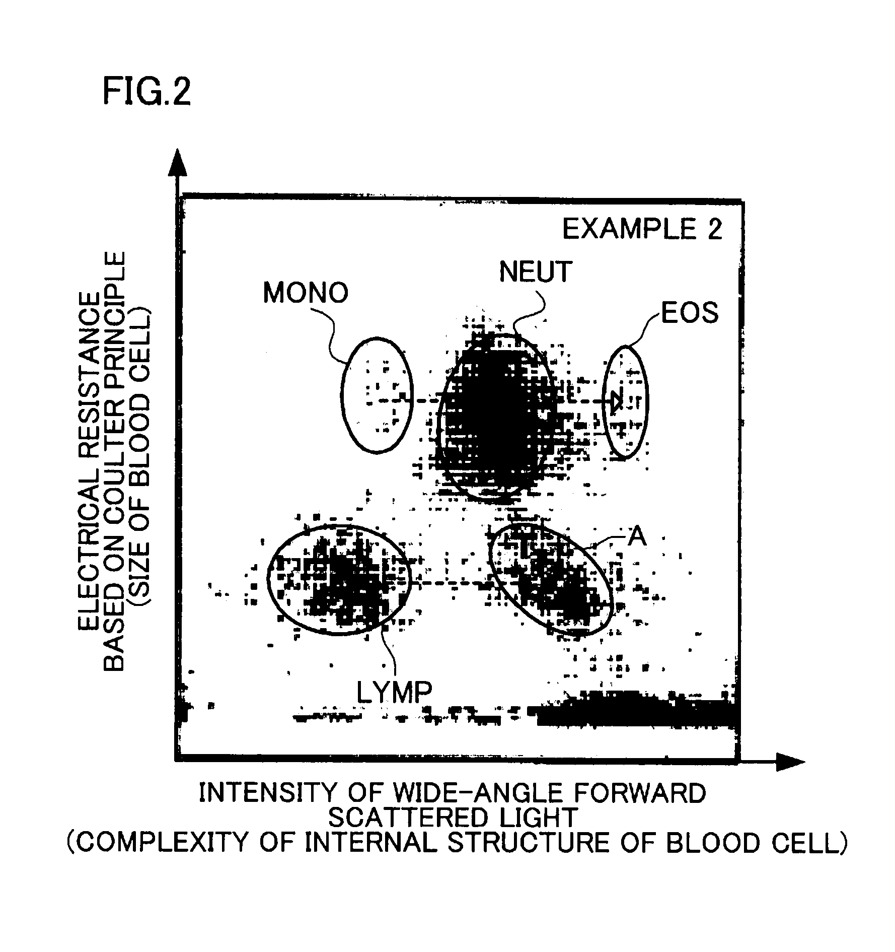
[0097]2 ml of blood collected from the vein of a healthy individual using a blood collection tube to which EDTA was added was put in a microtube. 200 microliters of the labeled particle dispersion obtained in each of Labeled Particle Production Examples 1 to 4 was added to the microtube to obtain a dispersion used in each example. The mixture was then mixed upside down 20 times, allowed to stand at room temperature (15 to 25° C.) for 20 minutes, and used in each example. The concentration of the labeled particles in the dispersion was 5×10⁷ particles per microliter. As a control, 100 microliters of blood was used.

2.3. Measurement

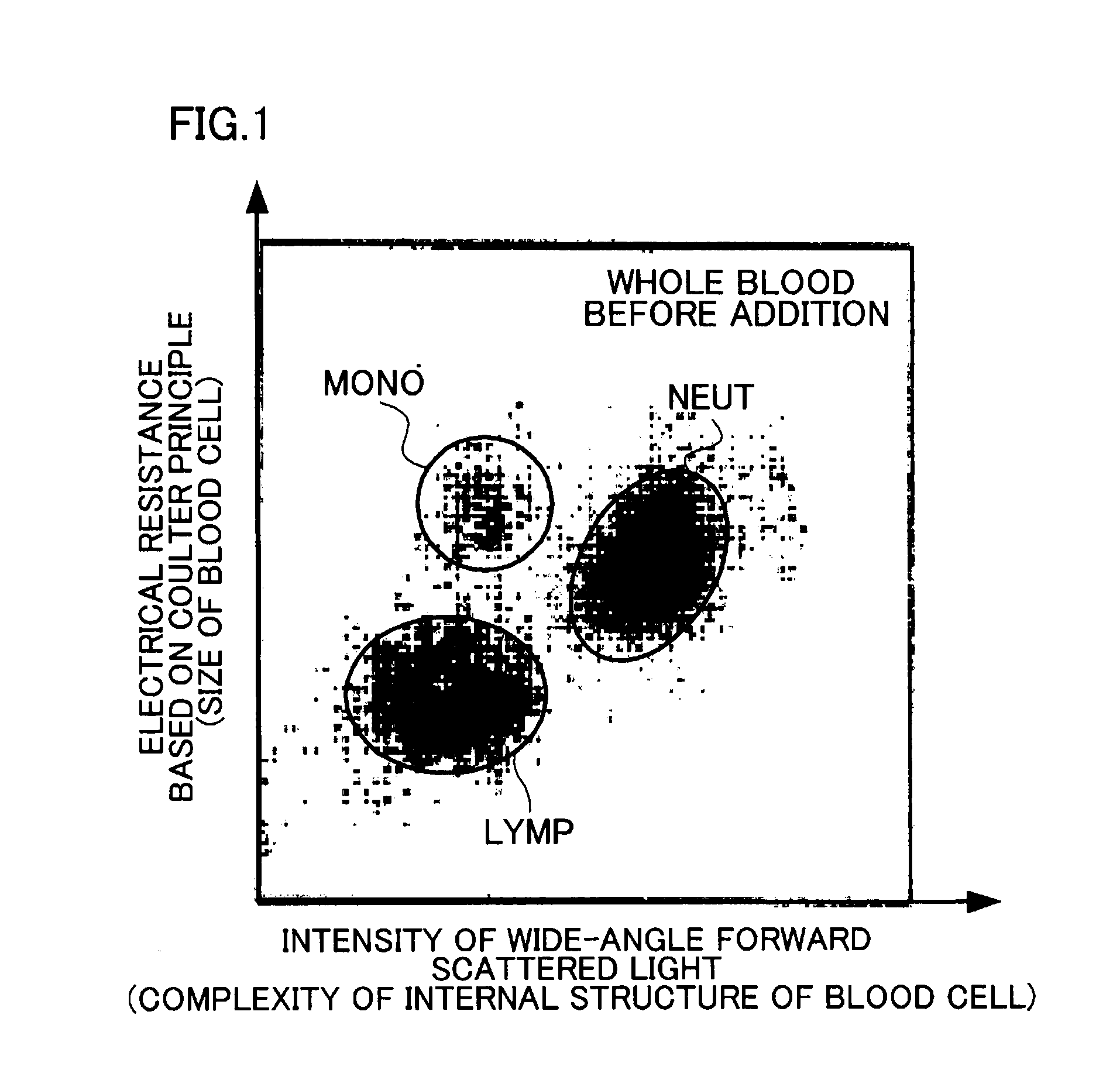
[0098]The intensity of wide-angle forward scattered light and the electrical resistance based on the Coulter principle were measured using a blood analyzer “LH750”(manufactured by Beckman Coulter, Inc.) (5-part WBC differential hematology analyzer). The measured values were two-dimensionally plotted for each blood cell. The...


PUM

PropertyMeasurementUnit
Particle sizeaaaaaaaaaa
Fractionaaaaaaaaaa
Fractionaaaaaaaaaa

Abstract

A target cell detection method includes performing a test measurement on a dispersion to obtain a measurement result 1, the test measurement being an optical or electromagnetic measurement, and the dispersion including labeled particles and target cells, the labeled particles being particles on each of which a substance that specifically binds to a specific molecule present on a surface of each of the target cells is immobilized, performing measurement that is identical with the test measurement on a dispersion that includes the target cells, but does not include the labeled particles to obtain a measurement result 2, and comparing the measurement result 1 with the measurement result 2.

Description

TECHNICAL FIELD[0001]The present invention relates to a target cell detection method.BACKGROUND ART[0002]A method that performs a test on cells contained in a body fluid (i.e., cell dispersion) (e.g., blood, bone marrow, or lymph), and screens specific cells (e.g., cells that relate to a specific disease) that may be present in the cell dispersion, has been widely used. For example, flow cytometry or the like utilizes the fact that test target cells include a specific molecule (marker) (hereinafter may be referred to as a cell surface marker, a cell surface antigen, or a CD antigen) on the surface thereof, and detects cells that include such a cell surface marker (see JP-A-2008-187932 and JP-T-2008-538609). When the cell dispersion is a body fluid, the analysis results are used as data for diagnosis of a disease, for example. Note that a test target cell that is included in a cell dispersion and includes a specific cell surface marker is hereinafter referred to as “target cell”.[000...

Claims


Application Information

Patent Timeline
IPC IPC(8): G01N21/75C12Q1/68G01N27/72
CPCG01N15/12G01N15/1459G01N33/5047G01N2015/1486G01N2015/008G01N2015/1062G01N2015/1477G01N33/54313G01N2015/016G01N2015/1024
Inventor IMAI, TOSHIHIKOMIYAJI, MASAAKI
Owner JSR CORPORATIOON
Learn More