Muscle function enhancing peptide
a peptide and muscle technology, applied in the field of peptides, can solve the problems of no clinical inotropic therapies available for skeletal muscle disorders, increased heart rate and life-threatening proarrhythmogenic potential, and severe side effects
- Summary
- Abstract
- Description
- Claims
- Application Information
AI Technical Summary
Benefits of technology
Problems solved by technology
Method used
Image
Examples
example 1
Inotropic effects of S100A1 Protein and S100A1 C-Terminal 20-mer Peptide on Permeabilized Cardiomyocytes and Skeletal Muscle Fibers
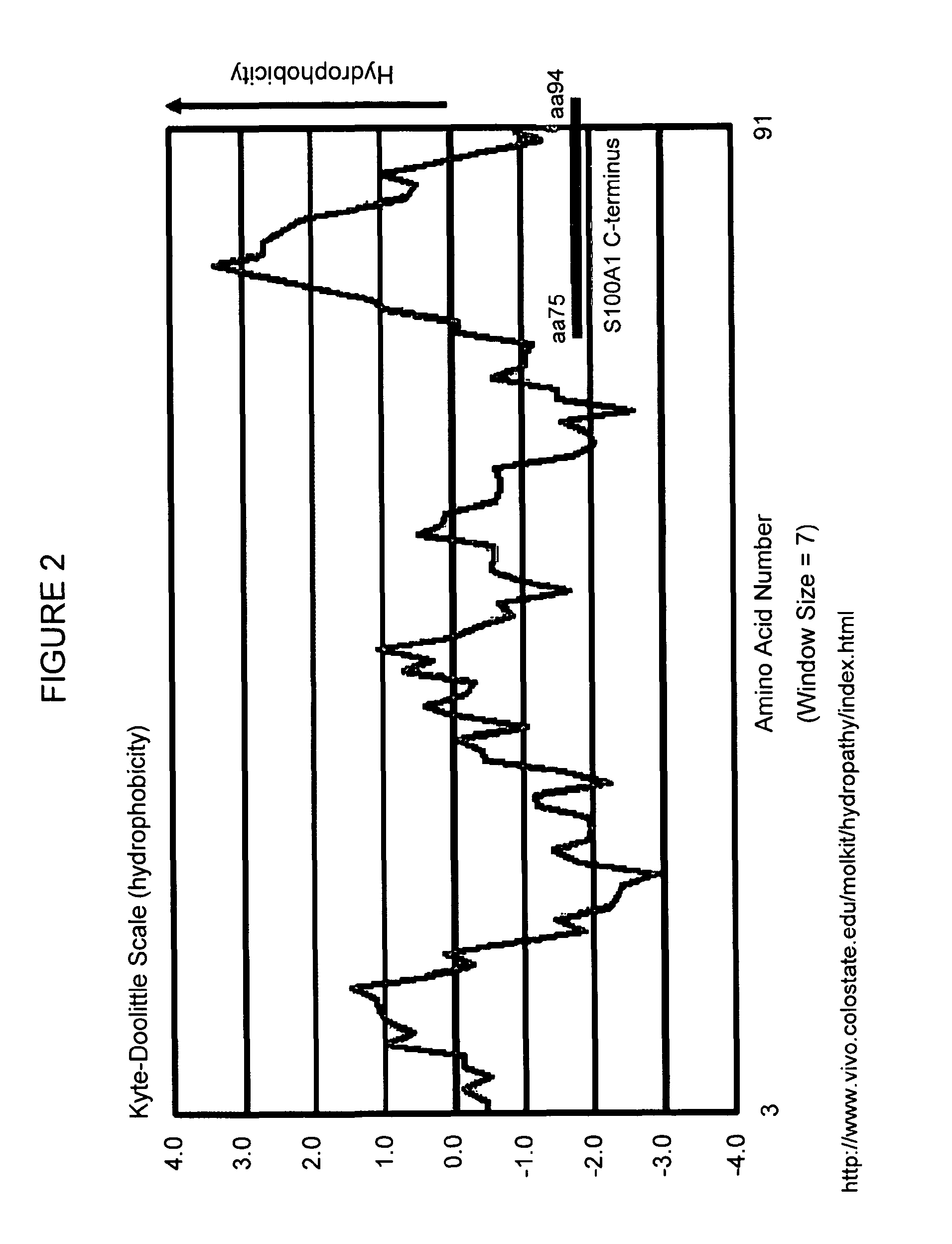
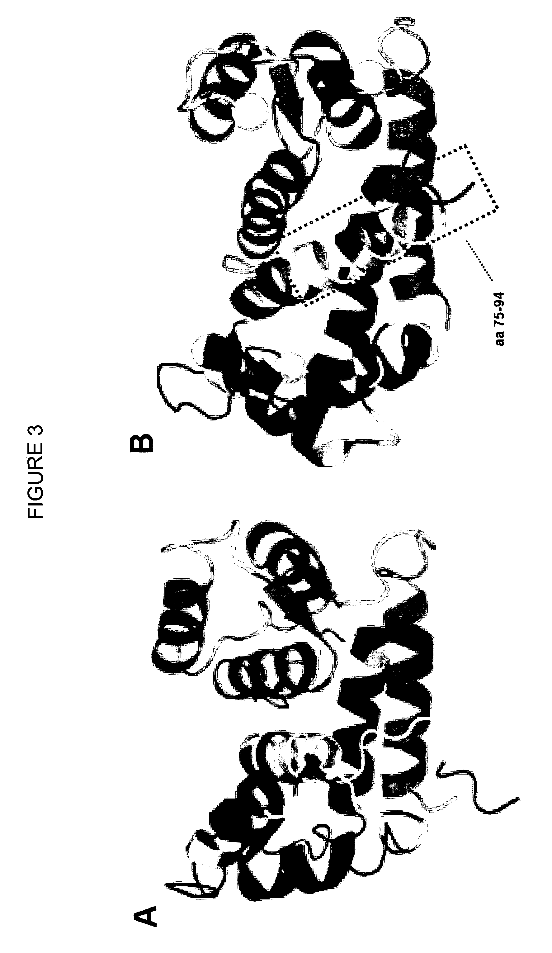
[0143]Adult ventricular rabbit cardiomyocytes were isolated from four different animals as previously described (Loughrey C. M. et al., 2004, J. Physiol. 556:919-934) and permeabilized using β-escin (0.1 mg / ml). The permeabilized cells were incubated for 1 minute with rhodamine-labeled human S100A1 protein (0.1 μM) or FITC-labeled S100A1 C-terminal 20-mer peptide (0.1 μM) fused to a hydrophilic linker (amino acids 75 to 94 of human S100A1 fused to D-K-D-D-P-P (SEQ ID NO: 354)). Cells were monitored using a Bio-Rad 2000 laser scanning confocal microscope (LSCM). A striated staining pattern can be observed that resembles the ryanodine staining pattern (FIG. 4).
[0144]Furthermore, Ca2+-spark frequency has been assessed in the permeabilized cells treated with S100A1 protein or the S100A1 C-terminal 20-mer peptide fused to a hydrophilic linker and it was shown...
example 2
Cell-Permeability of the S100A1ct6 / 11 Peptide
[0147]Neither rhodamine-labeled S100A1 protein nor the FITC-labeled S100A1 C-terminal 20-mer peptide with or without a hydrophilic motif such as D-K-D-D-P-P (SEQ ID NO: 354) are able to penetrate the cell membrane of adult intact cardiomyocytes. However, the present inventors surprisingly found that a peptide having the sequence D-K-D-D-P-P-Y-V-V-L-V-A-A-L-T-V-A (SEQ ID NO: 372) referred to as S100A1ct6 / 11 is cell permeable. FITC-labeled S100A1ct6 / 11 was incubated with intact rat ventricular cardiomyocytes for 15 minutes before the cells were monitored using confocal laser scanning microscopy. Endogenous S100A1 protein was stained using a conventional immunofluorescence protocol. The intracellular staining pattern of FITC-labeled S100A1ct6 / 11 resembles that of endogenous S100A1 (FIG. 8).
example 3
Functional Characterization of the S100A1ct6 / 11 peptide in cardiomyocytes
[0148]All experiments performed for the functional characterization of the S100A 1ct6 / 11 peptide were performed on intact, i.e., non-permeabilized cardiomyocates. It was shown that the S100A1ct6 / 11 peptide exerts positive inotropic effects on stimulated isolated ventricular cardiomyocytes (FIG. 9), while fragments thereof (FIG. 10) or corresponding peptides derived from the carboxy-terminus of S100A4 or S100B (FIG. 11) do not show this ability. Calcium transients were assessed in FURA2-AM field-stimulated cardiomyocytes employing epifluorescent digitalized microscopy and sarcoplasmic reticulum calcium load was determined (FIG. 12).
[0149]Calibration and Measurement of Ca2+ Transients and SR Ca2+ Load in Cardiomyocytes. Intracellular Ca2+ transients of mouse ventricular cardiomyocytes were calibrated and measured as previously described (Remppis A. et al., 2002, Basic Res. Cardiol. 97: I / 56-I / 62). Briefly, isolat...
PUM
| Property | Measurement | Unit |
|---|---|---|
| hydrophobic | aaaaa | aaaaa |
| length | aaaaa | aaaaa |
| spark frequency | aaaaa | aaaaa |
Abstract
Description
Claims
Application Information
 - R&D
- Intellectual Property
- Life Sciences
- Materials
- Tech Scout
- Unparalleled Data Quality
- Higher Quality Content
- 60% Fewer Hallucinations
Browse by: Latest US Patents, China's latest patents, Technical Efficacy Thesaurus, Application Domain, Technology Topic, Popular Technical Reports.
© 2025 PatSnap. All rights reserved.Legal|Privacy policy|Modern Slavery Act Transparency Statement|Sitemap|About US| Contact US: help@patsnap.com


