Endoscope system, processor of endoscope system, and image producing method

a technology which is applied in the field of endoscope system and endoscope system processor, and image producing method, can solve the problems of difficult diagnostic inspection of changes in mucosal properties, and difficult scaling of oxygen saturation by red hue gradation, so as to achieve superior and useful medical diagnosis

Active Publication Date: 2012-06-21
FUJIFILM CORP
View PDF2 Cites 32 Cited by
  • Summary
  • Abstract
  • Description
  • Claims
  • Application Information

AI Technical Summary

Benefits of technology

[0028]According to the present invention, oxygen saturation levels of a test subject are measured based on a first image signal that is captured under first illumination light of a first wavelength range, preferably a narrow blue wavelength range, in which light absorption coefficient of blood hemoglobin varies with oxygen saturation thereof, and an oxygen saturation image is produced by changing color properties of a subject image according to the measured oxygen saturation levels. Since the subject image is produced from a second image signal that is captured under second illumination light of a second wavelength range different from the first w...

Problems solved by technology

As a result, it is difficult to diagnostically inspect changes in mucosal properties.
However, because the pixel levels of the red image signal depend not only on the oxygen saturation but also on the blo...

Method used


Image

  • Endoscope system, processor of endoscope system, and image producing method
  • Endoscope system, processor of endoscope system, and image producing method
  • Endoscope system, processor of endoscope system, and image producing method

Examples


first embodiment

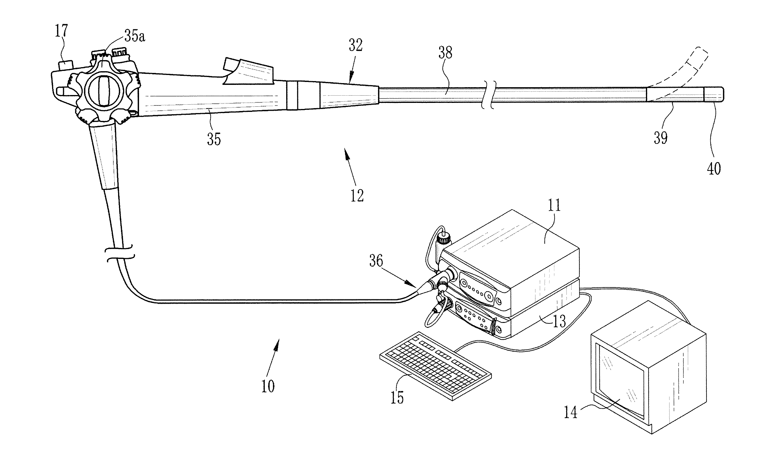
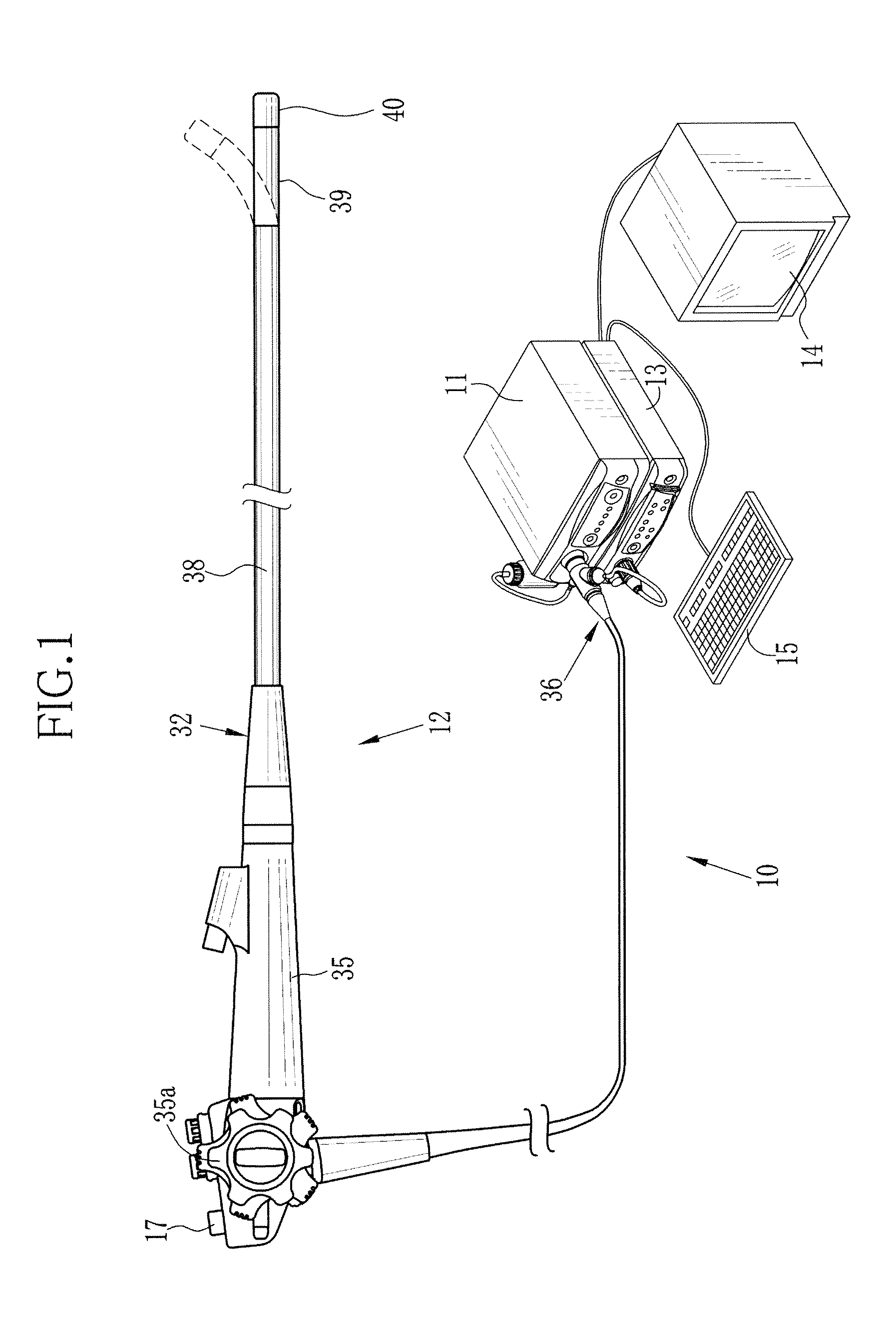
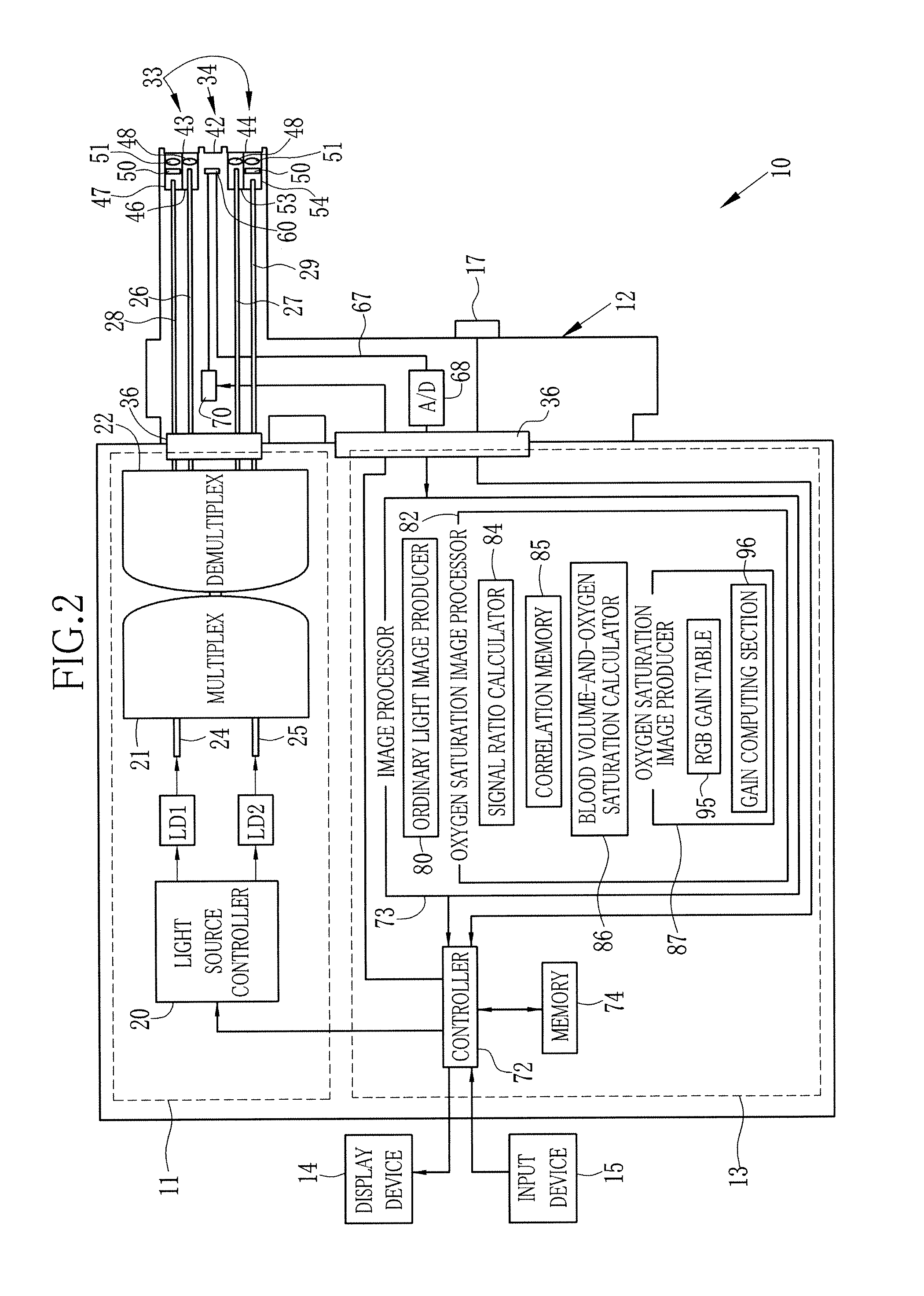
[0056]As shown in FIGS. 1 and 2, an endoscope system 10 according to the present invention includes a light source unit 11, an endoscope 12, a processor unit 13, a display device 14, and an input device 15 including a keyboard and the like. The light source unit 11 may emit light of a predetermined wavelength range. The endoscope 12 conducts the light from the light source unit 11 to project it into a target area inside a test subject, and captures images from light reflected from the target area. The processor unit 13 processes image signals captured by the endoscope 12 so that the display device 14 may display an endoscopic image using the processed image signals.

[0057]The endoscope system 10 is provided with an ordinary light inspection mode and an oxygen saturation inspection mode. In the ordinary light inspection mode, the display device 14 displays an ordinary light image of the test subject captured under the illumination of visible rays ranging from blue to red wavelength re...

second embodiment

[0102]Referring now to FIG. 16, an endoscope system 120 according to the present invention will be described. The endoscope system 120 adopts a light source unit 11 of a rotary filter type. The rotary filter type light source unit 11 is provided with a broadband light source 121 like a xenon lamp that emits white light having such spectral intensity characteristics as shown in FIG. 17, a rotary filter 122 that transmits either the entire white light or those wavelength components of the white light which correspond to the oxygen saturation measuring light, an optical fiber 123 for receiving and conducting the light transmitted through the rotary filter 122, and a rotary controller 124 for controlling rotation of the rotary filter 122, instead of the laser light sources LD1 and LD2, the light source controller 20 and the combiner 21. The light entering the optical fiber 123 is divided into two beams through a coupler 22. The divided beams are conducted through light guides 26 and 27 ...


PUM


Abstract

First illumination light of a first wavelength range, in which light absorption coefficient of blood hemoglobin varies with oxygen saturation thereof, is projected into a test subject body, to capture a first image signal from the first illumination light as reflected from inside the test subject body. Then second illumination light of a second wavelength range different from the first wavelength range is projected into a test subject body, to capture a second image signal from the second illumination light as reflected from inside the test subject body. A subject image of the test subject is produced from the second image signal. Oxygen saturation levels of the test subject are calculated using the first and second image signals. According to the calculated oxygen saturation levels, color properties of the subject image are changed to produce an oxygen saturation image.

Description

BACKGROUND OF THE INVENTION[0001]1. Field of the Invention[0002]The present invention relates to an endoscope system that is adapted to produce an image visualizing information about oxygen saturation of blood hemoglobin. The present invention also relates to a processor and an image producing method for the endoscope system.[0003]2. Description of Related Art[0004]In recent medical field, endoscopes are widely used for diagnoses and treatments. To inspect an interior of a test subject body through the endoscope, white or broadband light is ordinary projected into the test subject body for illumination, but also narrowband light of limited wavelength ranges come to be more frequently used as illumination light for the sake of making particular organs such as blood vessels conspicuous in subsequent endoscopic images displayed on a monitor screen.[0005]Beside the endoscopic inspection using the special narrowband light, it has also been practiced in the art to derive information about...

Claims


Application Information

Patent Timeline
IPC IPC(8): A61B1/04
CPCA61B1/00009A61B1/043A61B1/045A61B1/0638A61B5/14551A61B5/6852G06T2207/30101A61B1/063A61B1/0646A61B1/0653G06T7/0012G06T2207/10024G06T2207/10152G06T2207/10068A61B1/000094A61B1/0655
Inventor SAITO, TAKAAKI
Owner FUJIFILM CORP
Why Eureka
Try Eureka