Methods and apparatus for sub-retinal catheterization

a subretinal and catheterization technology, applied in the field of subretinal catheterization, can solve the problems of progressive decrease in visual acuity, eventual blindness, and difficulty in performing interventional procedures targeting tissues beneath the sensory retina

Inactive Publication Date: 2012-07-26
ISCI INTERVENTIONAL CORP
View PDF6 Cites 118 Cited by
  • Summary
  • Abstract
  • Description
  • Claims
  • Application Information

AI Technical Summary

Benefits of technology

[0007]the catheter having flexural rigidity in bending and a response to critical buckling load sufficient to allow flexing of the catheter in the eye without causing substantial tissue trauma or distension of local tissue.

Problems solved by technology

There are many diseases and conditions that affect the retina which can lead to a progressive decrease in visual acuity and eventual blindness.
Interventional procedures targeting tissues beneath the sensory retina are difficult to perform due to limited accessibility and the delicate structure of the retina which can be easily damaged during surgical manipulation.

Method used


Image

  • Methods and apparatus for sub-retinal catheterization
  • Methods and apparatus for sub-retinal catheterization
  • Methods and apparatus for sub-retinal catheterization

Examples


example 1

[0050]Two enucleated rabbit eyes and a human cadaver eye were prepared for testing. The pars plana region of the sclera was dissected with an approximately 4 mm incision to expose the choroid. A series of rounded steel probes were used to apply pressure on the choroid and retina to determine if a particular size range of an atraumatic catheter tip would help to prevent inadvertent penetration into the posterior chamber.

[0051]Rounded steel probes with the tip diameters as shown in Table 1 were tested during dissection of the choroid to the sub-retinal space. Table 1 also shows the resultant effects observed.

TABLE 1Probe tip diameters and dissection resultsProbe tip diameterEffect on choroid and retina115 micronsEasily penetrates into posterior chamber165 micronsEasily penetrates into posterior chamber220 micronsBlunt dissects the choroid, must be carefulnot to penetrate into posterior chamber275 micronsBlunt dissects the choroid330 micronsBlunt dissects the choroid360 micronsBlunt di...

example 2

[0053]Enucleated human cadaver eyes were used to determine the mechanical properties for atraumatic advancement in the sub-retinal space. The eyes were prepared in an “open sky” approach by dissecting off the anterior segment of the globe at the level of the ciliary body, and removing the lens. In a living eye, the retinal tissues are attached to the RPE by interdigitation of the cells and the fluid pumping mechanism of the RPE. Post-mortem, the retina no longer has strong attachment to the RPE, so a method was used to maintain the positioning of the retina during the experiments. A heavy fluid, perfluoromethylcyclopentane (Flutec PC1C, F2 Chemicals LTD), with a density of 1.707 Kg / L was injected into the vitreous cavity to displace the vitreous fluid and to hold the retina in place similar to the use of heavy fluids in retinal detachment repair. A notch was cut into the globe down to the level of the anterior insertion of the retina, in order to gain direct access to the retina fro...

example 3

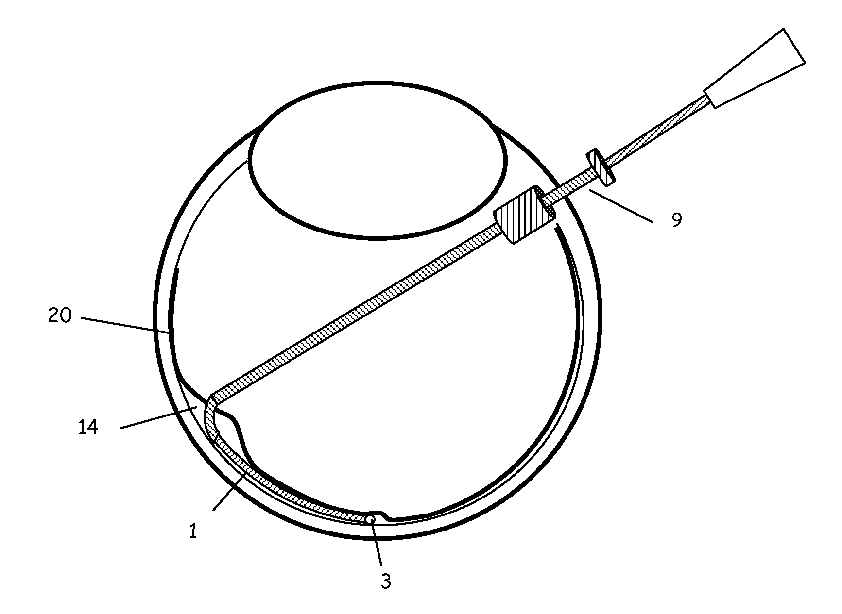
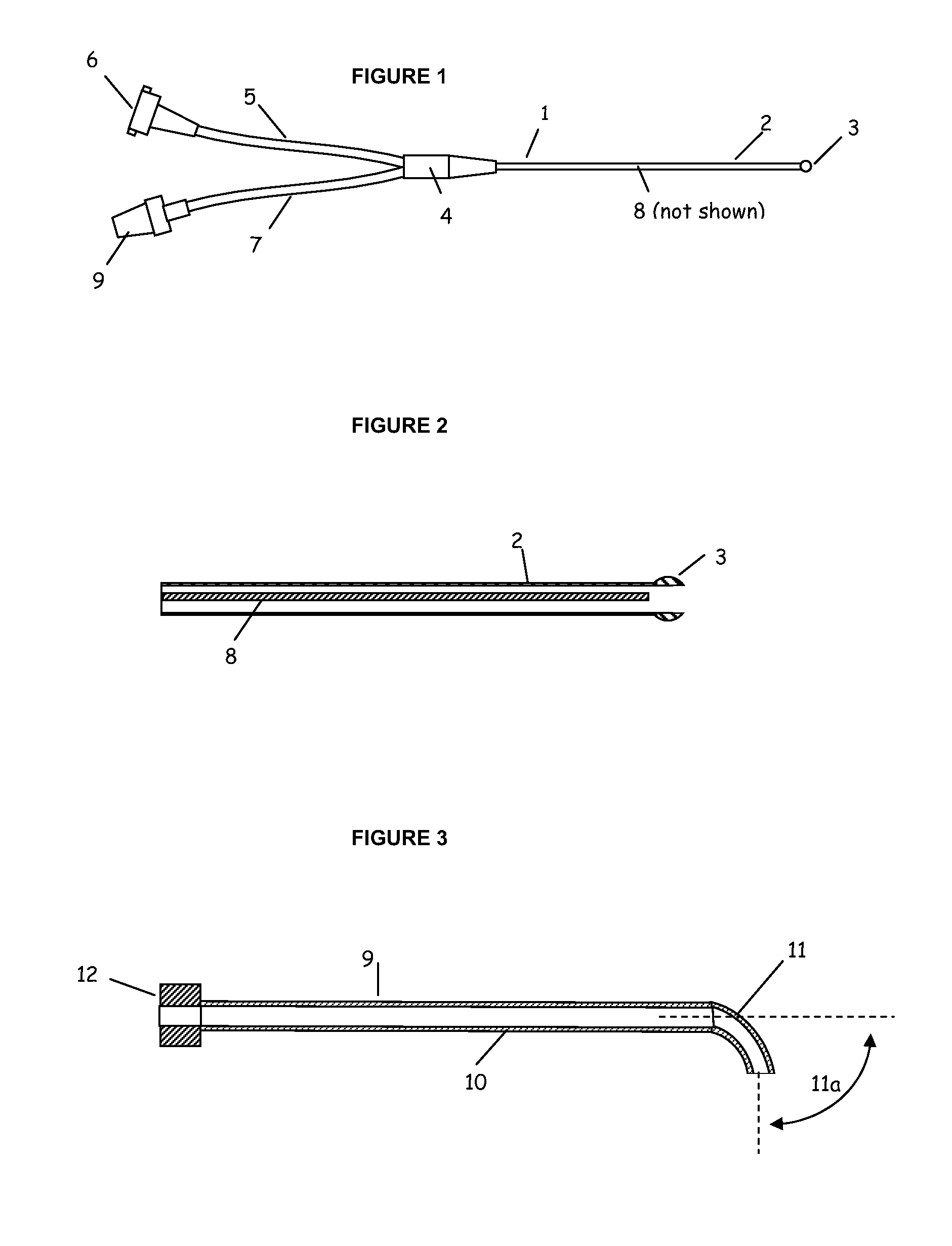
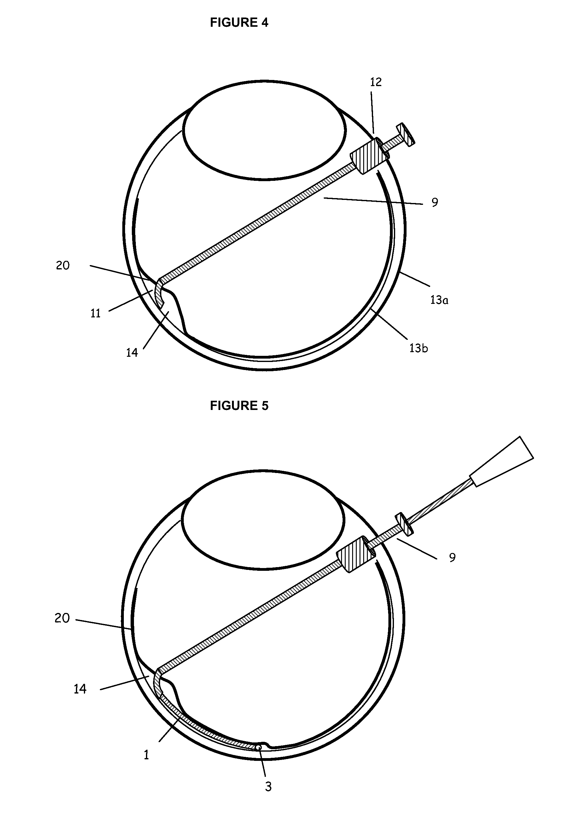
[0059]Enucleated human and rabbit cadaver eyes were used to evaluate catheter access to the sub-retinal space. The eyes were prepared by removing the muscles, conjunctiva, and tenons. Both ab-interno and ab-externo approaches as described herein were performed. A catheter device with distal shaft outer diameter of 200 microns and a bulbous distal tip with diameter of 275 microns was used. The distal shaft was comprised of a polyether block amide tube with a durometer of 63 Shore D (Pebax 6333, Arkema Inc). The catheter had a measured average flexural rigidity in bending of 1.49×10⁻¹⁰ kN*m2 and average critical load in buckling of 8.0 grams force. The distal shaft terminated proximally in a polymer hub with two proximal elements. The catheter device incorporated an 85 micron (0.003 inch) plastic optical fiber in the lumen extending to the distal tip which was connected to a 0.25 mm (0.010 inch) fiberoptic in one proximal element of the catheter. The larger fiberoptic terminated in a ...


PUM

PropertyMeasurementUnit
lengthaaaaaaaaaa
diameteraaaaaaaaaa
angleaaaaaaaaaa

Abstract

Devices and methods are provided for access to the sub-retinal space that lies between the retina and the choroid in order to introduce therapies to the retina and more specifically to the sensory retina and RPE, particularly in the region of the macula. The devices comprise a catheter that incorporates advantageous size, flexibility and tip features to properly, accurately and atraumatically access the sub-retinal space. Ancillary devices to assist in placing catheters into the sub-retinal space are also provided. The catheter devices incorporate a lumen for delivery of therapeutic substances or devices into the eye.

Description

[0001]This application claims the priority under 35 USC §119(e) and §§363-365 of U.S. Provisional Application No. 61 / 178,882, filed May 15, 2009, the disclosure of which is incorporated herein by reference in its entirety for all purposes.BACKGROUND OF INVENTION[0002]There are many diseases and conditions that affect the retina which can lead to a progressive decrease in visual acuity and eventual blindness. Deleterious consequences from disease processes or physiological defects can affect specific tissues of the retina such as the photoreceptors, ganglion cells and the retinal pigment epithelium (RPE). Diseases such as age-related macular degeneration, diabetic retinopathy, retinitis pigmentosa, Stargardt's disease and conditions such as macular holes, retinal detachments, epiretinal membranes, retinal or choroidal venous occlusions can all lead to vision loss that ranges from mild to total. Many of these ailments are treated through systemic or intravitreal injections of pharmace...

Claims


Application Information

Patent Timeline
Patent Type & Authority Applications(United States)
IPC IPC(8): A61M5/00A61L29/14A61M25/00
CPCA61F9/00727A61M5/00A61M25/01A61F9/008
Inventor CONSTON, STANLEY R.YAMAMOTO, RONALD
Owner ISCI INTERVENTIONAL CORP
Why Eureka
Try Eureka