Method and device for detecting bright brain regions from computed tomography images

a computed tomography and brain region technology, applied in image enhancement, instruments, applications, etc., can solve the problems of difficult diagnosis of ich from ct, morbidity, and ct-induced isch, and achieve the effects of increasing contrast, reducing morbidity, and increasing ct values

Active Publication Date: 2012-09-20
SHENZHEN INST OF ADVANCED TECH CHINESE ACAD OF SCI
View PDF2 Cites 14 Cited by
  • Summary
  • Abstract
  • Description
  • Claims
  • Application Information

AI Technical Summary

Benefits of technology

[0014]Bright brain regions (BBRs) vary in grayscales due to pathology or external imaging conditions. For example, hemorrhage in its acute stage will be brighter than the hemorrhage in non-acute stage, a CT scanner with bad calibration may have higher CT values than normal CT scanners. Judging the normal range of BBRs will be able to compensate these changes: for example, if the detected BBRs have a small average grayscale, it may correspond to hemorrhage in the non-acute stage, and thus needs a smaller window width to increase the contrast between the BBRs and its neighboring normal brain tissues; if the CT value is higher than normal, the detected BBRs may have a higher average, and thus it is desirable to increase the window width to decrease the contrast between the BBRs and its neighboring normal brain tissues.

Problems solved by technology

It accounts for 10-30% of all stroke admissions to hospital, and leads to catastrophic disability, morbidity, and a 6 month mortality of 30-50%.
Diagnosis of ICH from CT can be difficult when the lesion is small or is masked by normal structures or when the reader is inexperienced which is the case in emergency setting.
The technical challenges for automatic delineation of ICH include 1) existence of non-ICH voxels with similar intensity to ICH, such as skull, calcification and streaks; 2) ICH itself exhibits large variation in grayscales, i.e., voxels around the boundary of an ICH region are darker and the grayscale is smaller when ICH enters ventricles; 3) models such as brain atlas are difficult to be directly incorporated due to the large voxel size and pathological nature of the clinical CT data; 4) the mass effect, deformation of brain tissues and spontaneous movement during scanning making the segmentation and anatomical localization of ICH hard.
However, report on quantifying hemorrhage from CT is scarce.
This method has several limitations including 1) user intervention is needed for setting key parameters (seed point and intensity thresholds), which may yield different ICH for different users; 2) mathematical morphological operations with fixed structuring element may not be able to remove the skull due to the ICH; and 3) fixed thresholds to grow ICH may under-segment or over-segment.
This suffers from being tedious and laborious, requiring readers of CT to be an expert in neuron-radiology, and inability to control accuracy and to provide accurate 3D shape information of the ICH.

Method used


Image

  • Method and device for detecting bright brain regions from computed tomography images
  • Method and device for detecting bright brain regions from computed tomography images
  • Method and device for detecting bright brain regions from computed tomography images

Examples


Embodiment Construction

[0026]In describing example embodiments illustrated in the drawings, specific terminology is employed for the sake of clarity. However, the disclosure of this patent specification is not intended to be limited to the specific terminology so selected and it is to be understood that each specific element includes all technical equivalents that operate in a similar manner.

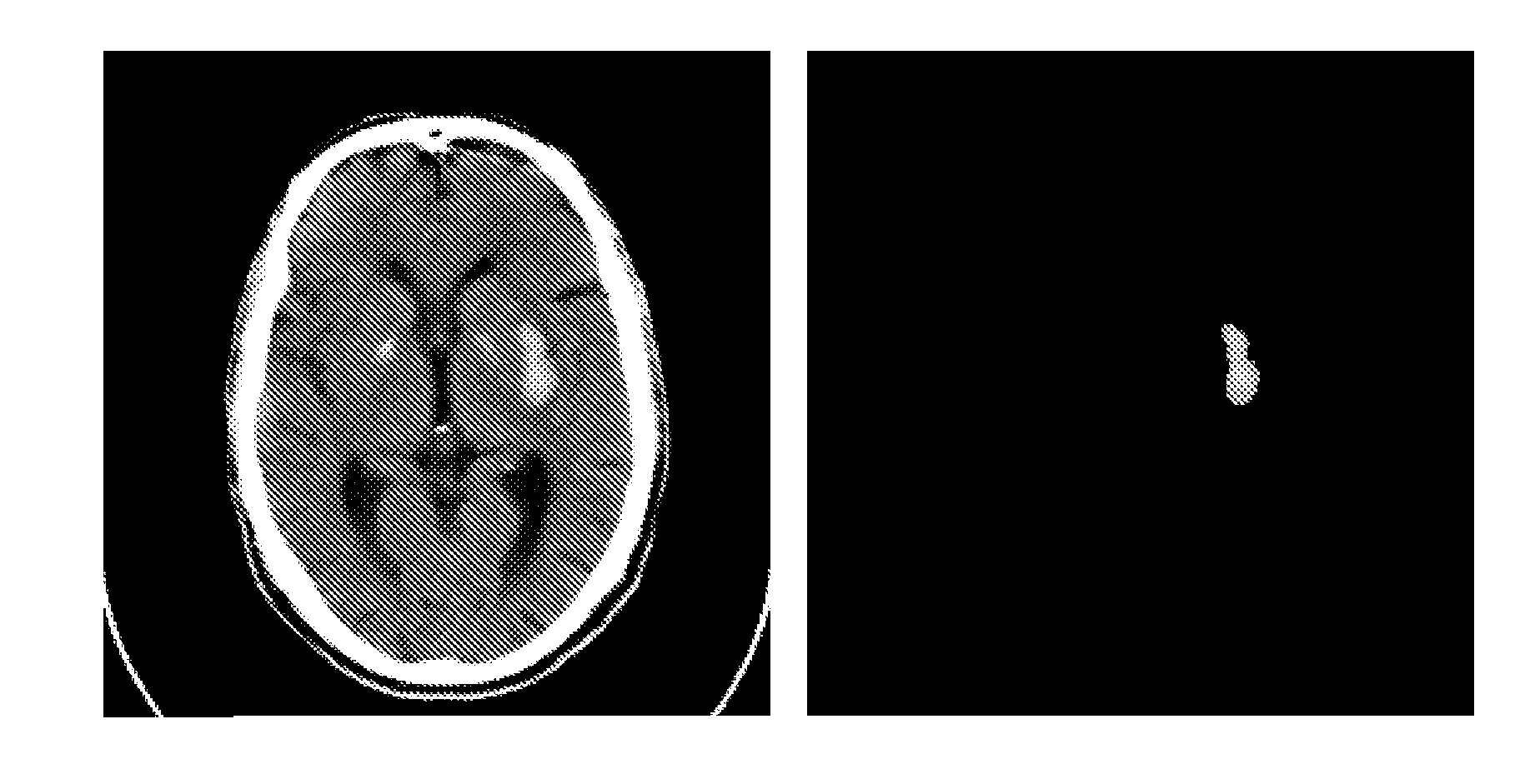
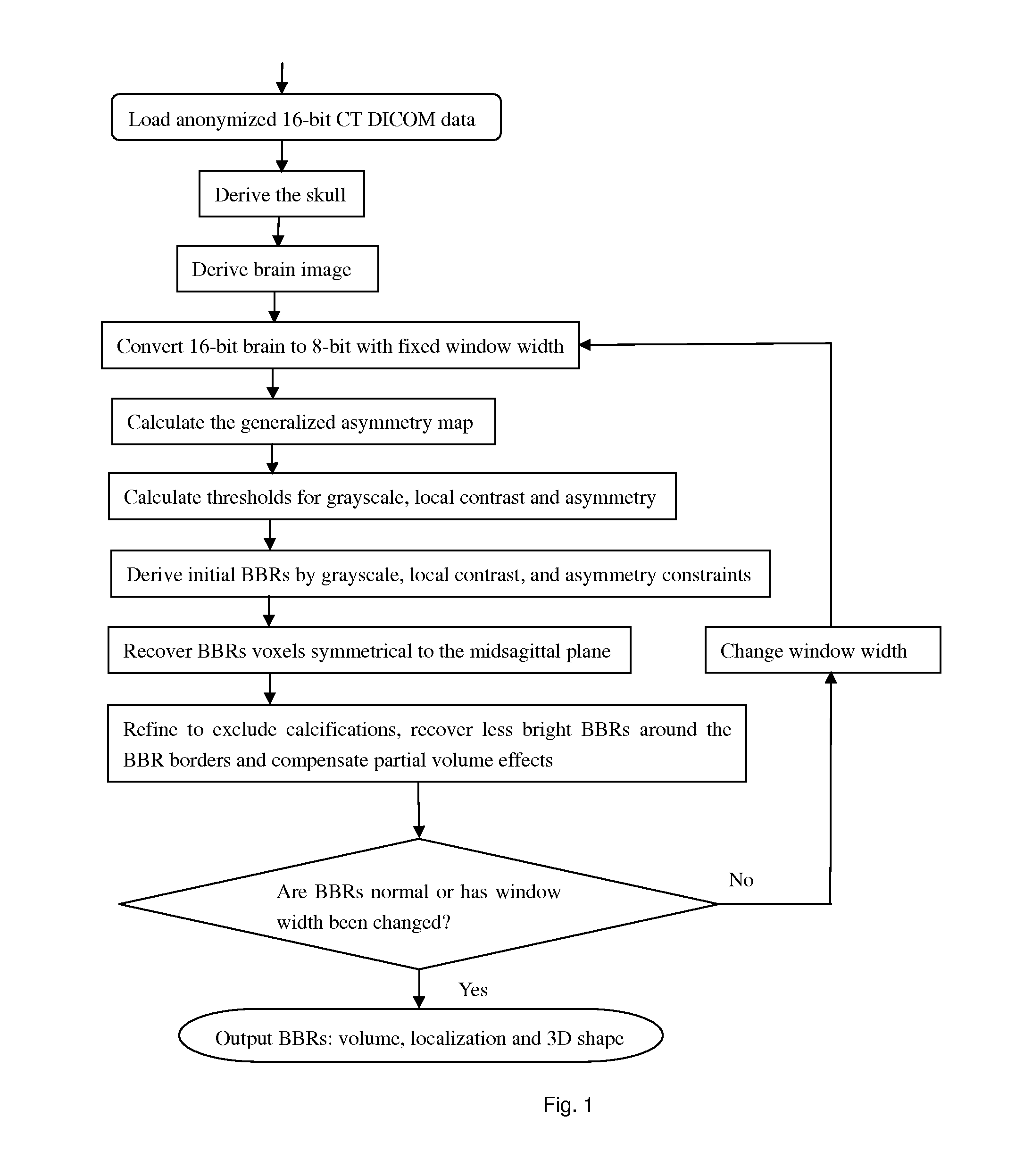
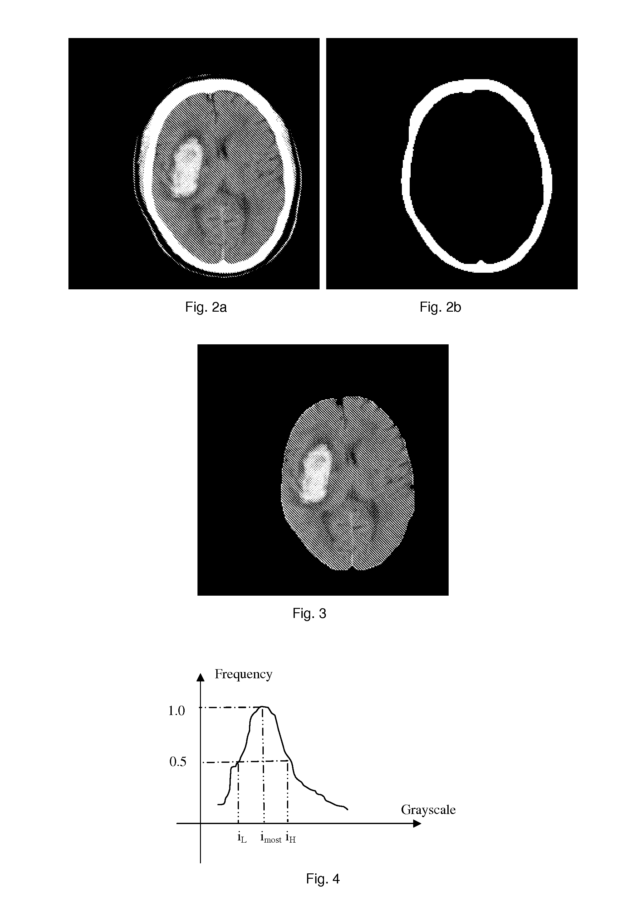
[0027]The method or device of the present invention takes anonymized DICOM (Digital Imaging and Communication in Medicine, 16-bits) CT images as input, removes the skull from the original 16-bit data, then converts the remaining images into 8-bit with default window width and window center. Subsequently, the average grayscale of the detected bright brain regions (BBRs) is checked to see if it falls within a reasonable range. If no, another round is iterated by changing the window width of the data conversion. The algorithm consists of: derivation of the skull; derivation of the brain to confine the locations of BBRs; ...


PUM


Abstract

A method and devices are disclosed to detect bright brain regions (BBRs) from clinical non-enhanced computed tomography images through large grayscale, large grayscale asymmetry with respect to the midsagittal plane (MSP), and large grayscale local contrast. An adaptive approach is disclosed to determine thresholds of the 3 features and adjust the window width for data conversion. The substantial grayscale variability of BBRs for a subject is addressed by finding the bright portion followed by recovering. Those BBR voxels symmetrical to the MSP are recovered, partial volume effects are compensated and the high grayscale regions which may not correspond to intracerebral hemorrhage are excluded. The disclosed method and system could be a useful tool to aid classifying stroke types, quantifying intracerebral hemorrhage and enhancing stroke therapy.

Description

FIELD OF THE INVENTION[0001]The present invention relates to a medical image processing technology, in particular, relates to a method and a device for detecting the bright brain regions of a patient utilizing computed tomography (CT) images.BACKGROUND OF THE INVENTION[0002]Strokes are either ischemic or hemorrhagic. Because the management of these subtypes is so different, the clinical distinction among the subtypes is one of the most important and urgentest steps in stroke management.[0003]Intracerebral hemorrhage (ICH) is an acute and spontaneous extravasation of blood into the brain parenchyma. It accounts for 10-30% of all stroke admissions to hospital, and leads to catastrophic disability, morbidity, and a 6 month mortality of 30-50%.[0004]The presentation of ICH is very variable and is indistinguishable from ischemic stroke on clinical grounds alone, making neuro-imaging absolutely necessary. CT has become part of the standard evaluation of ICH because of its wide availabilit...

Claims


Application Information

Patent Timeline
Patent Type & Authority Applications(United States)
IPC IPC(8): G06K9/00
CPCA61B6/03A61B6/501G06T7/0012G06T7/0081G06T2207/30016G06T7/602G06T7/608G06T2207/10081G06T2207/20148G06T7/0091G06T7/11G06T7/155G06T7/62G06T7/68G06T7/136
Inventor HU, QINGMAO
Owner SHENZHEN INST OF ADVANCED TECH CHINESE ACAD OF SCI
Why Eureka
Try Eureka