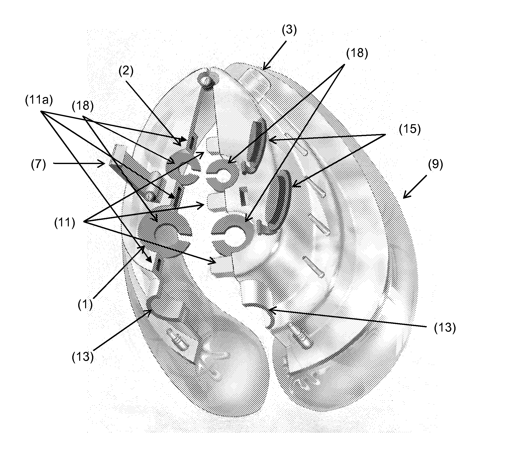
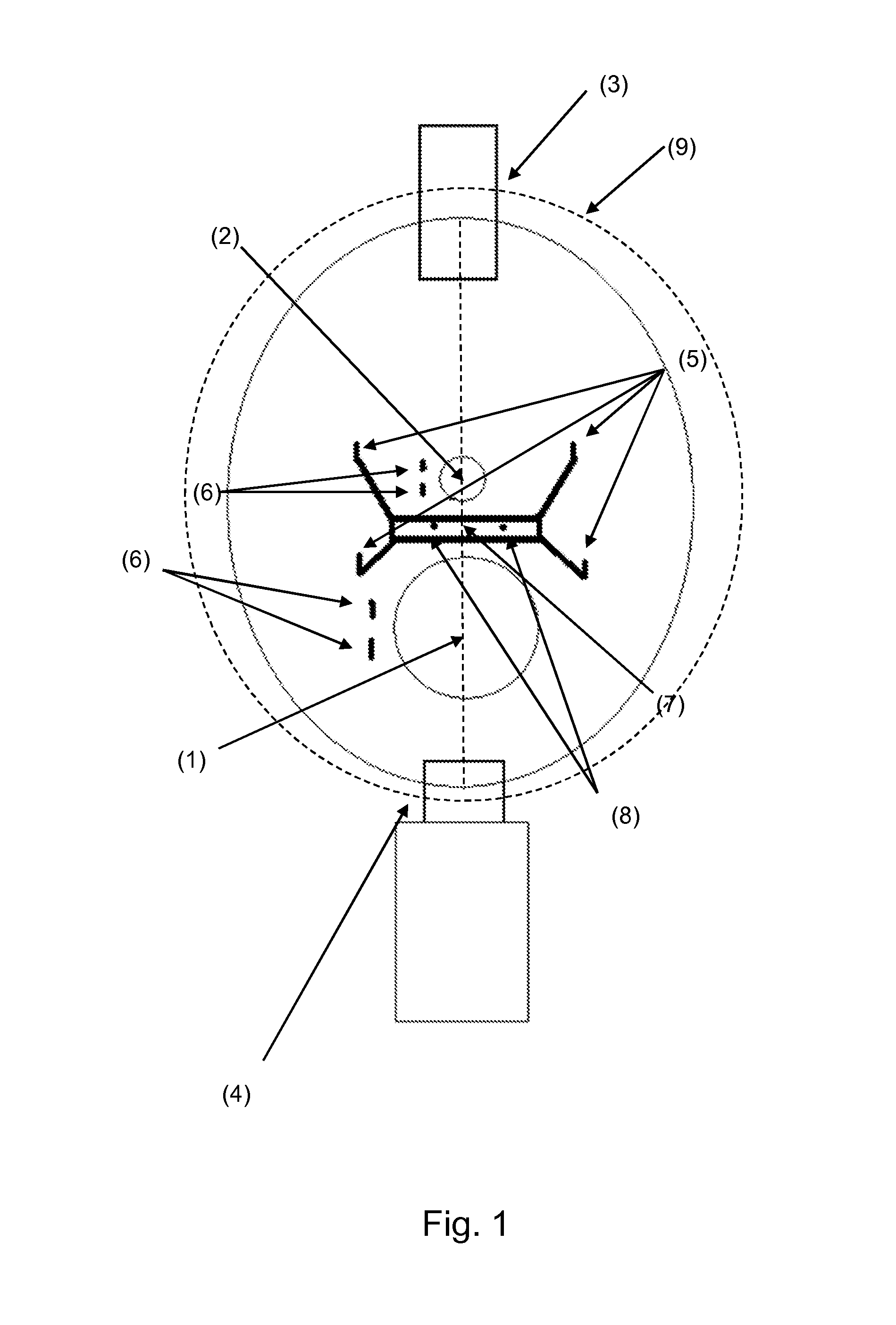
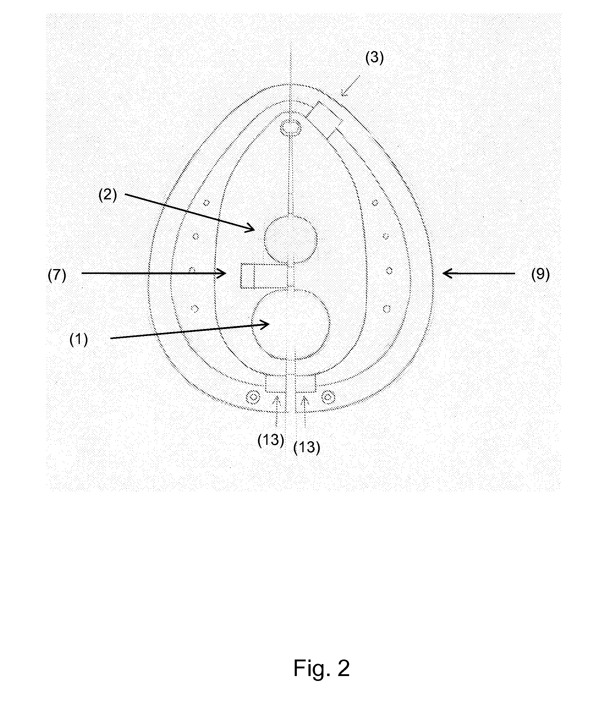
Non-invasive ventilation mask and use thereof

a ventilation mask and non-invasive technology, applied in the field of respiratory devices and methods, can solve the problems of unfitting for many probes, most masks will be wasted, and available masks can only be used for limited kinds of endoscopy

Inactive Publication Date: 2012-10-25
LANDONI GIOVANNI GUGLIELMO +1
View PDF12 Cites 5 Cited by
  • Summary
  • Abstract
  • Description
  • Claims
  • Application Information

AI Technical Summary

Problems solved by technology

Unfortunately, since it is not possible to foresee which patients will really develop respiratory failure, most masks will be wasted.
Furthermore, available masks can be used only for limited kinds of endoscopies, being unfitted for many probes both as port position and diameter.
Alternatively, there are masks with a port for endoscopic probes, but these masks should be always placed on the patient's face before starting the endoscopic procedures, without knowing if the patient will require to be ventilated, with increased costs and poor utilization.
Conversely, if the patient is under endoscopic examination and subsequently requires ventilation, the examination must be interrupted, the probe(s) must be removed and inserted through the port(s) and then again through the patients' nose or mouth, loosing a considerable amount of time and increasing the risks related to probe insertion since repeating the probe insertion can damage the patient's mucosa or perforate the larynx / pharynx.
Probe removal can be uneasy and the time (and risks) spent to reinsert it in the right position are wasted.
The same concepts apply to patients who are denied endoscopic procedures because considered at very high risk of ventilatory failure, or for the costs and organizing problems related to assisted ventilation during the procedures.
The transition from spontaneous breathing with the endoscopic probes to tracheal intubation and repositioning of the endoscopic probes is dangerous and wastes plenty of time: the insertion of the endotracheal tube can take too long and the patient can suffer hypoxia and ventilator associated pneumonia; the repeated insertion of the probes can be dangerous and wastes time.
This may require a significant amount of time and the patient may not be breathing sufficiently to maintain adequate blood oxygen levels.
In addition, the speed with which the transition process must be completed increases the chances of a mistake being made or unnecessary injury to the patient during the intubation procedure.
Even with a cooperative patient, probe insertion and keeping the probe in situ is very uncomfortable and can cause the patient to panic.
This procedure can also result in a choking or gagging response that makes the procedure dangerous or impossible.
These side effects are unpredictable and may be unacceptable when dealing with a patient who already suffers from cardiopulmonary complications.

Method used


Image

  • Non-invasive ventilation mask and use thereof
  • Non-invasive ventilation mask and use thereof
  • Non-invasive ventilation mask and use thereof

Examples


example 1

[0069]Endotracheal intubation under fiberoptic guide is the suggested technique for planned difficult endotracheal intubation in the operating room. Awake intubation is an extremely painful procedure.

[0070]PATIENT A didn't receive sedation and died of myocardial infarction during the procedures. Tachycardia and hypertension caused by the procedure initiated myocardial infarction in this patient.

[0071]PATIENT B received too much sedation for the procedure and died because of hypoxia and respiratory failure. The sedation caused respiratory arrest in this patient.

[0072]PATIENT C was anesthetized and but underwent endotracheal intubation with delay because of difficult airway. He died shortly thereafter (hypoxic brain death). The junior anesthesiologist wasn't confident with the procedure of fiberoptic intubation.

[0073]In patient A, the presence of the mask of the invention would have allowed the physician to administer sedation to the patient and prevent myocardial infarction. In patie...

example 2

[0074]Transoesophageal echocardiography (TEE) is widely used in intensive care units, arrhythmology departments (before electrical cardioversion and / or ablation procedures), cardiac surgery departments (before cardiac surgery), cardiology outpatients.

[0075]PATIENT A was denied TEE because suffering of chronic obstructive pulmonary disease and died shortly thereafter of severe aortic stenosis.

[0076]PATIENT B received general anesthesia to perform TEE in the suspect of severe aortic stenosis and died because of general anesthesia

[0077]PATIENT C was slowly recovering in intensive care unit after major surgery. He received TEE to diagnose pericardial effusion. His clinical condition deteriorated during the procedure, the patient required intubation and died 2 weeks later because of ventilator associated pneumonia.

[0078]PATIENT D was planned for percutaneous aortic valve replacement (too high risk for cardiac surgery). The procedure was lengthy and after three hours he became restless an...


PUM


Abstract

The present invention relates to a non-invasive-ventilation-mask to patients with and ongoing endoscopic procedure without interrupting the procedure, without removing the endoscopic probe and without requiring endotracheal intubation.

Description

FIELD OF THE INVENTION[0001]The present invention relates generally to the field of respiratory devices and methods. More specifically, the present invention discloses a method and apparatus for applying a non-invasive-ventilation-mask to patients with an ongoing endoscopic procedure without interrupting the procedure, without removing the endoscopic probe and without requiring endotracheal intubation.BACKGROUND OF THE INVENTIONEndoscopic Procedures[0002]Technical and medical progress continuously add new properties to endoscopic examinations, like ultra-sonography. Many endoscopic examinations require to pass through the mouth or the nose of the patient to make diagnosis or treatment in a partially non-invasive way.[0003]All hospitals daily perform endoscopic procedures. The most frequently performed procedures are:[0004]TEE (transoesophageal echocardiography) performed by intensive care specialists and cardiologists;[0005]Fiberoptic bronchoscopy with or without intubation performe...

Claims


Application Information

Patent Timeline
Patent Type & Authority Applications(United States)
IPC IPC(8): A61M16/06
CPCA61M16/0488A61M16/0683A61M16/06
Inventor LANDONI, GIOVANNI GUGLIELMOCABRINI, LUCA
Owner LANDONI GIOVANNI GUGLIELMO
Why Eureka
Try Eureka