Methods and reagents for analyzing riboswitches using fret
- Summary
- Abstract
- Description
- Claims
- Application Information
AI Technical Summary
Benefits of technology
Problems solved by technology
Method used
Image
Examples
example 1
General Methods and RNA Preparation
1. Solid-Phase Synthesis of Oligoribonucleotides
[0081]All oligonucleotides were synthesized on Pharmacia instruments (Gene Assembler Plus) following DNA / RNA standard methods.
[0082]Detritylation (2.0 min): dichloroacetic acid / 1,2-dichloroethane (4 / 96); coupling (3.0 min): phosphoramidites / acetonitrile (0.1 M×120 μL) were activated by benzylthiotetrazole / acetonitrile (0.3 M×360 μL); capping (3×0.4 min): A: Ac2O / sym-collidine / acetonitrile (20 / 30 / 50), B: 4-(dimethylamino)pyridine / acetonitrile (0.5 M), A / B=1 / 1; oxidation (1.0 min): I2 (10 mM) in acetonitrile / sym-collidine / H2O (10 / 1 / 5). For 5-aminoallyl-uridine sequences, mild capping solutions were used: A: 0.2 M phenoxyacetic anhydride in THF, B: 0.2 M N-methylimidazole and 0.2 M sym-collidine in THF. Acetonitrile, solutions of amidites and tetrazole were dried over activated molecular sieves overnight.
[0083]2′-O-TOM standard nucleoside phosphoramidites were obtained from GlenResearch or ChemGenes. 5′-...
example 2
Bulk Fret Detection of Pseudoknot Formation
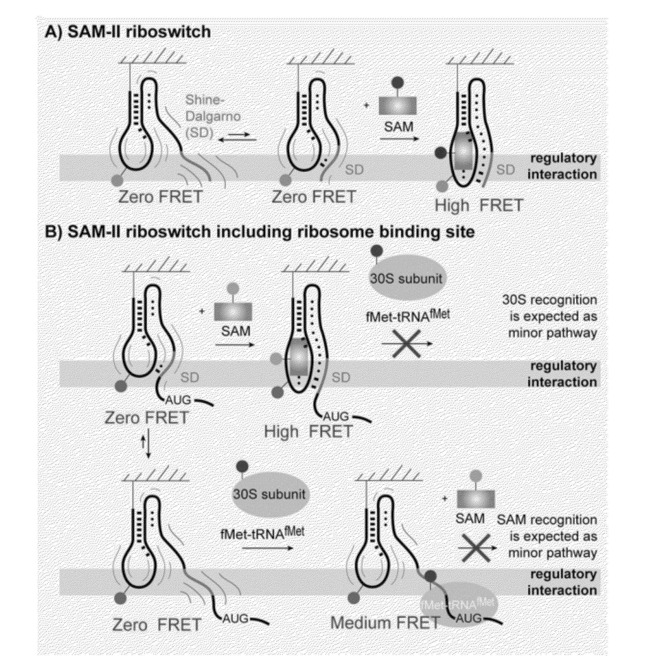
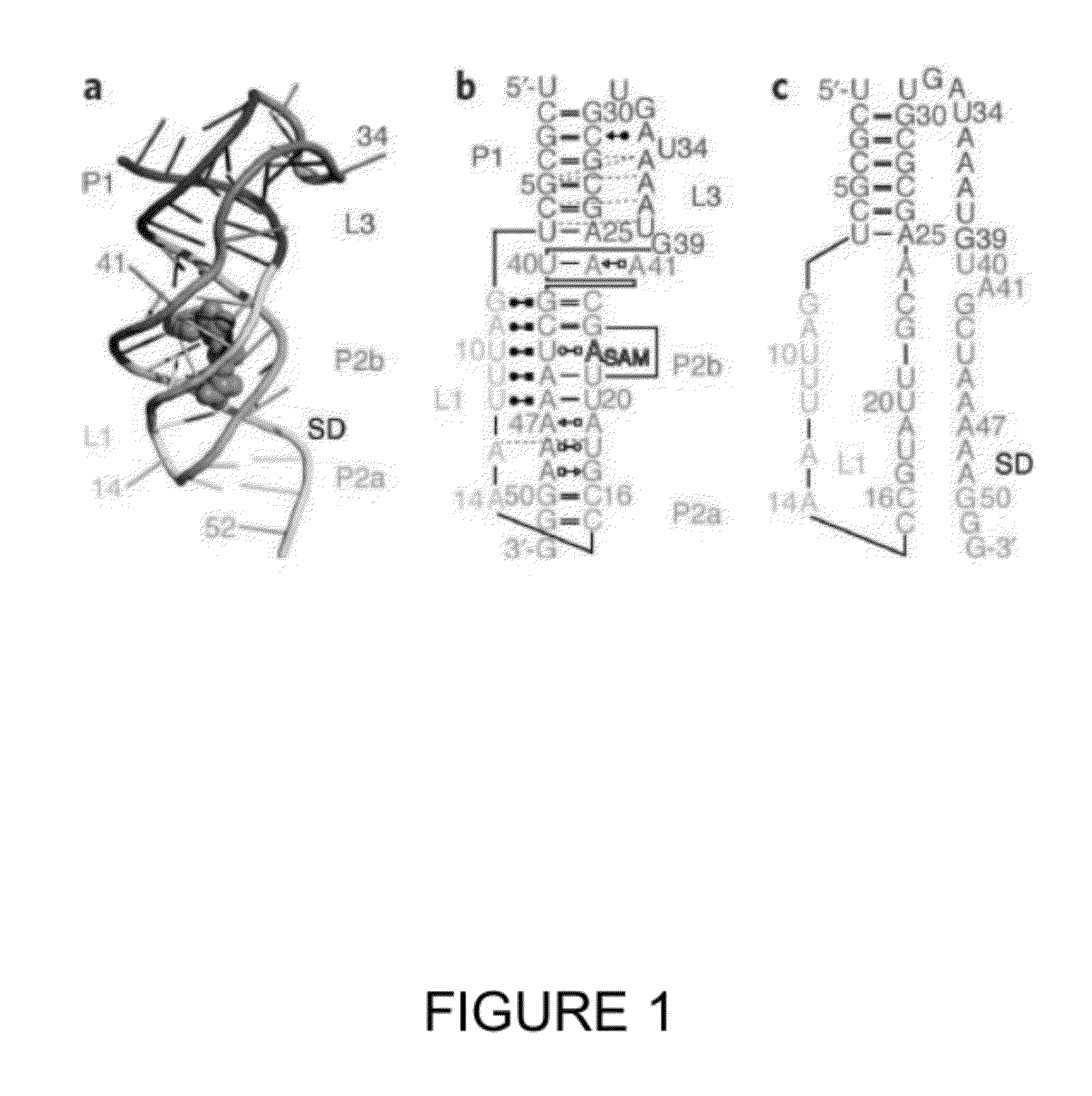
[0088]NMR studies (Haller 2011) provided structural insights for designing fluorescently-labeled SAM-II riboswitch constructs for following pseudoknot formation using a FRET-based approach. Through such experiments, the formation of P2a helix, reflecting the sequestration of the SD sequence, was followed.
1. Preparation of Cy3 / Cy5 Labeled Oligoribonucleotides
[0089]Cy3 and Cy5 NHS esters were purchased from GE Healthcare. DMSO was dried over activated molecular sieves.
[0090]Labeling was performed according to Solomatin 2009, with slight modifications as described below: Dye-NHS ester (1 mg; ˜1300 nmol) was dissolved in anhydrous DMSO (500 μL). Lyophilized RNA (20 nmol) containing the 5-aminoallyluridine modification was dissolved in labeling buffer (20 μL; 500 mM phosphate buffer pH=8.0) and nanopure water was added to reach a fraction of 55% (v / v) (122 μL) of the intended final reaction volume (222 μL) with a final concentration of cRNA of 9...
example 3
smFRET for Sensing Pseudoknot Formation
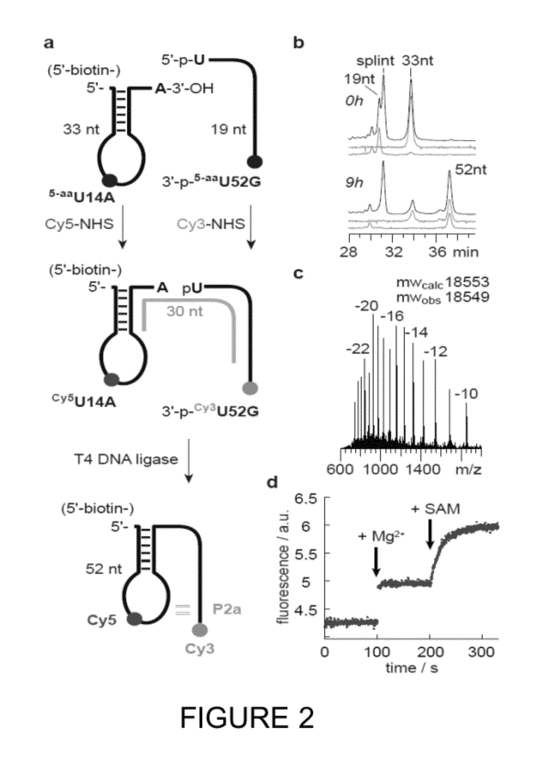
[0100]To reveal the underlying dynamics of SAM-II pseudoknot formation, single-molecule FRET (smFRET) (Aleman 2008; Lemay 2006; Brenner 2010) investigations were performed using the same labeling strategy as in Example 2 (i.e., A14Cy3U and G52Cy5U). To enable surface immobilization of the SAM-II riboswitch within a passivated microfluidic device and extended observation periods, a 5′-biotin moiety was included in the design (FIG. 2, FIG. 3).
1. Acquisition of smFRET Data
[0101]smFRET data were acquired using a prism-based total internal reflection (TIR) microscope, where the biotinylated SAM-II riboswitch was surface immobilized within PEG-passivated, strepatividin-coated quartz microfluidic devices (Munro 2007). The Cy3 fluorophore was directly illuminated under 1.5 kW / cm2 intensity at 532 nm (Laser Quantum). Photons emitted from both Cy3 and Cy5 were collected using a 1.2 NA 60× Plan-APO water-immersion objective (Nikon), where optical treatmen...
PUM
| Property | Measurement | Unit |
|---|---|---|
| Electrical conductance | aaaaa | aaaaa |
| Concentration | aaaaa | aaaaa |
| Cytotoxicity | aaaaa | aaaaa |
Abstract
Description
Claims
Application Information
 - R&D
- Intellectual Property
- Life Sciences
- Materials
- Tech Scout
- Unparalleled Data Quality
- Higher Quality Content
- 60% Fewer Hallucinations
Browse by: Latest US Patents, China's latest patents, Technical Efficacy Thesaurus, Application Domain, Technology Topic, Popular Technical Reports.
© 2025 PatSnap. All rights reserved.Legal|Privacy policy|Modern Slavery Act Transparency Statement|Sitemap|About US| Contact US: help@patsnap.com


