
Method of enhancing skin barrier function

a skin barrier and function technology, applied in the field of evaluating skin barrier function, can solve the problems of increasing the amount, skin barrier dysfunction, and liable to be recognized the expression of the above-mentioned skin barrier dysfunction, and achieve enhanced skin barrier function, and excellent ability

Inactive Publication Date: 2012-11-08
POLA CHEM INDS
View PDF6 Cites 2 Cited by
  • Summary
  • Abstract
  • Description
  • Claims
  • Application Information

AI Technical Summary

Benefits of technology

The present invention provides a technology for developing a material that can enhance skin barrier function and a cosmetic based on this technology. The method involves screening materials using the permeability of metal ions, specifically calcium ions, to evaluate their effectiveness in enhancing skin barrier function. The method involves culturing normal human epidermal cells in the presence and absence of a test substance, measuring the concentration of metal ions in each medium, and calculating the permeability of the metal ions in an epidermal cell layer based on the measured concentrations. The invention also provides a material for enhancing skin barrier function and an adjuster for calcium ion in epidermis. The cosmetic comprises the material for enhancing skin barrier function.

Problems solved by technology

In fact, there is a high incidence of the skin barrier dysfunction, so it is required to develop means for preventing a disease in an early stage from becoming severe or means for ameliorating an actual symptom.
However, in many cases, expression of the skin barrier dysfunction described above is liable to be recognized after developments of severe symptoms such as an occurrence of skin roughness and a rapid increase in amount of transepidermal water loss.
There are some proposals on animal models for skin barrier dysfunction (see, for example, Patent Documents 5 and 6), but in all the proposals, animals must be used and much time and efforts are required.
Further, there is a room for doubt about its reproducibility.
Moreover, in such screening using animal models, the number of test samples to be evaluated is limited.
There has not been known at all a method of evaluating skin barrier function using culture cells.
However, there has been also known that the excessive calcium ion impairs the skin barrier function (see, for example, Non-patent Documents 1, 2, and 3).

Method used


Image

  • Method of enhancing skin barrier function
  • Method of enhancing skin barrier function
  • Method of enhancing skin barrier function

Examples


example 1

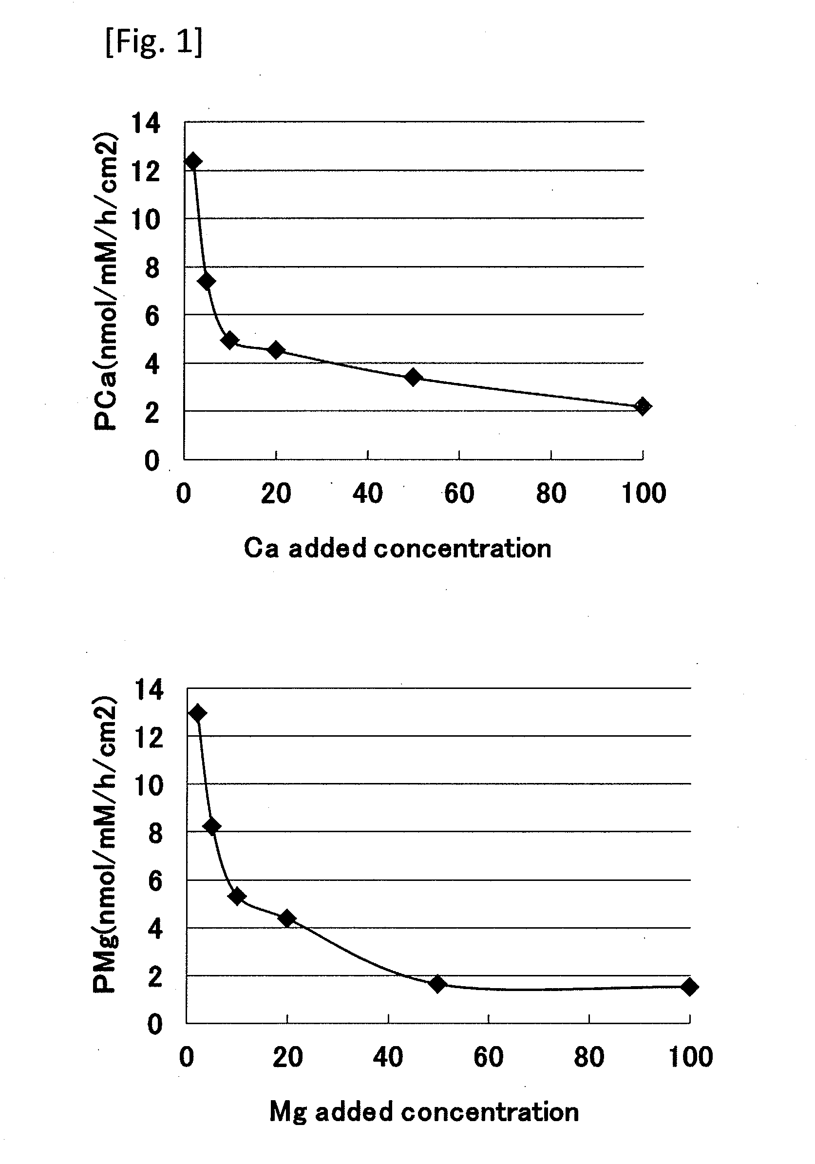
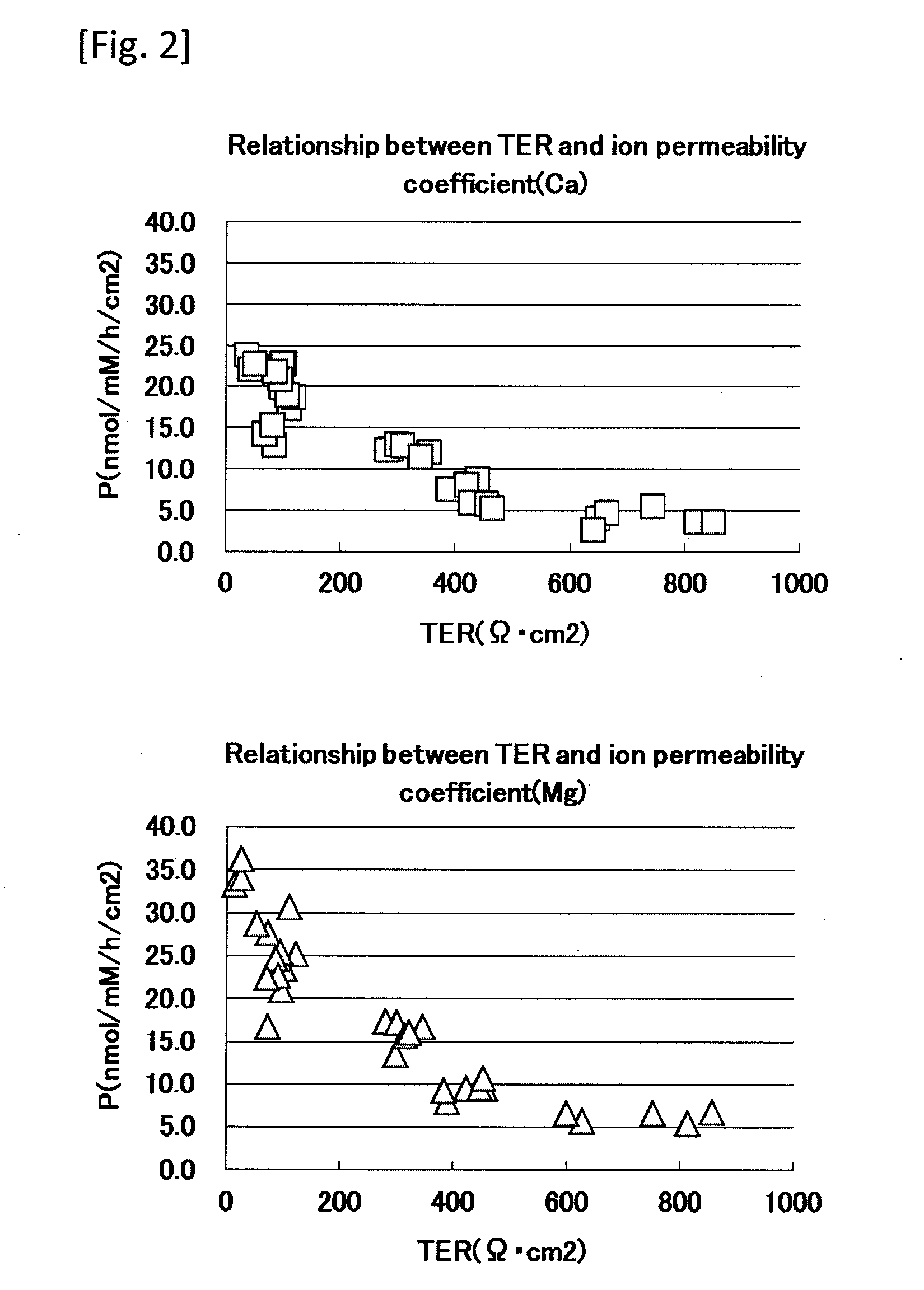
[0048]An ion permeability test using normal human epidermal cells was performed based on a method of evaluating skin barrier function of the present invention.

[0049]1) Cell Inoculation and Ion Permeability Experiment

[0050]Frozen normal human epidermal keratinocytes (NHEK) (manufactured by Kurabo Industries Ltd.) were thawed and cultured in a 0.15 mM Ca-containing culture medium (Humedia-KG2; manufactured by Kurabo Industries Ltd.) at 37° C. in 50% carbon dioxide atmosphere. A Transwell (registered trademark) manufactured by Corning Incorporated (diameter 12 mm, polyethylene terephthalate, 0.4 μm pore) was placed on a Millicell (registered trademark) tissue culture plate (manufactured by Millipore), and the above-mentioned culture medium was added in amounts of 1.5 ml to the lower layer and 0.5 ml to the upper layer. Then, the normal human epidermal keratinocytes (NHEK) were inoculated at 1×10⁵ / cm2 and cultured for additional 72 hours. After the cells were confirmed to be confluent, ...

example 2

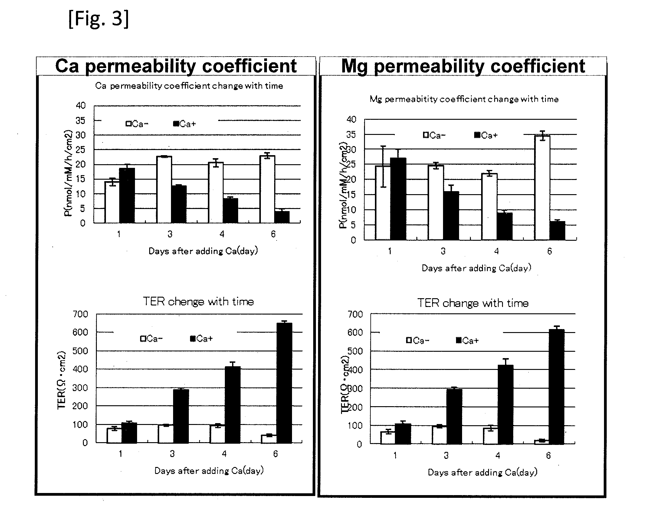
[0065]In the same way as in Example 1, calcium was selected as a metal ion to be added to compare calcium permeability coefficients and magnesium permeability coefficients in a low-calcium medium cultured cells (shown as Ca− in FIG. 3) (0.15 mM) and in a high-calcium medium cultured cells (shown as Ca+ in FIG. 3) (1.45 mM). That is, frozen normal human epidermal keratinocytes (NHEK) (manufactured by Kurabo Industries Ltd.) were thawed and cultured in a 0.15 mM Ca-containing culture medium (Humedia-KG2; manufactured by Kurabo Industries Ltd.) at 37° C. in 50% carbon dioxide atmosphere. A Transwell (registered trademark) manufactured by Corning Incorporated (diameter 12 mm, polyethylene terephthalate, 0.4 μm pore) was placed on a Millicell (registered trademark) tissue culture plate (manufactured by Millipore), and the above-mentioned culture medium was added in amounts of 1.5 ml to the lower layer and 0.5 ml to the upper layer. Then, the normal human epidermal keratinocytes (NHEK) we...

example 3

[0066]TERs and metal ion permeability coefficients in the case where claudin-1, one of tight junction proteins serving as adhesion factors for epidermal cells, was suppressed with siRNA were measured in accordance with the method of Example 1. That is, frozen normal human epidermal keratinocytes (NHEK) (manufactured by Kurabo Industries Ltd.) were thawed and cultured in a 0.15 mM Ca-containing culture medium (Humedia-KG2; manufactured by Kurabo Industries Ltd.) at 37° C. in 50% carbon dioxide atmosphere. Thereafter, siRNA was transfected into the cultured cells. A Transwell (registered trademark) manufactured by Corning Incorporated (diameter 12 mm, polyethylene terephthalate, 0.4 μm pore) was placed on a Millicell (registered trademark) tissue culture plate (manufactured by Millipore), and the above-mentioned culture medium was added in amounts of 1.5 ml to the lower layer and 0.5 ml to the upper layer. Then, the human epidermal keratinocytes transfected with siRNA were inoculated ...


PUM

PropertyMeasurementUnit
culture timeaaaaaaaaaa
diameteraaaaaaaaaa
volumeaaaaaaaaaa

Abstract

A method for adjusting calcium ion in epidermis is described. The method involves applying a material for enhancing skin barrier function to the skin of a subject. Appropriate materials include Pimpinella extract, Asteroidea extract, L-Carnitine, Royal jelly hydrolysate, Pellicer, Palmaria extract, ε, γ-Glutamyl lysine, Coptis japonica extract and citrus aurantium peel extract

Description

CROSS-REFERENCE TO RELATED APPLICATIONS[0001]This application is a divisional of U.S. application Ser. No. 12 / 516,483, filed May 27, 2009, which is incorporated herein by reference and which is the U.S. National Phase under 35 U.S.C. §371 of International Application PCT / JP2007 / 074070, filed Dec. 7, 2007, which claims priority to JP 2006-331294, filed Dec. 8, 2006 and JP 2007-133776, filed May 21, 2007.TECHNICAL FIELD[0002]The present invention relates to a method of evaluating skin barrier function, a method of screening material for enhancing skin barrier function using the evaluating method, the material for enhancing skin barrier function, and a cosmetic containing the material for enhancing skin barrier function.BACKGROUND ART[0003]At present days, patients who suffer from skin diseases due to skin barrier dysfunction, such as a high incidence of atopic dermatitis, have increased rapidly. This is considered to be caused by an excessive increase in stress (see, for example, Pate...

Claims


Application Information

Patent Timeline
Patent Type & Authority Applications(United States)
IPC IPC(8): A61K8/97A61Q19/00
CPCG01N33/84G01N33/5044A61P3/14A61P17/00
Inventor KURODA, SHOHEIYAMAMOTO, TAKUYASAEKI, YUKOKURASAWA, MASUMI
Owner POLA CHEM INDS
Learn More