

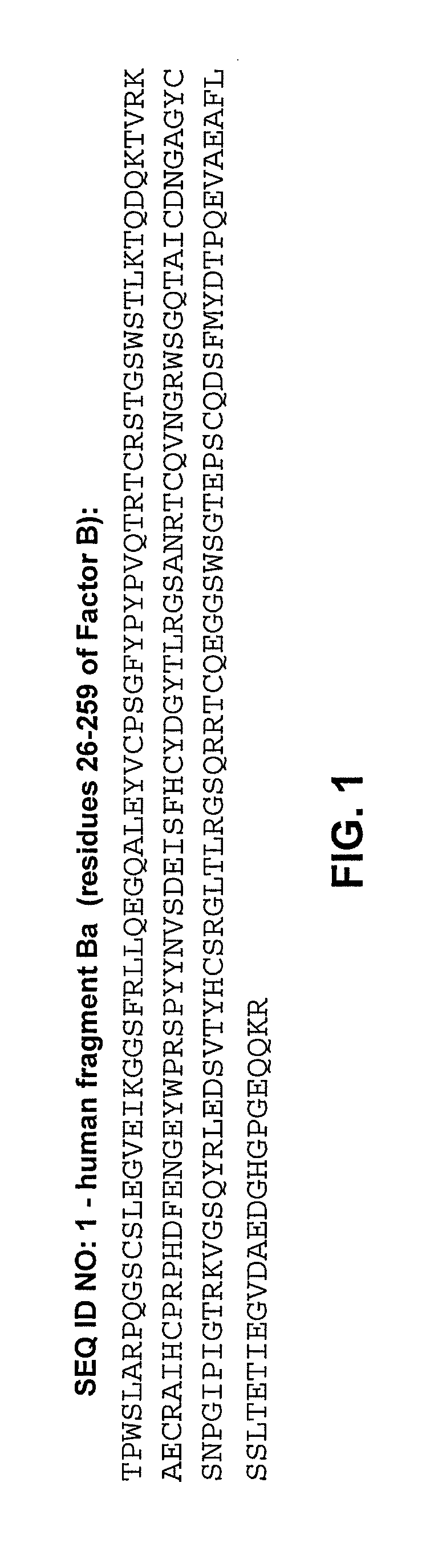
METHODS OF USE FOR AN IMMUNOASSAY DETECTING FRAGMENT Ba
a technology of immunoassay and fragment, applied in the field of new techniques for prenatal diagnosis, can solve the problems of adverse pregnancy outcomes, uncontrolled complement activation, and inflammation of the placenta, and achieve the effects of reducing the risk of infection, and reducing the effect of immunoassay
- Summary
- Abstract
- Description
- Claims
- Application Information
AI Technical Summary
Benefits of technology
Problems solved by technology
Method used
Image

Examples
example 1
Basic Assay Protocol 1
[0123]100 μL standards and / or samples were added to anti-Ba coated plates, incubated 1 hour at 24° C., and washed five times in 20× wash buffer. 100 μL diluted conjugate was added to wells, and plates were then incubated 30 minutes at 24° C. Wells were washed five times in 20× wash buffer. 100 μL TMB substrate was added to wells and the plates were incubated 15 minutes at 24° C. 100 μL 1N HCl was added to wells to stop the reaction. Optical density was read at 450 nm.
Basic Assay Protocol 2
[0124]100 μL standards and / or samples were added to anti-Ba coated plates, incubated 1 hour at 24° C., and washed five times in 20× wash buffer. 100 μL diluted conjugate was added to wells, and plates were then incubated 1 hour at 24° C. Wells were washed five times in 20× wash buffer. 100 μL TMB substrate was added to wells and the plates were incubated 15 minutes at 24° C. 100 μL 1N HCl was added to wells to stop the reaction. Optical density was read at 450 nm.
example 2
Purification of mAb Anti-Human Ba Neo Antibody
[0125]Ascites used to express the monoclonal antibody anti-human Ba (mAb anti-Ba) were produced by Strategic Diagnostics Inc. (SDI). 150 ml of ascites were processed on a 50 ml ProSep®-A affinity column with a yield of 407 mg (2.7 mg of antibody / ml ascites). The final purity was >95% by SDS-PAGE. The final concentration was 1.1 mg / ml and it was stored at −80° C.
example 3
Proof of Principle and Feasibility for the MicroVue® Ba Assay
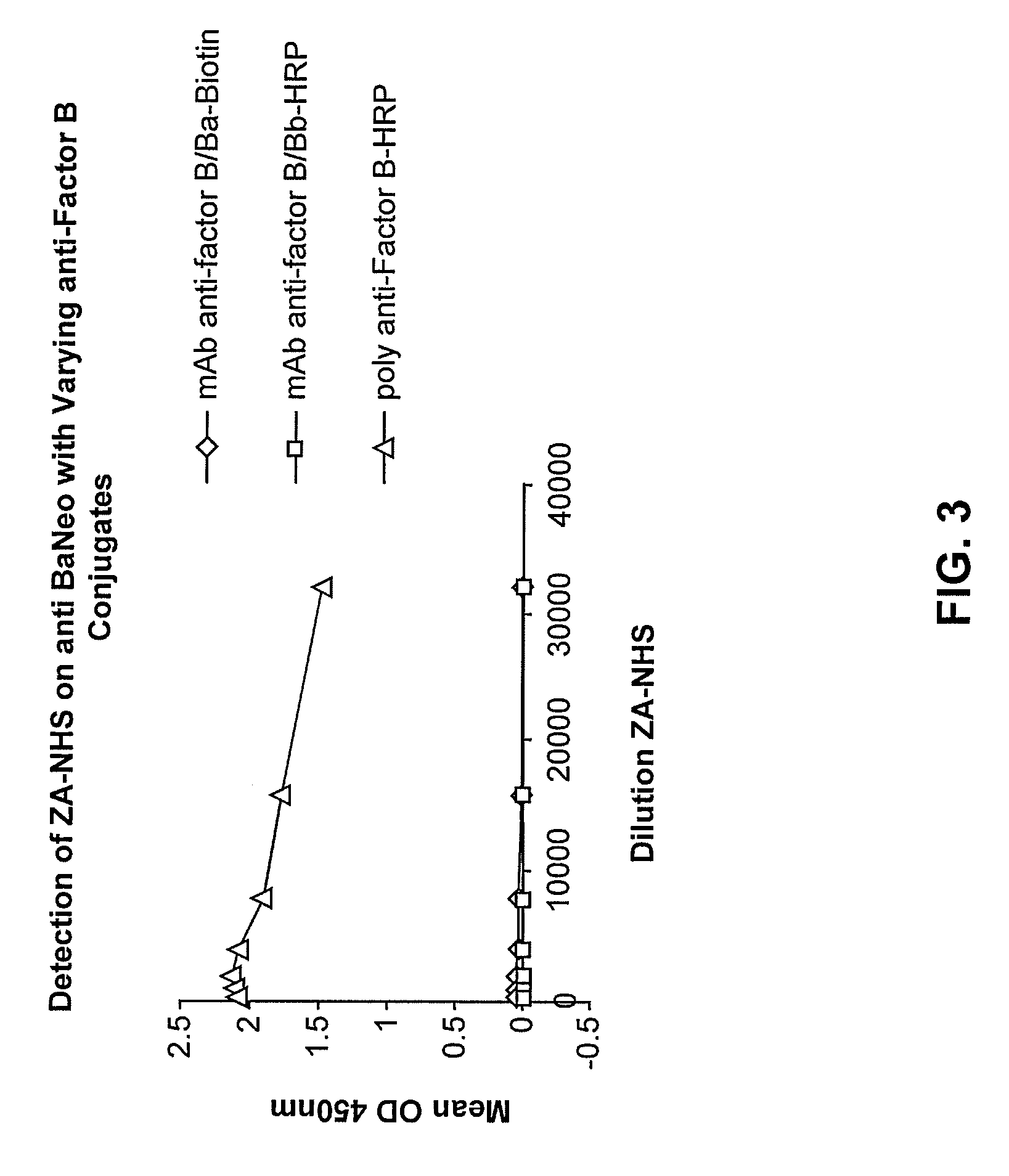
[0126]Nunc 96 well plates were coated with 10 μg / ml of the anti-Ba neo antibody overnight at 4° C. They were then incubated for one hour with a titration of zymosan activated normal human serum (ZA-NHS). The plate was subsequently washed with wash buffer and then incubated with biotinylated mAb anti Factor B / Ba, and mAb anti-Factor B / Bb-HRP as a control, and goat polyclonal Ab anti-Factor B-HRP. Excess conjugate was then removed, the biotinlyated mAb anti-Factor B / Ba was probed further with a streptavidin-horseradish peroxidase (SA-HRP) conjugate then washed and developed with 3,3′,5,5′-Tetramethylbenzidine (TMB) substrate and stopped with 1N HCl. See FIG. 3, showing that the antibody pair of mAb anti-human Ba neo and the polyclonal Factor B-HRP conjugated antibody are a highly sensitive matched pair and can detect Ba in a dilution series of ZA-NHS. The anti-Factor B-HRP is conjugated by Jackson Immunodiagnostics and is al...
PUM
| Property | Measurement | Unit |
|---|---|---|
| Time | aaaaa | aaaaa |
| Time | aaaaa | aaaaa |
Abstract
Description
Claims
Application Information