Imaging agents of fibrotic diseases

a technology of fibrotic diseases and imaging agents, which is applied in the field of organic chemistry, pharmaceutical chemistry, biochemistry, molecular biology and medicine, can solve the problems of no option other than transplantation for recovering such tissue, no satisfactory methods, and so as to facilitate the early detection of fibrotic diseases, reduce the burden of a subject to be examined, and broaden the range of subjects

Inactive Publication Date: 2013-01-10
NITTO DENKO CORP
View PDF9 Cites 15 Cited by
  • Summary
  • Abstract
  • Description
  • Claims
  • Application Information

AI Technical Summary

Benefits of technology

[0081]Although the accurate mechanism of the imaging agent of the present invention has not been completely elucidated, it is surmised that retinoid functions as targeting agent for alpha-SMA (smooth muscle actin)-positive ECM (extracellular matrix)-producing cells such as activated stellate cells, and makes it possible to detect said cells by delivering thereto a labeling substance.
[0082]Since the imaging agent of the present invention allows to nondestructively, preferably noninvasively detect fibrotic diseases in vivo, the risk of complications due to conventional biopsy is eliminated, and therefore, it is possible to considerably reduce the burden of a subject to be examined. In addition, since this may broaden the range of subjects to be examined, early detection of fibrotic diseases is facilitated, which allows an effective slowing of progression of said diseases. Thus, the present invention makes a considerably large contribution in human and veterinary medicines.
[0083]Furthermore, the imaging agent of the present invention allows to nondestructively, preferably noninvasively observe a fibrotic disease in vivo over time in a same individual, which leads to evaluate the treatment of said disease with high accuracy.
[0084]These and other embodiments are described in greater detail below.

Problems solved by technology

However, a method for recovering fibrotic tissue still does not exist, and when a large part of a tissue has been substituted with fibrotic tissue to the extent that the affected tissue cannot normally function, there are actually no options other than transplantation for recovering such a tissue.
However, since the biopsy is a highly invasive technique which may cause complications such as infection, hemorrhage, pain, and injury of other tissue, various studies relating to non-invasive diagnostic methods of fibrotic diseases are done.
However, none of such methods are satisfiable, and development of further diagnostic techniques is needed.

Method used


Image

  • Imaging agents of fibrotic diseases
  • Imaging agents of fibrotic diseases
  • Imaging agents of fibrotic diseases

Examples


example 1

Synthesis of Retinoid-PGA-DOTA

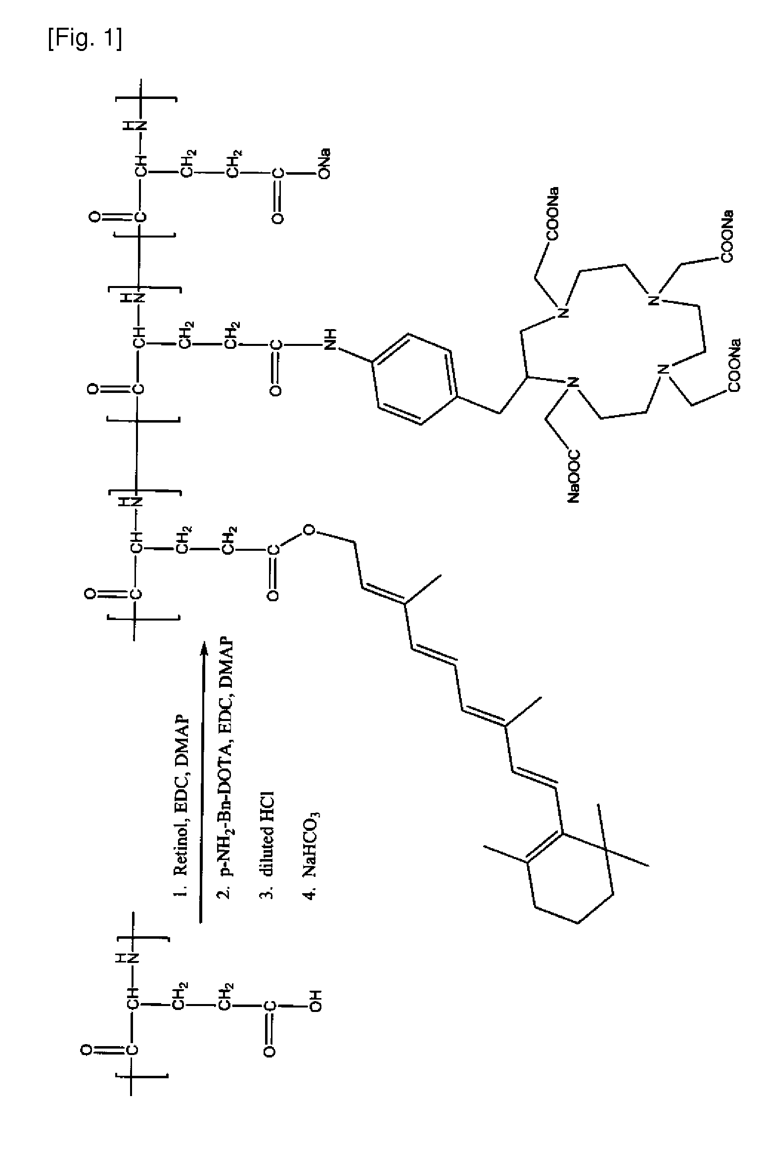
[0263]A Retinoid-PGA-DOTA polymer conjugate is prepared according to the general scheme illustrated in FIG. 1 as follows: PGA (150 mg) is dissolved in DMF (15 mL). Retinol (10 mg), EDC (50 mg), and DMAP (50 mg) are added. The mixture is stirred for 24 hours. pNH2-Bn-DOTA (10 mg), EDC (50 mg), and DMAP (50 mg) are then added. The resulting mixture is stirred for 24 hours. Diluted HCl solution (0.2M) is then added to induce precipitation. The mixture is stirred for 2 minutes and centrifuged at 10,000 rpm for 15 minutes. A solid precipitate is collected, washed with water, and redissolved with sodium bicarbonate solution (0.5 M). The mixture is dialyzed in water for 24 hours. The product, Retinoid-PGA-DOTA polymer conjugate, is freeze-dried. The product's identity is confirmed by 1H-NMR.

example 2

Synthesis of Retinoid-PGA-DTPA

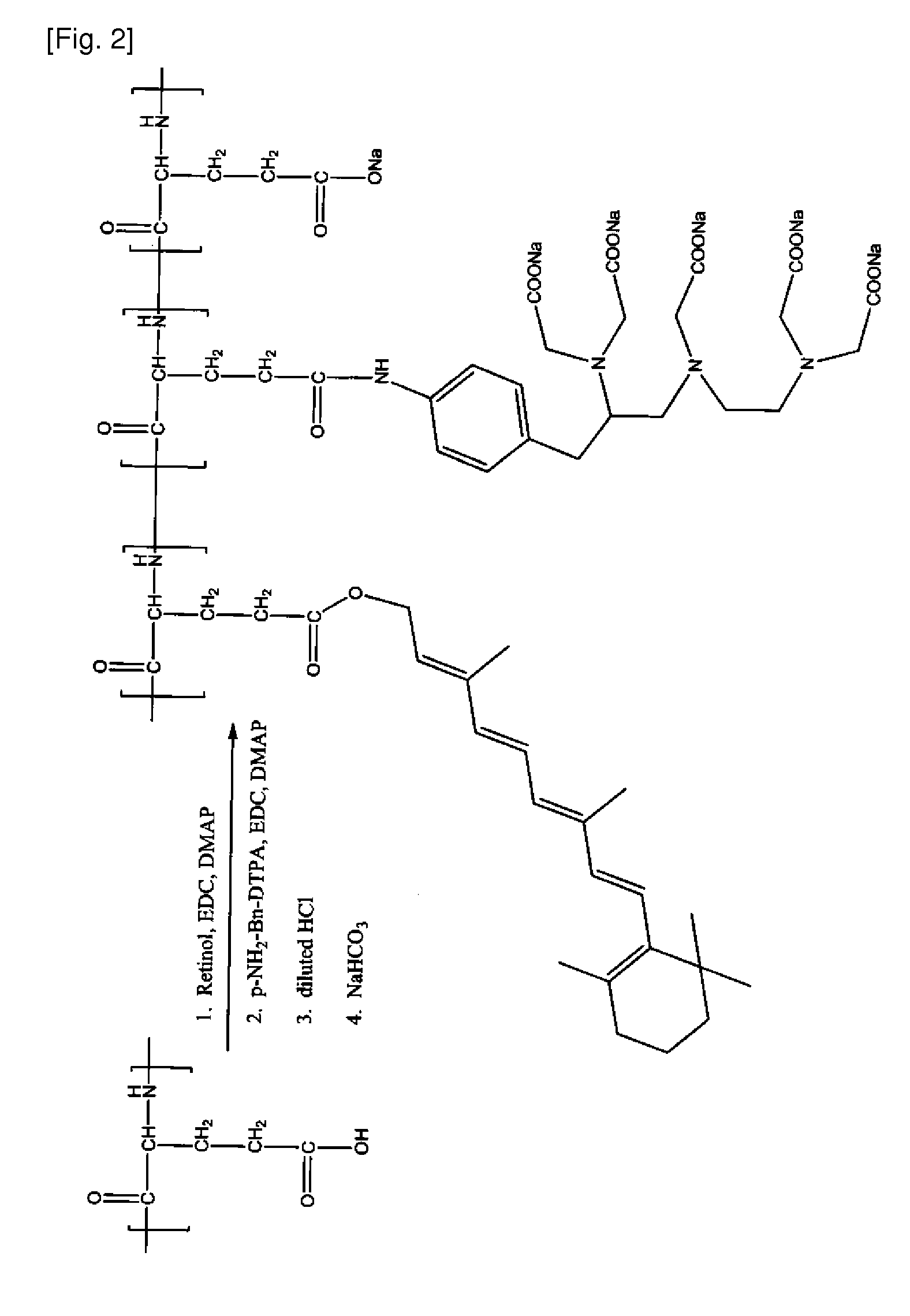
[0264]A Retinoid-PGA-DTPA polymer conjugate is prepared according to the general scheme illustrated in FIG. 2 as follows: PGA (150 mg) is dissolved in DMF (15 mL). Retinol (10 mg), EDC (50 mg), and DMAP (50 mg) are added. The mixture is stirred for 24 hours. pNH2-Bn-DTPA (10 mg), EDC (50 mg), and DMAP (50 mg) are then added. The resulting mixture is stirred for 24 hours. Diluted HCl solution (0.2M) is then added to induce precipitation. The mixture is stirred for 2 minutes and centrifuged at 10,000 rpm for 15 minutes. A solid precipitate is collected, washed with water, and redissolved with sodium bicarbonate solution (0.5 M). The mixture is dialyzed in water for 24 hours. The product, Retinoid-PGA-DTPA polymer conjugate is freeze-dried. The product's identity is confirmed by 1H-NMR.

example 3

Synthesis of Retinoid-PGGA-DOTA

[0265]A Retinoid-PGGA-DOTA polymer conjugate is prepared according to the general scheme illustrated in FIG. 3 as follows: Poly(L-gamma-glutamylglutamine) (PGGA, 150 mg) is dissolved in DMF (15 mL). Retinol (10 mg), EDC (50 mg), and DMAP (50 mg) are added. The mixture is stirred for 24 hours. pNH2-Bn-DOTA (10 mg), EDC (50 mg), and DMAP (50 mg) are then added. The resulting mixture is stirred for 24 hours. Diluted HCl solution (0.2M) is then added to induce precipitation. The mixture is stirred for 2 minutes and centrifuged at 10,000 rpm for 15 minutes. A solid precipitate is collected, washed with water, and redissolved with sodium bicarbonate solution (0.5 M). The mixture is dialyzed in water for 24 hours. The product, Retinoid-PGGA-DOTA polymer conjugate is freeze-dried. The product's identity is confirmed by 1H-NMR.


PUM

PropertyMeasurementUnit
temperatureaaaaaaaaaa
weight average molecular weightaaaaaaaaaa

Abstract

Agents and methods for imaging a cell and/or a portion of tissue characterized by fibrosis, as well as to agents and methods for determining and/or diagnosing fibrotic diseases are disclosed herein. Also disclosed herein are polymer conjugates that can include a detectable label, a retinoid and a polymer. The polymer conjugates can be used to image a portion of tissue, deliver a detectable label to a portion of tissue or a cell and/or diagnosis a condition or disease.

Description

CROSS-REFERENCE TO RELATED APPLICATIONS[0001]This application claims benefit of U.S. Provisional Patent Application Ser. No. 61 / 096,488, filed Sep. 12, 2008, and Japanese Patent Application No. 2009-184806, filed Aug. 7, 2009, the contents of which are hereby incorporated herein by reference in its entirety.TECHNICAL FIELD[0002]Disclosed herein are compositions and methods related to the fields of organic chemistry, pharmaceutical chemistry, biochemistry, molecular biology and medicine. In particular, embodiments disclosed herein relate to agents and methods for imaging a cell and / or a portion of tissue, for example, a cell and / or tissue characterized by fibrosis, as well as to agents and methods for determining and / or diagnosing fibrotic diseases.BACKGROUND ART[0003]Fibrosis, or the development of excess fibrous connective tissue within the body, has been associated with a number of diseases and disorders such as hepatic fibrosis, pancreatic fibrosis, vocal cord scarring, and numer...

Claims


Application Information

Patent Timeline
Patent Type & Authority Applications(United States)
IPC IPC(8): A61K49/00A61K49/04A61K49/10G01N21/62A61K51/06A61K49/22A61P1/16C08G69/48C07C403/08A61K51/04
CPCA61K49/0002A61K49/0041A61K49/0052A61K49/0054C08G69/48A61K49/128A61K51/065C08G69/10A61K49/085A61P1/16A61P43/00
Inventor NIITSU, YOSHIROYU, LEIZHAO, GANGVAN, SANGWANG, XINGHELIU, JIANKUMAR DAS, SANJIBTANAKA, YASUNOBUKAJIWARA, KEIKOTAKAHASHI, HIROKAZUMIYAZAKI, MIYONO
Owner NITTO DENKO CORP
Why Eureka
Try Eureka