Systems and Methods for Monitoring Organ Activity

a technology of organ activity and monitoring system, applied in the field of systems and methods for detecting urinary tract conditions, can solve the problems of ineffective primary treatment of approximately 40% of patients, difficult to treat patients in time to effect timely therapy, and still not well understood

Active Publication Date: 2013-01-17
BOSTON SCI SCIMED INC
View PDF9 Cites 51 Cited by
  • Summary
  • Abstract
  • Description
  • Claims
  • Application Information

AI Technical Summary

Benefits of technology

[0013]In various embodiments, the medical device may include one or more of the following additional features: the wireless sensor may include at least one of a pressure sensor, a flow sensor, a displacement sensor, a vibration sensor, a voltage sensor, a Doppler sensor, a chemical sensor, or a biosensor; the wireless sensor may be biodegradable; the wireless sensor may include an anchoring element for securing the wireless sensor to a bladder wall; the wireless sensor may include a first portion configured to be secured to an internal surface of a bladder wall and a second portion configured to be secured to an external surface of a bladder wall; the first and second portions may be configured to measure a thickness of the bladder wall; the wireless sensor may include a band sensor configured to surround a portion of the bladder; a urea sensor configured to detect bladder filling; the wireless sensor may be disposed within a mechanical casing capable of shielding the wireless sensor; and the wireless sensor may be coupled to a medical device configured to deliver therapy to the urinary system.
[0014]A further aspect of the present disclosure describes a method for monitoring a urinary bladder. The method may include the step of inserting a medical device within a vesicle of a urinary tract. The inserted medical device may include an elongate member having a proximal end and a distal end and an end-effector assembly extending distally from the distal end of the elongate member. The end-effector assembly may include a plurality of sensing elements for detecting abnormal organ function, wherein the sensing elements may include one or more of mechanical, chemical, and biological sensors. The method may further include the steps of positioning the medical device adjacent a wall of the bladder, and measuring at least one of a physical, chemical, or biological property of the bladder.
[0015]In various embodiments, the method may further include one or more of the following additional features: the end-effector assembly may include a plurality of steerable legs; and the plurality of sensing elements may include at least one of a pressure sensor, a flow sensor, a displacement sensor, a vibration sensor, a voltage sensor, a Doppler sensor, a chemical sensor, or a biosensor.

Problems solved by technology

Often patients and physicians discover ailments or diseases too late, making it difficult to treat a patient in time to effect timely therapy.
For example, the etiology of an overactive bladder (OAB) syndrome, which affects approximately 50 million patients worldwide, is still not well understood, and the commonly prescribed non-invasive primary treatment (e.g., drugs) is ineffective for approximately 40% of those patients.
But these treatments are not readily prescribed due to the high invasiveness and other adverse effects related with these procedures.
They are, however, unable to quickly assess whether a drug favorably affects the bladder, and to what extent.
For example, it is difficult to discern whether a drug has affected urothelium sensitization, detrusor over-activity, nerve firing, and other such events.
Further, if drug treatments are ineffective, selective screening may be required before patients undergo more invasive treatments such as neural stimulation.
Presently, physicians are unable to closely monitor patients' progress or their disease stage.
In addition, physicians are unable to readily determine the efficacy of certain treatments, such as drug therapies on the patient.

Method used


Image

  • Systems and Methods for Monitoring Organ Activity
  • Systems and Methods for Monitoring Organ Activity
  • Systems and Methods for Monitoring Organ Activity

Examples


Embodiment Construction

[0028]Reference will now be made in detail to embodiments of the present disclosure, an example of which is illustrated in the accompanying drawings. Wherever possible, the same reference numbers will be used throughout the drawings to refer to the same or like parts.

Overview

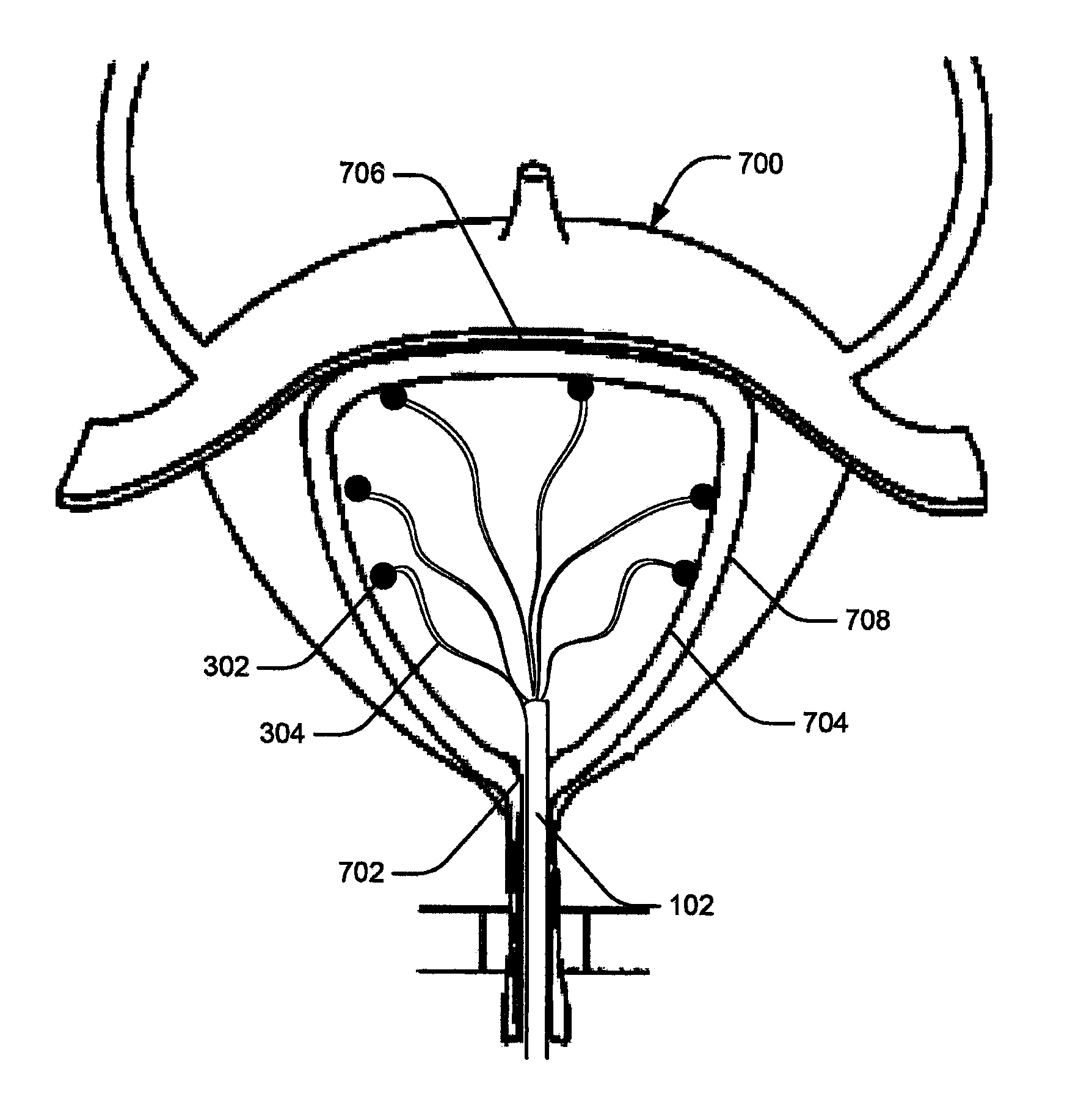
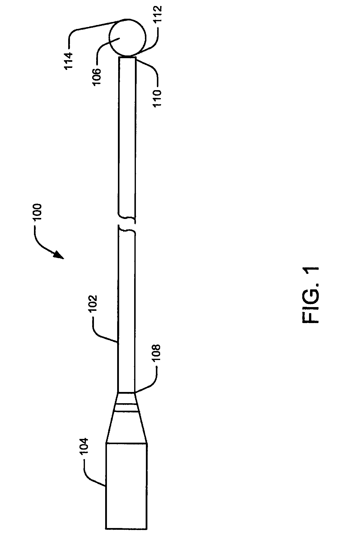
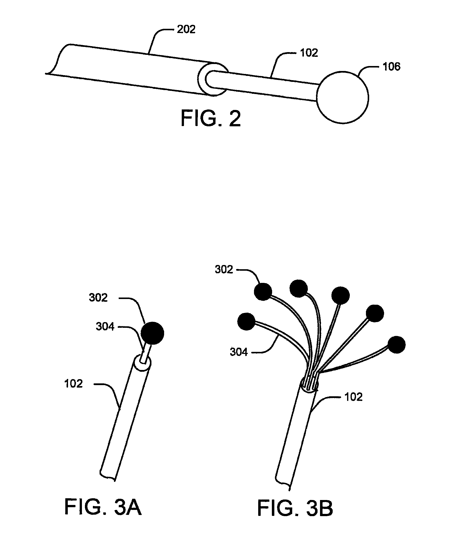
[0029]Embodiments of the present disclosure relate to systems and methods for monitoring abnormal organ activity. To this end, some embodiments introduce a sensing element that may be positioned inside, outside, or around an organ, or implanted in organ walls to measure characteristics such as physical, mechanical, or biochemical changes associated with certain diseases. The various sensing element embodiments described here may be used to measure the characteristics of any body organ, such as the stomach, bladder, intestines, bowel, or urinary tract, without departing from the scope of the present disclosure. Moreover, the sensing elements may be inserted during medical procedures or they may be implanted to co...


PUM


Abstract

A medical device for monitoring physical, chemical, or biological characteristics of an organ, such as the urinary bladder. The medical device may include an elongate member having a proximal end and a distal end. The device may further include an end-effector assembly extending distally from the distal end of the elongate member. The end-effector assembly may also include a plurality of sensing elements for detecting abnormal organ function. Each of the plurality of sensing elements may include one or more of mechanical, visual, chemical, and biological sensors.

Description

CROSS-REFERENCE TO RELATED APPLICATIONS[0001]This patent application claims the benefits of priority under 35 U.S.C. §§119-120 to U.S. Provisional Application No. 61 / 508,169, filed on Jul. 15, 2011, the entirety of which is incorporated herein by reference.FIELD OF THE INVENTION[0002]This disclosure relates generally to systems and methods for detecting urinary tract conditions and the effects of therapy thereon. More particularly, embodiments of the claimed invention relate to sensing elements for detecting conditions symptomatic of conditions such as bladder overactivity.BACKGROUND OF THE INVENTION[0003]Often patients and physicians discover ailments or diseases too late, making it difficult to treat a patient in time to effect timely therapy. Monitoring body organs regularly and effectively, therefore, can make the difference between saving and losing a life. Even for diseases that have positive prognosis, monitoring the disease over its lifetime can help administer appropriate t...

Claims


Application Information

Patent Timeline
Patent Type & Authority Applications(United States)
IPC IPC(8): A61B5/20
CPCA61B5/205A61B5/4848A61B5/4839A61B5/0036A61B5/0002A61B5/036A61B5/103A61B5/107A61B5/1076A61B5/1473A61B5/202A61B5/204A61B5/208A61B5/42A61B5/6858A61B5/6859A61B5/6874A61B5/6885A61B8/488A61B2562/04A61B2562/046A61B2562/066A61N1/36007
Inventor NAGALE, SANDRACHENG, RUTH
Owner BOSTON SCI SCIMED INC
Why Eureka
Try Eureka