Injectable bioartificial tissue matrix

a bioartificial tissue and injection technology, applied in the field of injectable bioartificial tissue matrix, can solve the problems of insufficient study of their potential to survive, differentiate in vivo, and improve organ function, and the morphological evidence of in vivo dividing and diffencing hesc that assumes the target organ-specific properties is still missing, so as to achieve the effect of not distorting the heart's architecture or structur

Inactive Publication Date: 2005-10-20
BOARD OF REGENTS THE LELAND STANFORD JR UNIV THE +1
View PDF11 Cites 19 Cited by
  • Summary
  • Abstract
  • Description
  • Claims
  • Application Information

AI Technical Summary

Benefits of technology

[0035] The use of a liquid bioartificial tissue offers significant advantages over the previously known compositions and methods. The liquid bioartificial tissue can be injected into an injured or damaged myocardium, such as following myocardial infarction, using minimally invasive procedures, such as using an endoscope device, without distorting the heart's architecture or structure. In addition, preparation of the liquid bioartificial tissue can be performed within a few minutes, and thus ov

Problems solved by technology

Even though scientific protocols hold promise for their future use, their potential to survive, differentiate in vivo, and thereby improve organ function has not been sufficiently studied.
There are no successful reports of hESC transfer into myocardium.
Furthermore, morphological evidence of in vivo dividing and differentiating hESC that assume the target organ-specific properties is still missing.
Cell transfer for restorative purposes is primarily limited by early cell death.
There is general agreement that at least 95% of the transplanted cells die following transplantation into the host target organ and therefore they cannot develop the desired effect.
Myocardial cell necrosis is an irreversible process that can ultimately lead to heart failure.
Innovative tissue engineering techniques developed to reconstitute organ function after a severe insult promise to diversify our approach to this condition but are associated with significant challenges.
The scaffold's physical condition, its in vivo kinetics, and its suitability as an adequate microenvironment for the inoculated cells, are another limitations.
Furthermore, most of the utilized biomaterials constitute single-component isotropic matrices, in which cells are seeded according to the rules of gravity, resulting in non-homogenous distribution and therefore inconsistent performance throughout the graft; the periphery of the graft which lies in culture medium or borders the host tissue is privileged in its blood or nutrient supply, as opposed to the core of the graft which is exposed to severe undersupply conditions (Robinson, K. A. and Matheny, R. G. (2002) He

Method used


Image

  • Injectable bioartificial tissue matrix
  • Injectable bioartificial tissue matrix
  • Injectable bioartificial tissue matrix

Examples


examples

[0063] The invention will be more readily understood by reference to the following examples, which are included merely for purposes of illustration of certain aspects and embodiments of the present invention and not as limitations.

example i

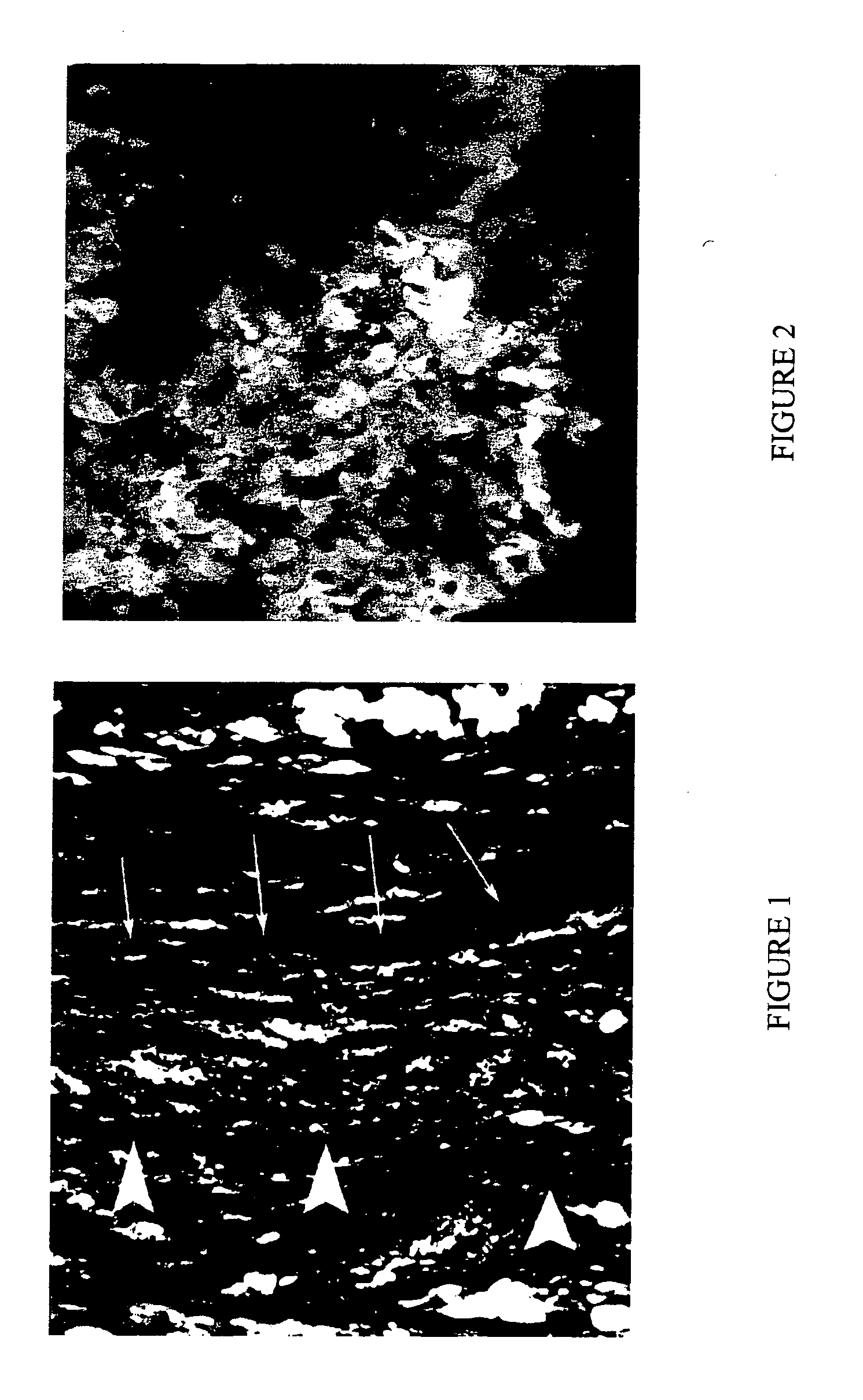
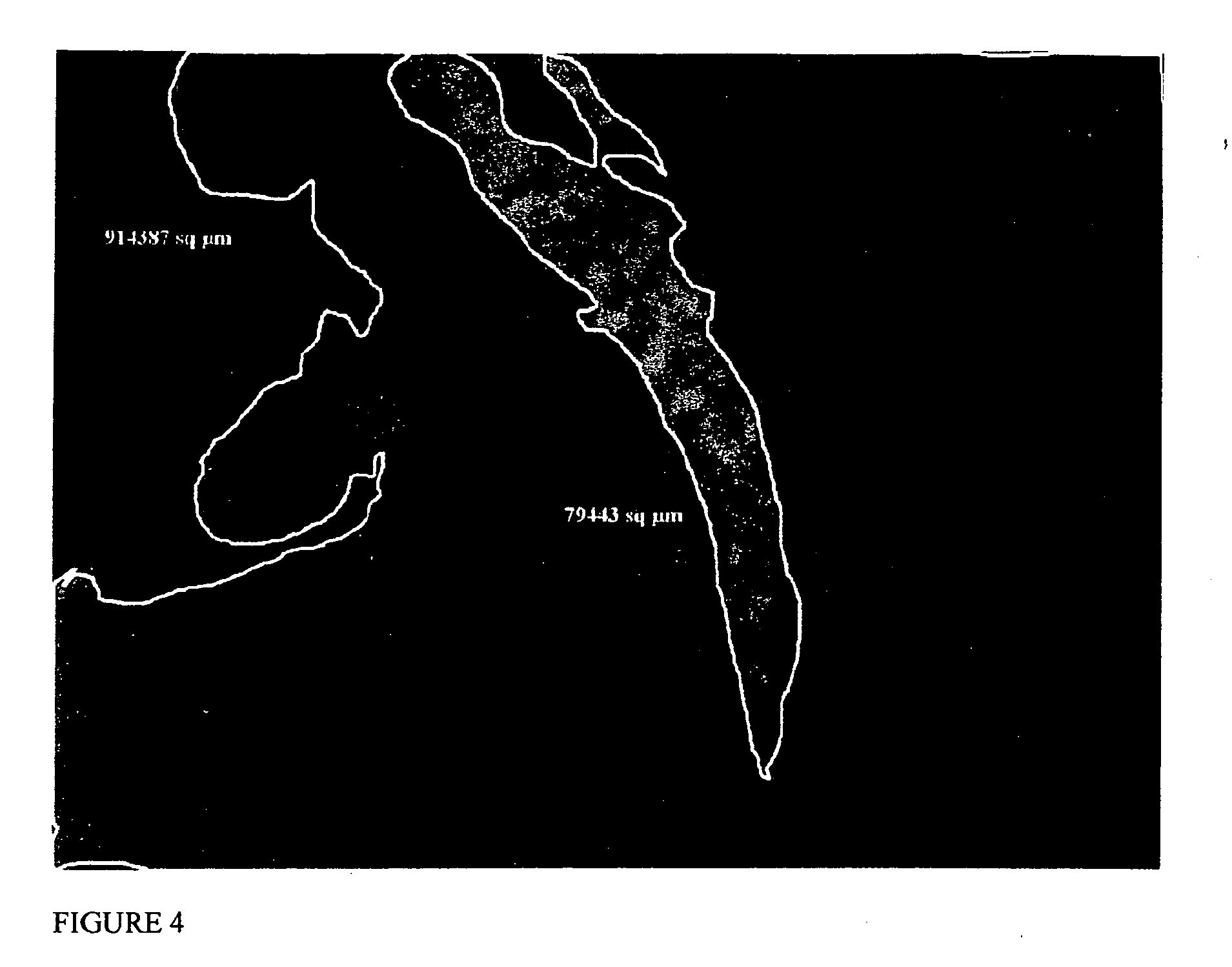
Preparation and Injection of ES-Derived Cardiomyocytes into Small Animal Model of Myocardial Infarction

[0064] Undifferentiated Green Fluorescent Protein (GFP)-labeled mouse ES cells (2×10⁶) were seeded in BD MATRIGEL matrix (BD Biosciences, Bedford Mass.). The ES cell suspension in MATRIGEL was maintained at a constant 37° C. The resulting cell suspension was the liquid bioartificial tissue.

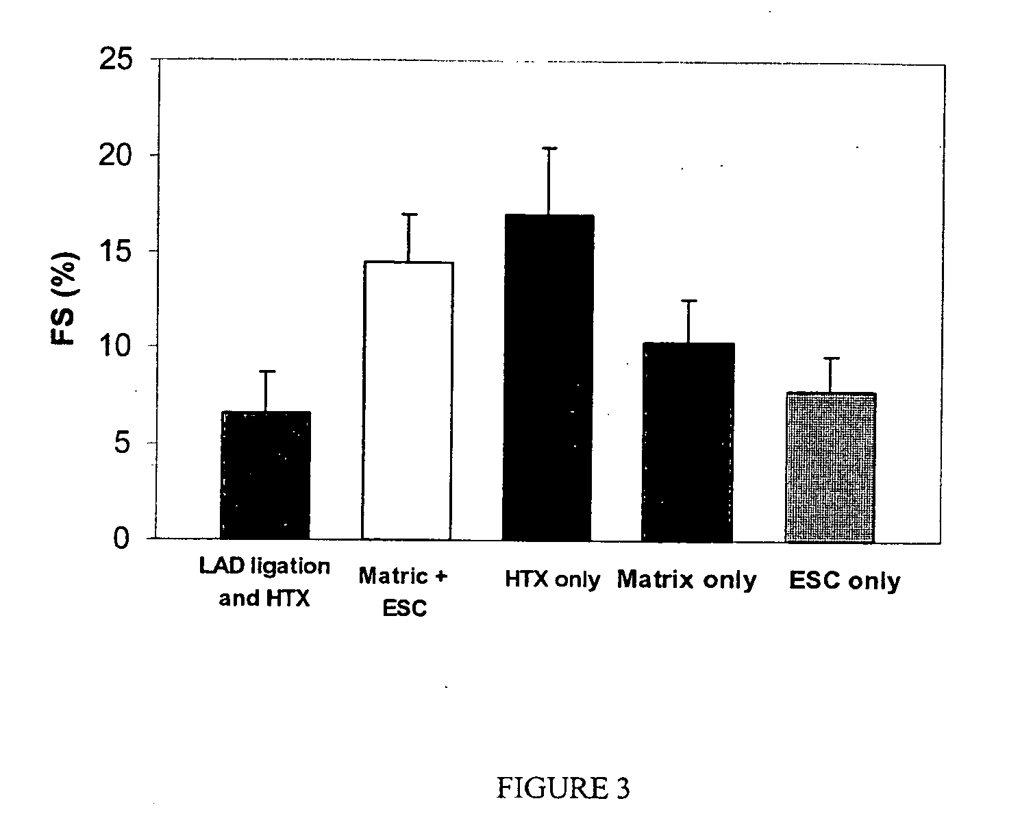
[0065] Lewis rats (150-200 g) were used in all experimental procedures. The Lewis rat is used as a heterotopic heart transplant model. In this example, the left anterior descending coronary artery (LAD) was ligated to create an intramural left ventricular pouch (infarcted area of the myocardium). The ES cells suspended in 0.125 ml MATRIGEL were injected in the resulting infarcted area within the pouch and the suspension became solid within a few minutes after transplantation. Five recipient groups were studied: transplanted healthy hearts (Group I), infarcted control animals (Group II), matrix ...

example ii

Preparation and Introduction of ES-Derived Cardiomyocytes into a Large Animal Model of Myocardial Infarction

[0077] The ES cells are prepared as described in Example I. A suitable large animal that is routinely used to study clinical procedures used to alleviate myocardial infarction is obtained. An example of such an animal is the pig.

[0078] A pig, with a mass of between 30 and 35 kg, is anaesthetized, paralyzed, and prepared for the procedure. The LAD is ligated to create an intramural left ventricular pouch (infarcted area of the myocardium). The ES cells (between 2×10⁶ and 109 cells) are suspended in between 0.125 ml and 1.0 ml MATRIGEL and injected in the resulting infarcted area within the pouch. Five recipient groups are studied: transplanted healthy hearts (Group I), infarcted control animals (Group II), matrix recipients alone (Group III), the study group which receives matrix plus cells (Group IV), and a group which receives ES cells alone (Group V). Two weeks later, the ...


PUM

PropertyMeasurementUnit
Timeaaaaaaaaaa
Molar densityaaaaaaaaaa

Abstract

The present invention encompasses a liquid bioartificial tissue for restoring tissue and organ function to an injured or damaged organ in a human subject. The liquid bioartificial tissue is injected into a target organ and can significantly restore organ function within two weeks. The invention also encompasses a cell culture medium comprising ascorbic acid (or other free-radical scavengers and/or anti-oxidants) that is used for pre-treating transplantable cells prior to organ transplantation. Pre-treatment with ascorbic acid increases transplanted cell viability and colonization by nearly fifty-fold compared with untreated cells. The invention is particularly useful for treating ischemic heart damage following myocardial infarction.

Description

[0001] The present application claims priority to U.S. Provisional Patent Application Ser. No. 60 / 563,095 entitled “Injectable Biosynthetic Tissue Matrix”, filed Apr. 17, 2004, which is herein incorporated by reference in its entirety for all purposes.FIELD OF THE INVENTION [0002] The present invention relates to compositions and methods used to create an injectable bioartificial tissue matrix; more specifically to compositions and methods used to treat and restore heart myocardium following an infarction event. The invention also relates to compositions and methods used to increase the viability of cells that are injected into an organ, more specifically to using ascorbic acid as an adjuvant. BACKGROUND [0003] The recent progress in harvest, culture and differentiation of embryonic stem (ES) cells has spurred research activity for tissue and organ restoration by cell transfer (Mummery, C., et al. (2002) J. Anat. 200: 233-242; Kehat, I., et al. (2001) J. Clin. Invest. 108: 407-414)....

Claims


Application Information

Patent Timeline
IPC IPC(8): A61K31/375A61K35/12A61K35/28A61K35/34A61K35/48A61K35/545A61K38/39A61K45/00A61K45/06C12N5/077
CPCA61K31/375A61L2430/20C12N5/0657C12N2500/30C12N2500/38C12N2506/02A61K45/06A61L2400/06A61L27/22A61L27/225A61L27/24A61L27/3834A61K35/34A61K35/545A61K2300/00
Inventor KOFIDIS, THEODOROS
Owner BOARD OF REGENTS THE LELAND STANFORD JR UNIV THE
Why Eureka
Try Eureka