Method and device for evaluating evolution of tumoural lesions

a tumour and evolution technology, applied in the field of methods and devices for evaluating the evolution of tumours, can solve the problems of losing residual hot spots, affecting so as to improve the prognosis of patients, reduce side effects, and shorten the hospital stay

Active Publication Date: 2013-04-18
DR K COENEGRACHTS
View PDF21 Cites 11 Cited by
  • Summary
  • Abstract
  • Description
  • Claims
  • Application Information

AI Technical Summary

Benefits of technology

[0080]The invention further is useful for early, reproducible and accurate evaluation of the progression, regression or stability of disease for each individual patient, i.e. patient-tailored evaluation. Such early evaluation allows an accurate use of expensive anti-tumoural therapies and allows and early switch-over to other, more efficient anti-tumoural therapy.
[0081]The invention will also enable improvement of patient prognosis, a decrease of side-effects, shorter hospital stay and earlier return to the work environment. Merely morphologic criteria like for instance RECIST

Problems solved by technology

The detection of such hot spots through improved image acquisition and post-processing is important because patients with residual hot spots are not tumour-free as a result of which their prognosis has not improved meaningfully.
Low resolution analysis averages the perfusion information resulting in loss of very high and very low values, thereby losing residual hot spots.
Such an approach results in loss of hot spots, and US 2003/0211036 fails to seek further improvements to this problem.
In summary, the method known from US 2003/0211036 recognizes but does not adequately solve the resolution problem.
As a result, residual malignant tumour cells that are therapy-resistant are not detected and efficient prognosis of therapy response by the physician remains difficult or impossible.
Although the article addresses the problem of whole tumour region-of-interest averaging, th

Method used


Image

  • Method and device for evaluating evolution of tumoural lesions
  • Method and device for evaluating evolution of tumoural lesions
  • Method and device for evaluating evolution of tumoural lesions

Examples


Embodiment Construction

)

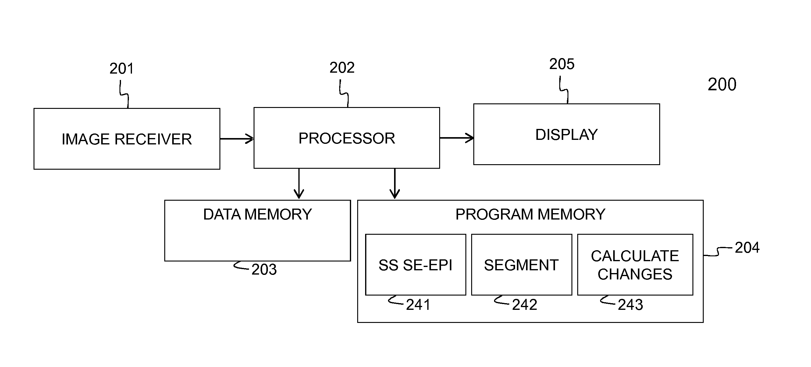
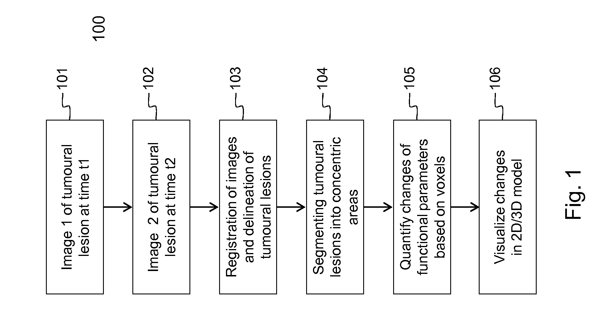
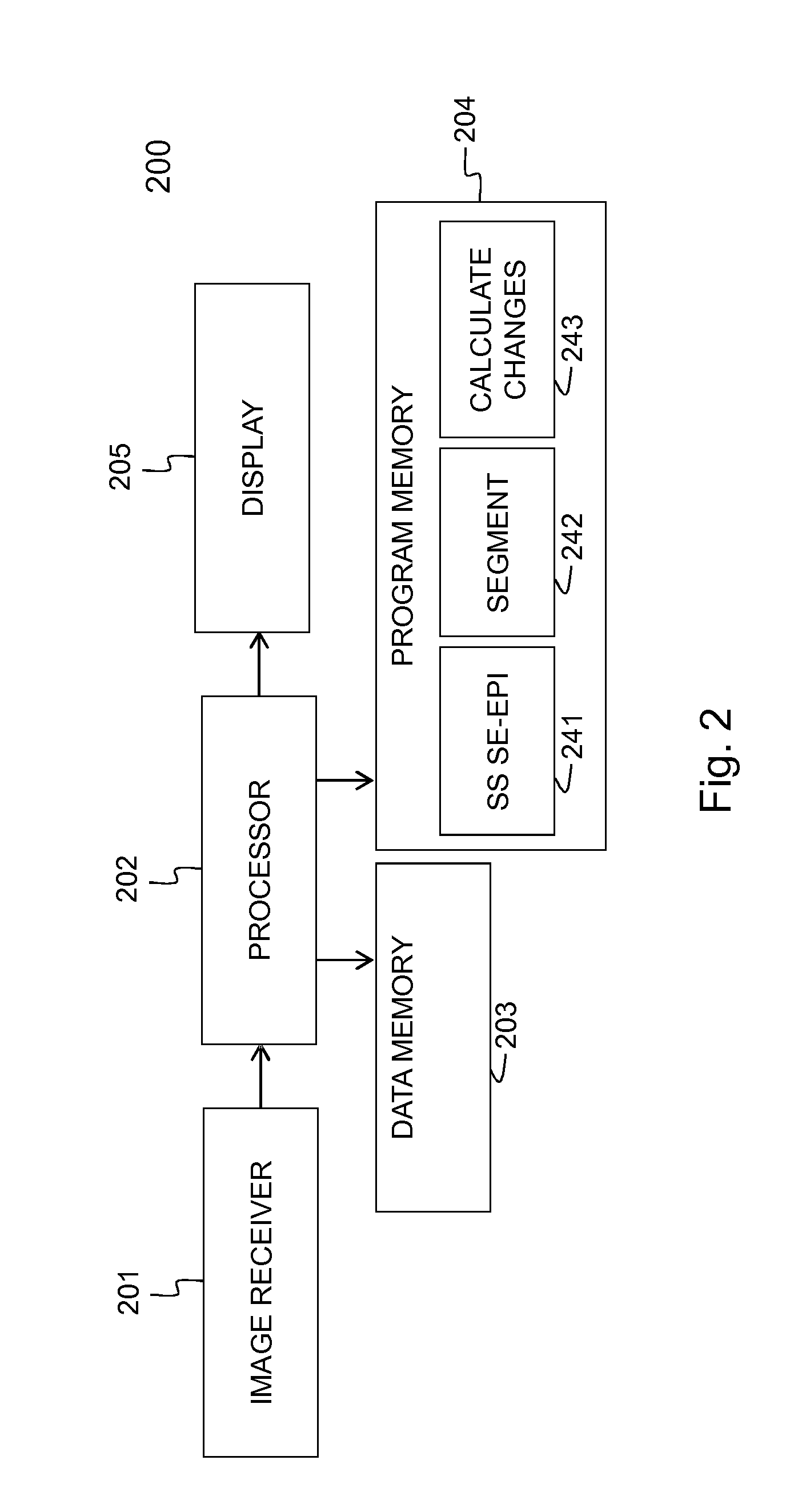
[0104]Embodiments of the method 100 and device 200 for evaluating the evolution of tumour lesions over a period of time t1-t2 are illustrated respectively by FIG. 1 and FIG. 2.In step 101, a first image taken at time t1 is received via the interface 201, e.g. a network connection or connection to an MRI scanner. Similarly, in step 102, a second image taken at time t2, is received via the interface 201. The images are temporarily stored in data memory 203 for being processed by processor 202. In the images, the tumour lesions are delineated using the SS SE-EPI algorithm. The delineation followed by registration of the images, i.e. step 103 in FIG. 1, is executed by the processor 202 that is software controlled. The SS SE-EPI algorithm 241 may be one out of several delineation algorithms that is available in the program memory 204 of the device 200 according to the invention. The delineation of tumour lesions may be fully automated, i.e. the algorithm also detects the tumour lesions ...


PUM


Abstract

A new method (100) for evaluating evolution of tumoural lesions includes: —providing (101) a first image of the tumoural lesions, the first image being made at a first time instant; —providing (102) a second image of the tumoural lesions, the second image being made at a second time instant that is later than the first time instant; —delineating (103) a border of the tumoural lesions in the first image and the second image; —registration (103) of the tumoural lesions in the first image the second image; —segmenting (104) the tumoural lesions in the first image and the second image into concentric areas; —quantifying (105) changes of at least one functional parameter between the concentric areas in the first image and respective corresponding concentric areas in the second image; and —visualizing (106) the changes in a two-dimensional or three-dimensional model of the tumoural lesions.

Description

FIELD OF THE INVENTION[0001]The present invention generally relates to a method and device for evaluating the evolution of tumour lesions through imaging systems, e.g. during oncology therapy like for instance radio-therapy or chemo-therapy. Through temporal imaging, plural 2D-digital images (pixel-based) or 3D-digital images (voxel-based) of the same tumour-affected organ may be acquired over a period in time, for instance a three month period during which the tumour is treated with a certain therapy. Advanced digital image acquisition systems such as scanners generating ultrasound (US) images, computed tomography (CT) images, magnetic resonance (MR) images, positron emission tomography (PET) images, positron emission tomography-computed tomography (PET-CT) images and single photon emission computed tomography (SPECT) images, allow detecting and visualizing tumour lesions. Through post-processing of the temporal images, changes in the tumour lesions may be visualized enabling the p...

Claims


Application Information

Patent Timeline
IPC IPC(8): G06K9/00
CPCG06T7/0016G06T7/2053G06K9/00362G06T2207/10132G06T2207/30096G06T2207/10072G06T7/254G06V40/10
Inventor COENEGRACHTS, KENNETH LOUIS STEFAN
Owner DR K COENEGRACHTS
Why Eureka
Try Eureka