Method for detection of urothelial cancer

a urothelial cancer and cancer technology, applied in the field of detecting urothelial cancer, can solve the problems of limited detection, achieve the effects of reducing the burden of patients, rapid and simple detection, and reducing national medical expenses

Active Publication Date: 2013-04-18
SBI PHARMA CO LTD +1
View PDF3 Cites 1 Cited by
  • Summary
  • Abstract
  • Description
  • Claims
  • Application Information

AI Technical Summary

Benefits of technology

[0013]According to the present invention, no particular culture apparatus is needed, and a determination just after urine collection is possible. Further, as it can be determined that there is an abnormality when fluorescence in cells in the urine is detected, it can be immediately transferred to diagnosis through an endoscope or to transurethral resection. For example, by performing the fluorescence detection test in the cells in the urine before an endoscopic examination for determining the presence or absence of relapse, if there is no suspicion, there is no need to go through an endoscopic examination. This significantly reduces burden of patients, which will also lead to reduction of national medical expenses. As such, the method of the present invention allows rapid and simple detection with high accuracy of not only bladder cancer but all urothelial cancers, which is a remarkable technique in the medical services of this field. Further, as the detection is possible with a small dosage amount of ALAs, it is advantageous economically, and there is also an effect in security that photolesion is substantially not caused.

Problems solved by technology

In the in vivo incubation, there was a problem to cause burden to the patient when injecting ALA solution in the bladder, and that the detection was limited to bladder cancer.

Method used


Image

  • Method for detection of urothelial cancer
  • Method for detection of urothelial cancer
  • Method for detection of urothelial cancer

Examples


example 1

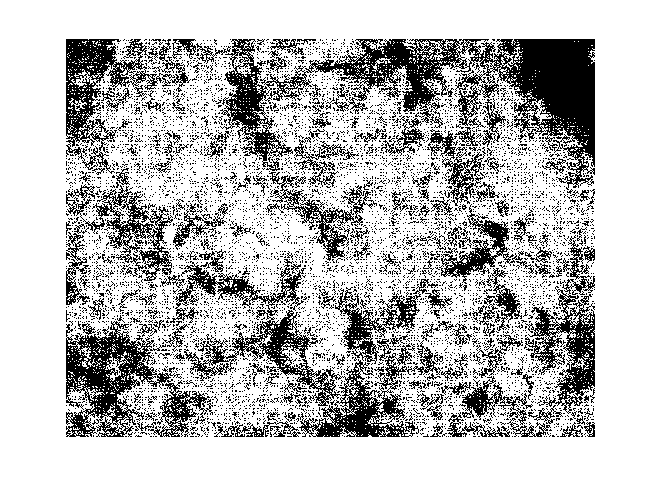
[0028]1 g of ALA hydrochloride was dissolved in 50 ml of orange juice, and was given to a patient suspected of having bladder cancer (body weight about 60 kg). Urine was collected after about 4 hours, which was immediately centrifuged (3000 rpm / min., 15 min.) in a light shielded state, and was confirmed by microscopic visualization using sediments. The results are shown in FIG. 1. The microscope used was OLYMPUS BX50CCD mounted with camera OLYMPUS DP70. The mirror set was Exciter: XF1076 400AF30 Dichroic:XF2007 475DCLP, Emitter: XF3090 585ALP (OMEGA OPTICAL), all of which being a standard fluorescence microscope system.

[0029]As it is clear from FIG. 1, a clear PPIX fluorescence derived from cancer cell was observed. From a normal pathological diagnosis of the cells, it was shown to be cancer cells.

[0030]From the above, it has been revealed that according to the method of the present invention, urothelial cancer can be detected by detecting fluorescence of the cells in the urine, wit...

example 2

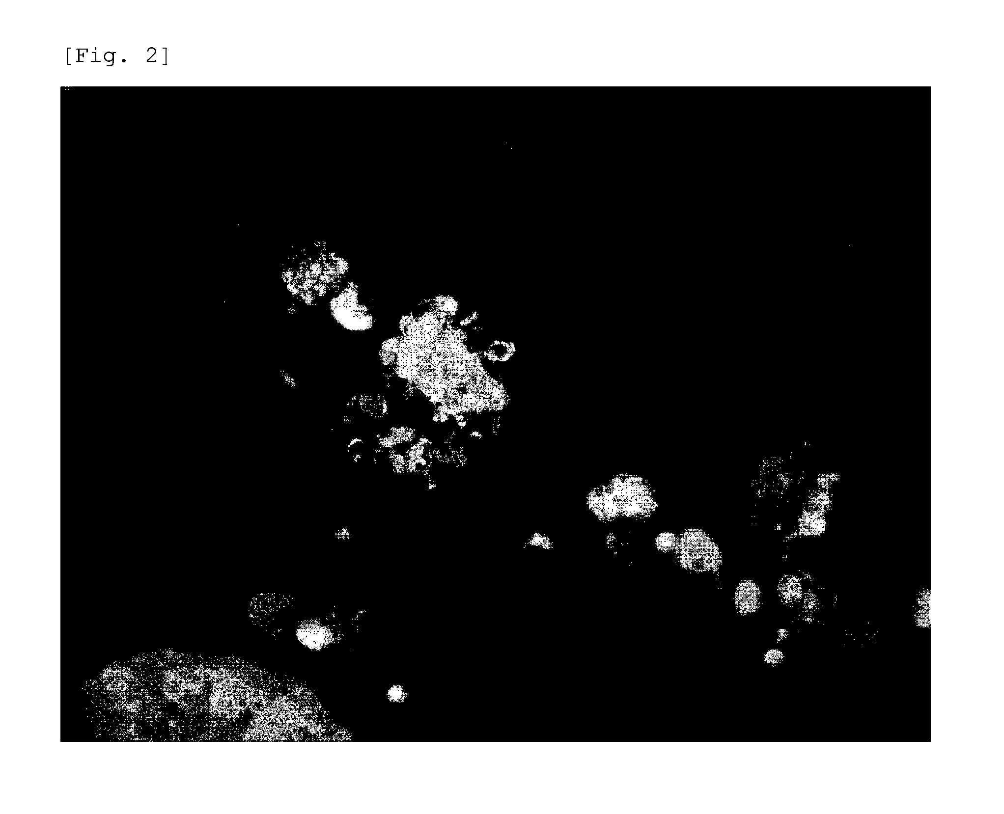
[0031]The detection method was performed in the same way as Example 1, except that the dosage amount was changed to 500 mg, which is the half amount of Example 1. As a result, the picture of FIG. 2 was obtained. From a normal pathological diagnosis of the cells, it was shown to be cancer cells.


PUM

PropertyMeasurementUnit
concentrationaaaaaaaaaa
body weightaaaaaaaaaa
fluorescenceaaaaaaaaaa

Abstract

It is to provide a method for detecting urothelial cancer simply and with high accuracy. It is a method for detecting urothelial cancer comprising administering 5-aminolevulinic acid (ALA), a derivative thereof, or a salt of these to a test subject, collecting urine from the test subject, and detecting the presence of fluorescence or amount of fluorescence in the collected urine.

Description

TECHNICAL FIELD[0001]The present invention relates to a method for detecting urothelial cancer, specifically to a method for detecting urothelial cancer using 5-aminolevulinic acid (ALA), a derivative thereof, or a salt of these (hereinafter may be referred to as “ALAs”).BACKGROUND ART[0002]Examination of urothelial cancer is usually performed by staining cells dropped out in the urine by Papanicolaou stain and observing them with a microscope. However, proficiency is required to diagnosis, and there are often oversights, which is a problem. The difficulty of detection leads to the delay in cancer detection, and thus to miss relapse.[0003]On the other hand, in the endoscopic operation for a bladder cancer patient, it is known to inject ALAs into bladder, observe the fluorescence derived from cancer cells in the bladder to determine the excision site (see for example, Nonpatent Document 1). It is also known that the observation of fluorescence in the bladder is possible by administer...

Claims


Application Information

Patent Timeline
Patent Type & Authority Applications(United States)
IPC IPC(8): G01N21/64
CPCG01N33/57407G01N33/57434G01N33/57438G01N21/6458G01N21/6486G01N21/6428G01N33/582G01N2800/34A61K49/0036A61K41/0061A61K49/0021
Inventor INOUE, KEIJISHUIN, TAROFURIHATA, MUTSUOHIRAO, YOSHIHIKOTANAKA, TOHRU
Owner SBI PHARMA CO LTD
Why Eureka
Try Eureka