
Access system for femoral vasculature catheterization and related method

a technology for accessing systems and vasculature, which is applied in the field of medical devices, can solve problems such as significant morbidity, significant morbidity, and inconvenient use of catheters, and achieve the effects of preventing damage to the vasculature, rapid and easy attachmen

Inactive Publication Date: 2013-04-18
UNIV OF VIRGINIA ALUMNI PATENTS FOUND
View PDF7 Cites 25 Cited by
  • Summary
  • Abstract
  • Description
  • Claims
  • Application Information

AI Technical Summary

Benefits of technology

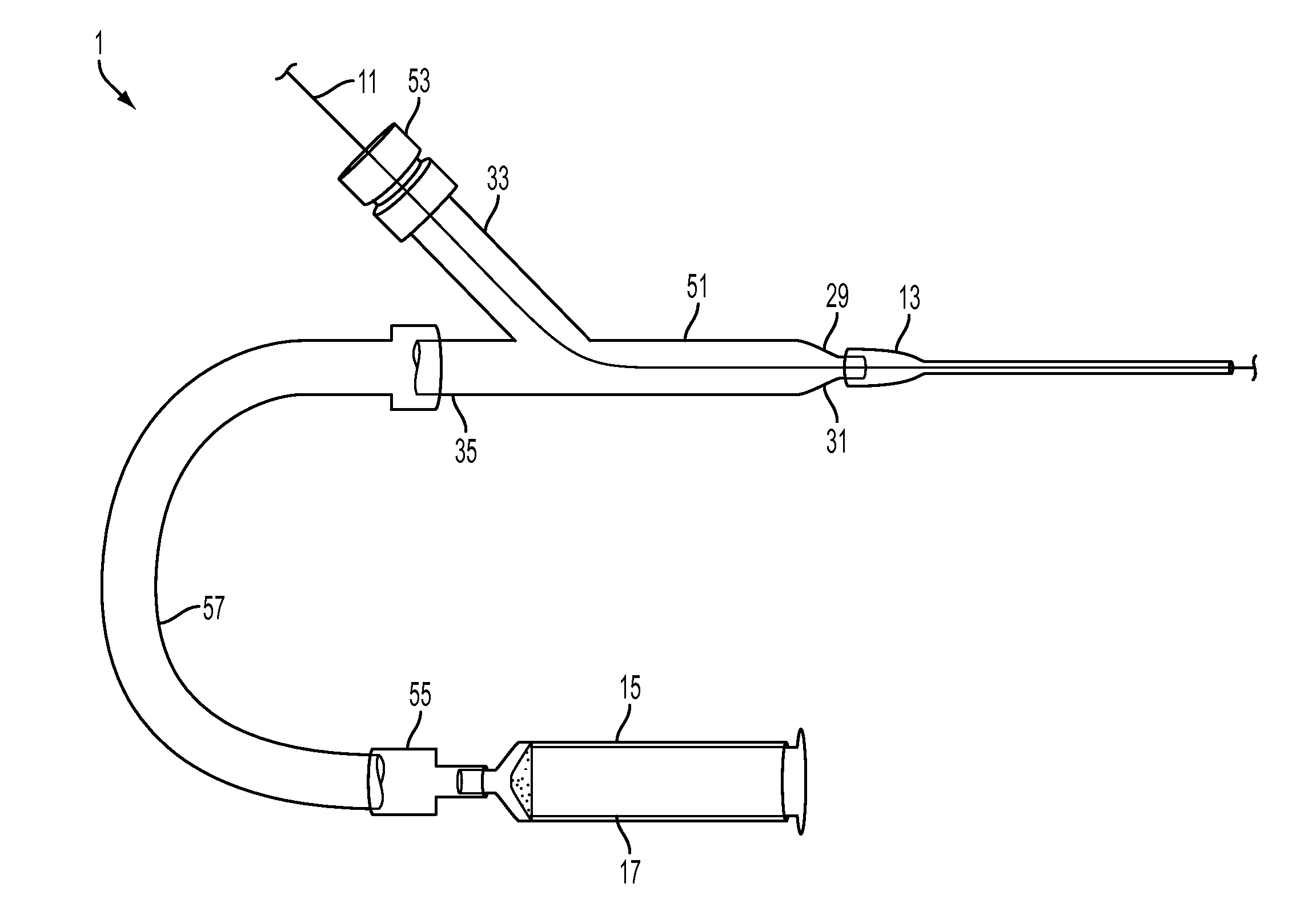
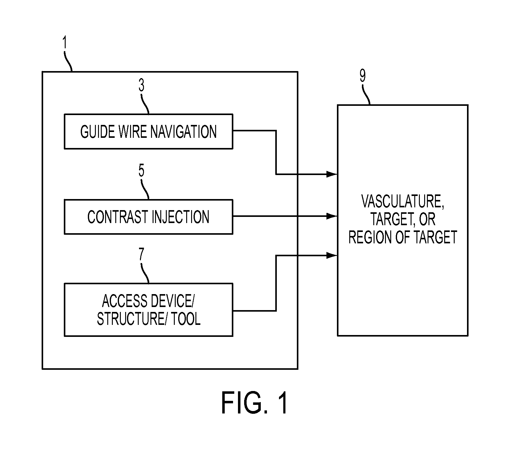
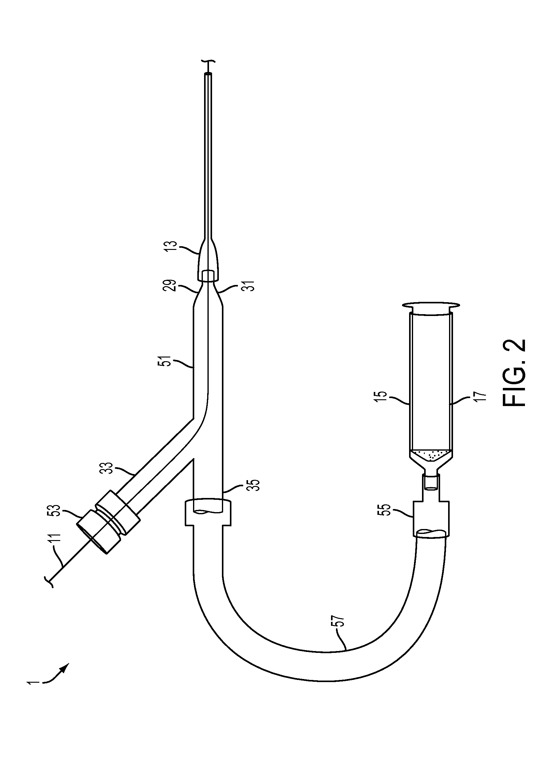
The present invention provides a way to inject contrast material and pass a guide wire through the same introducer device simultaneously, without moving any parts. The device is designed to prevent the contrast material from flowing back through the guide wire port during the injection process. A needle or related components of the access system may have markings that can be easily visualized during the insertion process. This allows the user to measure the insertion depth by reference to fluoroscopic imaging. The invention also allows for safely and reliably puncturing a certain part of the common femoral artery.

Problems solved by technology

Several palpable anatomic landmarks are used to guide the operator, however, these landmarks are notoriously imprecise and the exact puncture site is not known until after a sheath is inserted and an angiogram performed.
Failure to properly puncture the common femoral artery may result in several important and potentially life-threatening complications.
All together, these events complicate up to 5% of cardiac procedures and cause significant morbidity, patient discomfort, increased mortality, greater hospital costs and prolonged length of stay.
However, despite a myriad of prior art, access to the femoral vein and artery are complicated in 5% of all cases by hematoma or other vascular complication, especially during cardiac interventions when large doses of anticoagulants (blood thinners) are used.
This is often because the prior needle systems routinely access the femoral vein or artery either too high (leading to retroperitoneal bleeding) or too low (leading to femoral vein or artery bleeds, pseudoaneurysms, or arteriovenous fistulae).
However, a major limitation of the prior art is that it does not allow for an easy method to inject a contrast material into the artery to confirm the needle position in the common femoral artery or vein while simultaneously maintaining a method for introducing a guide wire.
Introducing this wire would be difficult since the needle opening is so small.
A further limitation of the prior art is that such introducer means are generally not small enough to easily reposition the needle during the process of insertion of it into and passage through the body tissues.
However, in the prior art the smaller needle requires an even smaller guide wire to be loaded.
Still another limitation of the prior art is that such introducer means can cause unnecessary and dangerous bleeding during usage or may led to catheter placement either into one of the smaller branches of the common femoral artery or in a deeper, more proximal artery (e.g. the external iliac artery), potentially resulting in an increased rate of serious vascular complications or bleeding.
An additional limitation of the prior art is that after the chamber has been loaded with contrast agent, the chamber cannot be cleared of air in the chamber.
The injection of contrast fluid with air bubbles can cause serious medical complications.
An additional limitation of the prior art is that the needle means incorporated into the introducer system kits generally do not have markings on them to indicate depth of insertion, nor do the needles have markings that are usually visible on fluoroscopic means for monitoring the needle insertion process.

Method used


Image

  • Access system for femoral vasculature catheterization and related method
  • Access system for femoral vasculature catheterization and related method
  • Access system for femoral vasculature catheterization and related method

Examples


example set no.67

Example Set No. 67

[0190]An aspect of an embodiment of the present invention solves the clinical need to improve the method used to obtain femoral access and precisely guide the operator to puncture the common femoral artery. This device and methodology allows the operator to locate the initial puncture site using fluoroscopically defined bony landmarks (e.g., middle of the femoral head) followed by puncture of the artery with a small, 21 gauge needle while allowing the operator to confirm the location within the common femoral artery by injecting contrast under fluoroscopy. If the operator confirms the proper site in the common femoral artery, a small guide wire can then be advanced through a channel installed in the plunger of the contrast-delivery syringe, into the barrel of the syringe, and then out the distal tip of the needle. Thereafter, the needle and syringe can be removed and replaced by a sheath. However, if the angiogram demonstrates improper position, the needle can be w...


PUM


Abstract

An access system and method for obtaining access to the interior vascular structures or other regions or collections of fluid or fluid-filled cavities inside the body. The system and method provides for injection of contrast agents (to confirm ideal position or condition), the passage of guide wires, and the eventual catheterization of the heart and other parts of the body via the pathway established through the puncture of a femoral artery. The system and method provides the ability to inject a contrast material and pass a guide wire through the same introducer device simultaneously (without necessarily moving it or removing any parts), with the device designed to prevent the backflow of the contrast material through the guide wire port during the contrast injection process The ideal location of access in the vein or artery can be seen by injecting contrast from a needle inside the structure and then using fluoroscopy.

Description

RELATED APPLICATIONS[0001]The present application claims priority from U.S. Provisional Application Ser. No. 61 / 305,152, filed Feb. 17, 2010, entitled “Access Device for Femoral Vasculature Catheterization and Method of Use;” the disclosure of which is hereby incorporated by reference herein in its entirety.FIELD OF INVENTION[0002]The present invention relates generally to the field of medical devices to be used for cardiological procedures. More specifically, the invention is in the subfield of vascular access devices and other tools to be used for cardiac and endovascular catheterizations, including, for example, ablation and in electrophysiological procedures.BACKGROUND OF THE INVENTION[0003]The femoral artery is the predominant site of vascular access for the performance of cardiac catheterizations, percutaneous coronary interventions and many other vascular related procedures. The operator's goal is to safely and reliably puncture a certain part of the common femoral artery. Se...

Claims


Application Information

Patent Timeline
Patent Type & Authority Applications(United States)
IPC IPC(8): A61M25/01
CPCA61M5/3148A61M5/31511A61M25/065A61M25/09041A61M25/01A61M39/22A61M2025/0008G01R33/28G01R33/5601A61M31/005A61B5/15003A61B5/150236A61B5/150244A61B5/150389A61B5/150503A61B5/153A61B5/155A61B5/150755
Inventor GILLIES, GEORGE T.MAHAPATRA, SRIJOYLIM, D. SCOTTRAGOSTA, MICHAEL
Owner UNIV OF VIRGINIA ALUMNI PATENTS FOUND
Learn More