Method and Apparatus for Generating Proton Therapy Treatment Planning Images

a proton therapy and treatment planning technology, applied in the field of tumor radiation therapy treatment planning, can solve the problems of 3 to 5 percent error in practice using the best known conversion method, and the incongruity of converting x-ray photon attenuation to proton stopping power using a conventional x-ray ct image, etc., to achieve the effect of greatly reducing the inaccuracy of converting x-ray photon attenuation to proton stopping power

Active Publication Date: 2013-05-02
WISCONSIN ALUMNI RES FOUND
View PDF1 Cites 21 Cited by
  • Summary
  • Abstract
  • Description
  • Claims
  • Application Information

AI Technical Summary

Benefits of technology

[0012]It is thus a feature of at least one embodiment of the invention to overcome tissue thickness limitations on proton imaging by enabling accurate conversion of x-ray attenuation data to proton stopping power in projection space. It is another feature of at least one embodiment of the invention to provide accurate proton stopping power data using readily obtainable photon imaging.
[0014]It is thus a feature of at least one embodiment of the invention to further improve the accuracy of the conversion process by using a photon energy range showing less tissue type variation.
[0018]It is thus a feature of at least one embodiment of the invention to blend the converted proton projection data with actual proton projection data for improved accuracy.
[0020]It is thus a feature of at least one embodiment of the invention to provide a system eliminating the need for proton imaging.
[0022]It is thus a feature of at least one embodiment of the invention to provide a method of calibrating the conversion functions to the particular patient for improved accuracy.

Problems solved by technology

This drawback has suggested the use of heavy charged particles, such as protons alpha particles or carbon ions, as a substitute for x-ray photon radiation.
Further, the dose deposited by a proton beam is not uniform along the entrance path of the beam, but rises substantially to a “Bragg peak” near a point where the proton beam stops within the tissue.
A conventional x-ray CT image can provide information about the attenuation of x-ray photons but is a relatively poor proxy for proton stopping power leading to errors of 3 to 5 percent in practice employing the best known conversion methods.
A drawback to this approach arises from the limited energy of therapeutic proton beams of approximately 200-250 MeV.
This proton energy is insufficient for the protons to traverse more than approximately 25 to 38 centimeters of tissue before entering the Bragg peak region at which point the either deposit significant energy or stop in the tissue, and therefore, for many patients, the image sinogram provided by the proton beam will be incomplete, leading to severe image artifacts that obscure the quantitative data that must be extracted for treatment planning.
Methods of reconstructing tomographic images when the underlying sinogram is incomplete are known in the x-ray CT art, however, the large and continuous regions of missing data likely in a proton sinogram, and the need for quantitative accuracy in the proton sinogram, make these x-ray CT techniques generally inapplicable.

Method used


Image

  • Method and Apparatus for Generating Proton Therapy Treatment Planning Images
  • Method and Apparatus for Generating Proton Therapy Treatment Planning Images
  • Method and Apparatus for Generating Proton Therapy Treatment Planning Images

Examples


Embodiment Construction

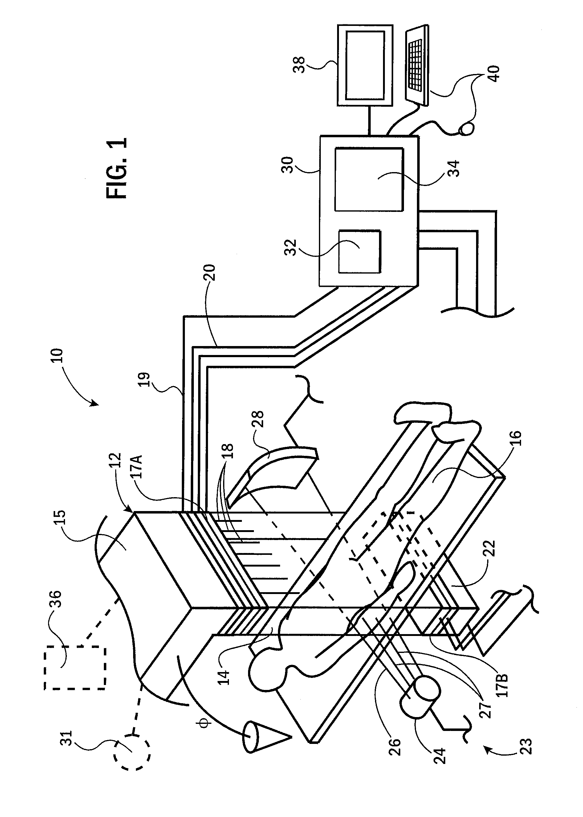
[0029]Referring now to FIG. 1, a combined proton therapy and imaging system 10 suitable for use of the present invention may provide a proton source 12, for example, being a repositionable accelerator (such as, but not limited to a dielectric wall accelerator) or an exit port of a conduit conducting a proton beam from a stationary cyclotron, for example using a series of quadrupole or other suitable magnetic directors.

[0030]The proton source 12 may be rotatable on a gantry 15 to direct a proton beam 14 at a patient 16 at a range of gantry angles φ about the patient 16, driven by a gantry drive 36. The proton beam 14 may be an areal beam or a scanned pencil beam and may be controlled in energy at a variety of different locations within the beam 14 according to an energy signal 19 controlling an accelerating voltage of the accelerator or material places in the beam such as a range shifter used to provide a pencil beam or the placement of an energy reducing filter into an areal beam 14...


PUM


Abstract

A method of producing a patient image indicating proton stopping power of the tissue may employ photon attenuation information converted to proton stopping power. The conversion uses different conversion functions for particular tissue types to account for a strong atomic number dependency in the conversion process. Megavoltage x-rays may be used for improved accuracy.

Description

STATEMENT REGARDING FEDERALLY SPONSORED RESEARCH OR DEVELOPMENTCROSS REFERENCE TO RELATED APPLICATIONBACKGROUND OF THE INVENTION[0001]The present invention relates to treatment planning for radiation therapy of tumors and the like and in particular for treatment planning system providing an improved assessment of the proton stopping power of tissue being treated.[0002]External-beam radiation therapy may treat a tumor within a patient by directing high-energy radiation in one or more beams toward the tumor. Selective disruption of tumor tissue is obtained both by concentrating radiation at the site of the tumor and by taking advantage of a greater susceptibility of tumor tissue to radiation, for example, resulting from its higher cell division rate.[0003]Photon external-beam radiation systems, for example, as manufactured by Accuray, Inc., of Sunnyvale Calif., treat a tumor with multiple x-ray fan beams directed at the patient over an angular range of 360° . Each of the beams is comp...

Claims


Application Information

Patent Timeline
Patent Type & Authority Applications(United States)
IPC IPC(8): A61N5/10A61B6/00
CPCA61N5/103A61N2005/1061A61N2005/1087A61B6/032A61B6/50A61B6/583A61B6/5217A61B6/544A61B5/4869A61B6/4258A61B6/505G16H50/30
Inventor TOME, WOLFGANG AXELWANG, DONGXUMACKIE, THOMAS R.
Owner WISCONSIN ALUMNI RES FOUND
Why Eureka
Try Eureka