Antibodies to human programmed death receptor pd-1

a technology of human programmed death receptor and antibodies, which is applied in the field of immunoinhibitory receptors to achieve the effects of increasing tumor antigen release, reducing tumor antigen release, and facilitating initial clinical respons

Active Publication Date: 2013-05-02
MERCK SHARP & DOHME BV
View PDF0 Cites 122 Cited by
  • Summary
  • Abstract
  • Description
  • Claims
  • Application Information

AI Technical Summary

Benefits of technology

The invention provides isolated antibodies and fragments that target PD-1, which can enhance T cell activation and improve immune response to cancer. These antibodies can be used alone or in combination with other therapies, such as hormone deprivation or targeted agents, to further improve cancer treatment and create durable immune control of cancer.

Problems solved by technology

In contrast, chronic infections are often characterized by varying degrees of functional impairment (exhaustion) of virus-specific T-cell responses, and this defect is a principal reason for the inability of the host to eliminate the persisting pathogen.

Method used


Image

  • Antibodies to human programmed death receptor pd-1
  • Antibodies to human programmed death receptor pd-1
  • Antibodies to human programmed death receptor pd-1

Examples


example 1

Immunization and Selection of Anti PD-1 Antibodies

[0184]Immunization of Mice with hPD-1 cDNA

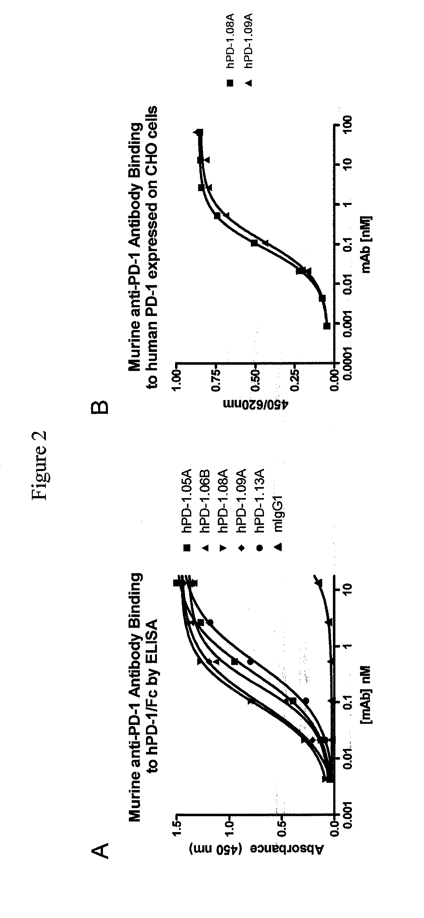
[0185]To generate antibodies against the human PD-1 ('hPD-1′) receptor, a cDNA encoding the open reading frame of the hPD-1 receptor was obtained by PCR and subcloned into vector pcDNA3.1 (Invitrogen, Carlsbad, Calif.). Next, CHO-K1 cells were stably transfected with hPD-1, and expression was monitored using flow cytometry. CHO-K1 clones were isolated expressing human PD-1 on their membranes and named CHO-hPD1.

[0186]Mice were immunized by gene gun immunization using a Helios gene gun (BioRad) and DNA coated gold bullets (BioRad) following manufacturers instructions. Briefly, 1 μm gold particles were coated with hPD-1 cDNA (cloned into pcDNA3.1) and, where indicated, commercial expression vectors for mouse Flt3L and mouse GM-CSF in a 2:1:1 ratio (both from Aldevron, Fargo N. Dak.). A total of 1 μg of plasmid DNA was used to coat 500 μg of gold bullets.

[0187]Specifically, 7-8 week-old female BA...

example 2

Purification and Characterization of Murine Anti-PD-1 Antibodies

Stabilization of Anti-PD-1 Producing Hybridomas and Purification of Anti-PD-1 Antibodies

[0192]Clonal cell populations were obtained for each of the hybridomas by subjecting them to multiple rounds (>4) of limiting dilution. Stable hybridoma cells were then cultured under serum-free conditions using CELLine bioreactors (Integra-biosciences) for six to eight days. Cells were seeded in the inner chamber in serum-free media at a density of 3×10⁶ c / mL in 15 mL and expanded to approximately 4×10⁷ c / mL over eight days. The outer chamber was filled with media supplemented with up to 10% BCS (bovine calf serum). On day six to eight, the inner chamber culture was harvested, washed with 15 mL SF media and re-innoculated with hybridoma cells. Bioreactor supernatant and wash were combined and clarified by centrifugation. The resulting supernatant was filtered through a 0.22 μM filter membrane. For antibody purification, supernatants...

example 3

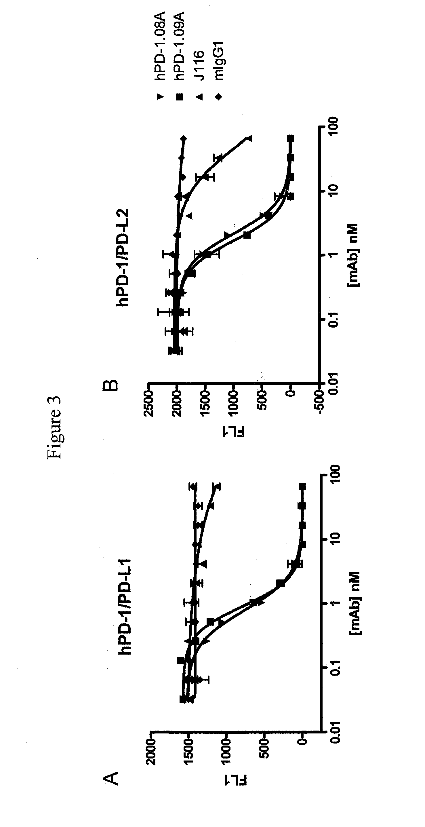
Functional Profiling of Anti-PD-1 Antibodies

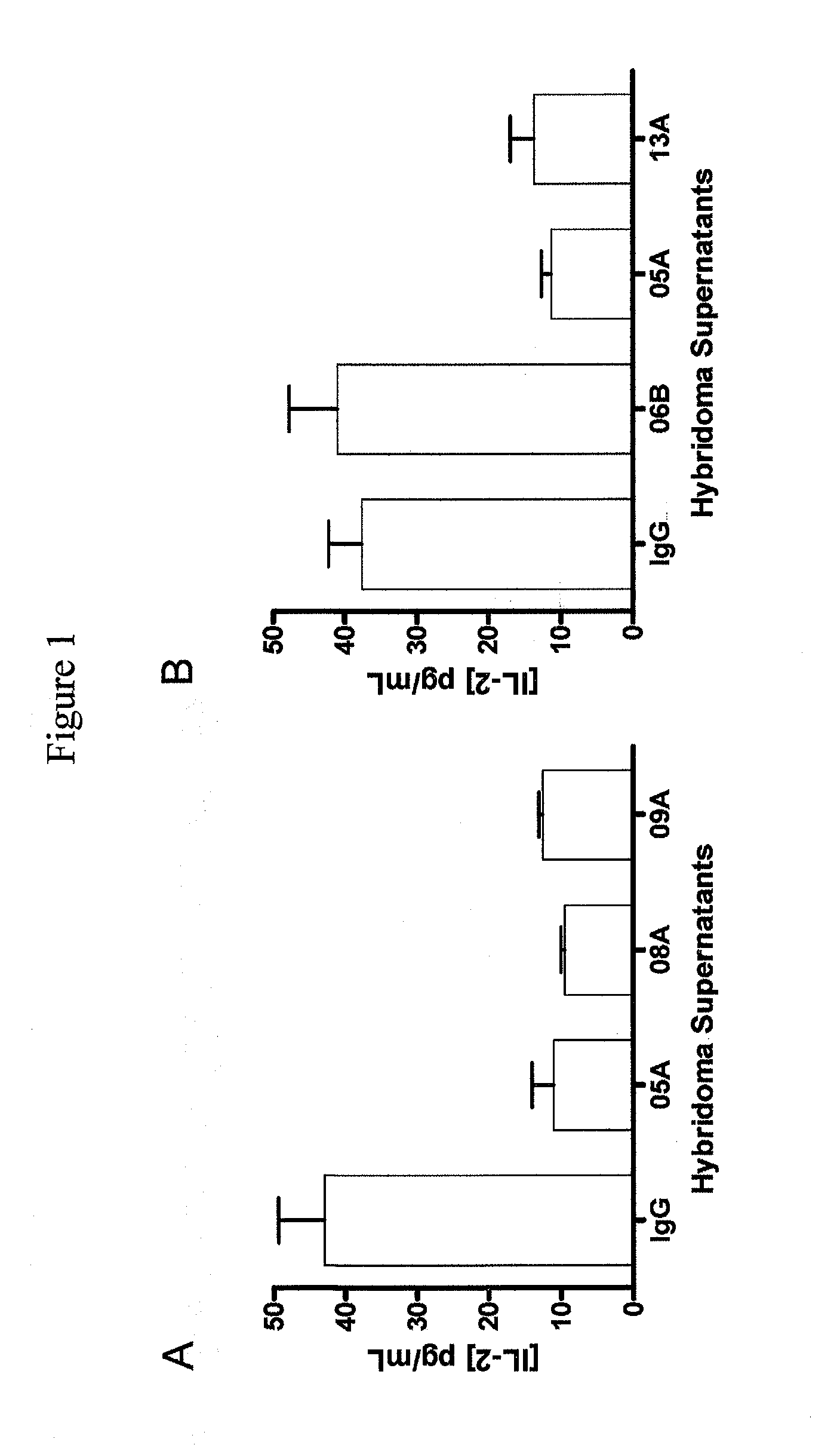
[0204]Human T Cell Response to SEB is Enhanced by hPD-1.08A and hPD-1.09A

[0205]Anti-PD-1 antibodies were tested for their capacity to enhance T cell activity in vitro using blood cells from healthy volunteers. One assay used to characterize the functional consequence of blocking human PD-1 receptor utilized Staphylococcus enterotoxin B (SEB) to engage and activate all T cells expressing the Vβ3 and Vβ8 T cell receptor chain. Healthy human donor blood was obtained and diluted 1:10 into culture medium. Diluted whole blood was plated (150 μl per well) in 96-well round-bottom plates and pre-incubated for 30-60 min with mAb and varying concentrations. SEB was then added at various concentrations ranging from 10 ng / mL to 10 μg / mL. Supernatants were collected after 2 to 4 days of culture and the amount of IL-2 produced was quantified using ELISA (described in Example 1) or using standard multiplex technology (Luminex platform—Biosource cytokine d...


PUM


Abstract

Antibodies which block binding of hPD-1 to hPD-L1 or hPD-L2 and their variable region sequences are disclosed. A method of increasing the activity (or reducing downmodulation) of an immune cell through the PD-1 pathway is also disclosed.

Description

BACKGROUND OF THE INVENTION[0001]Programmed death receptor 1 (PD-1) is an immunoinhibitory receptor that is primarily expressed on activated T and B cells. Interaction with its ligands has been shown to attenuate T-cell responses both in vitro and in vivo. Blockade of the interaction between PD-1 and one of its ligands, PD-L1, has been shown to enhance tumor-specific CD8+ T-cell immunity and may therefore be helpful in clearance of tumor cells by the immune system.[0002]PD-1 (encoded by the gene Pdcd1) is an Immunoglobulin superfamily member related to CD28, and CTLA-4. PD-1 has been shown to negatively regulate antigen receptor signaling upon engagement of its ligands (PD-L1 and / or PD-L2) The structure of murine PD-1 has been solved as well as the co-crystal structure of mouse PD-1 with human PD-L1 (Zhang, X. et al., Immunity 20: 337-347 (2004); Lin et al., Proc. Natl. Acad. Sci. USA 105: 3011-6 (2008)). PD-1 and like family members are type I transmembrane glycoproteins containing...

Claims


Application Information

Patent Timeline
Patent Type & Authority Applications(United States)
IPC IPC(8): C07K16/28
CPCC07K16/2818C07K2316/96C07K16/2803C07K2317/565C07K2317/92C07K2317/24C07K2317/76A61P31/00A61P35/00A61P37/04G01N33/57488G01N33/6869G01N2333/55G01N2800/52
Inventor CARVEN, GREGORY JOHNVAN EENENNEEM, HANSDULOS, GRADUS JOHANNES
Owner MERCK SHARP & DOHME BV
Why Eureka
Try Eureka