Medical imaging method and system

a medical imaging and system technology, applied in the field of medical imaging methods and devices, can solve the problems of difficult to determine a sufficiently high number of registration points, user-defined landmark establishment can be time-consuming, and user-defined establishment can be imprecise or error-prone, and achieve the effect of reliable registration of image data sets acquired

Inactive Publication Date: 2013-05-09
SIEMENS HEALTHCARE GMBH
View PDF10 Cites 9 Cited by
  • Summary
  • Abstract
  • Description
  • Claims
  • Application Information

AI Technical Summary

Benefits of technology

[0009]An object of the invention is to provide a method, a device and a system for imaging that allows a reliable registration of image data sets acquired with different imaging modalities. Furthermore, an object of the invention is to enable such registration using a number of registration points without the measurement of a temperature profile being required.

Problems solved by technology

A user-defined landmark establishment can be time-consuming.
Depending on the experience of the user, a user-defined establishment can be imprecise or error-prone if the user cannot reliably identify anatomical landmarks.
It can be difficult to determine a sufficiently high number of registration points in order to be able to produce a nonlinearity for registration in three dimensions.
The number of anatomical landmarks that can easily be recognized in images may be too few for such purposes.

Method used


Image

  • Medical imaging method and system
  • Medical imaging method and system
  • Medical imaging method and system

Examples


Embodiment Construction

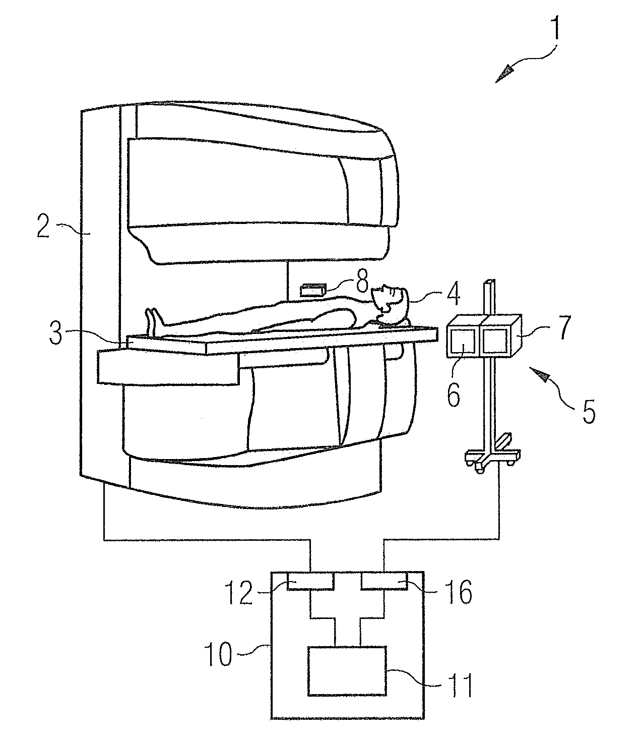
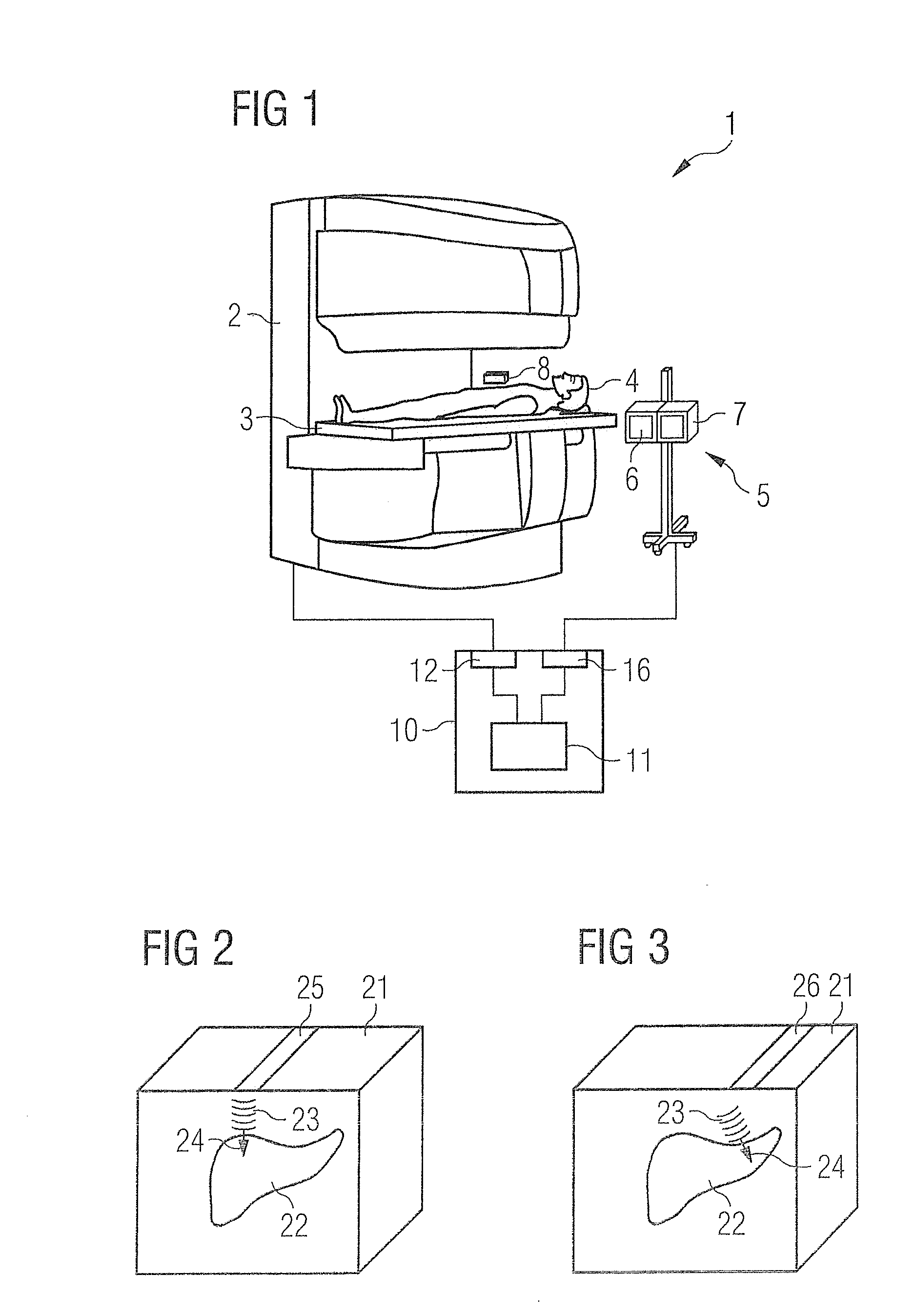
[0040]FIG. 1 shows an imaging system 1 according to an exemplary embodiment of the invention. The imaging system 1 has a magnetic resonance (MR) imaging device 2 and an ultrasound system 5. The MR imaging device 2 has a support device 3 (for example an examination bed) on which an examination subject 4 is borne. The ultrasound system 5 can have an ultrasound (US) imaging device 6 and a US treatment device 7 that can be arranged in a common housing. The US treatment device 7 generates an ultrasound pulse with an amplitude so that the resulting sound pressure leads to a deformation of soft tissue of the examination subject 4. The amplitude can be selected so that the displacement of soft tissue in a patient amounts to one or a few micrometers. The displacement of soft tissue can be between one and twenty micrometers. Given the use of ultrasound pulses, such displacements of soft tissue can be achieved by operation in which conventional acoustic energies are not exceeded. Alternatively...


PUM


Abstract

In an imaging procedure, sound pressure is generated by applying ultrasound to the region to be imaged in order to cause a deformation of tissue therein from at least one acquired ultrasound image data set, first data are determined that represent, with spatial resolution, the deformation of the tissue as a reaction to the sound pressure. At least one magnetic resonance image data set is acquired, from which second data are determined that represent, with spatial resolution, the deformation of the tissue as a reaction to the sound pressure. The at least one ultrasound image data set and the at least one magnetic resonance image data set are brought into registration with each other by a comparison of the first data and the second data.

Description

BACKGROUND OF THE INVENTION[0001]1. Field of the Invention[0002]The invention concerns methods and devices for medical imaging. The invention in particular concerns such methods and devices with which a registration of images that were acquired with different imaging modalities is possible.[0003]2. Description of the Prior Art[0004]Several different imaging methods are used in medical engineering. Among these are, for example, computed tomography, magnetic resonance tomography, ultrasound and positron emission tomography. These imaging modalities deliver spatially resolved information about different physical properties of an examined region of an examination subject. Since different imaging modalities can provide information about different properties of the examined region, it is desirable to acquire image data with different imaging modalities.[0005]In imaging of an examined region with multiple imaging modalities, the registration of different acquired images with respect to eac...

Claims


Application Information

Patent Timeline
Patent Type & Authority Applications(United States)
IPC IPC(8): A61B8/08A61B5/055A61B8/13A61B5/00
CPCA61B5/0035A61B5/055A61B8/14A61B8/485A61B8/5261A61B8/13
Inventor GROSS, PATRICK
Owner SIEMENS HEALTHCARE GMBH
Why Eureka
Try Eureka