
Animal Model Expressing Luciferase under Control of the Myelin Basic Protein Promoter (MBP-luci) and Use of the Model for Bioluminescence In Vivo Imaging

a technology of myelin and luciferase, which is applied in the field of manufactured myelin basic proteinluciferase (mbpluci) bioimaging model, can solve the problems of cumbersome animal models, impaired sensation, movement, cognition, etc., and achieves less time and resources, and avoids the sacrifice of animals at specific time points.

Inactive Publication Date: 2013-05-09
SANOFI SA
View PDF0 Cites 4 Cited by
  • Summary
  • Abstract
  • Description
  • Claims
  • Application Information

AI Technical Summary

Benefits of technology

The present invention provides a new way to test the effects of drugs on demyelination and remyelination in living animals. This helps to screen and validate the effectiveness of drugs and other therapies in real-time, high-resolution data. It also helps to assess potential toxicity and the therapeutic window, which makes it easier to determine the best ways to treat diseases in humans.

Problems solved by technology

Animal models traditionally have been cumbersome because of the difficulty in quantifying physiological events in real-time.
Diseases that affect myelin integrity result in impaired conduction of axonal signals in the affected neurons, and can result in impaired sensation, movement, cognition, or other functions, depending on which neurons are involved.
Current methods using animal models to assay changes in myelin in vivo are time-consuming, typically running between 4 to 8 weeks, and require a large number of animals.
Previous assays have to use separate groups of animals for each time point and this limits the number of data points that can be collected during a study.

Method used


Image

  • Animal Model Expressing Luciferase under Control of the Myelin Basic Protein Promoter (MBP-luci) and Use of the Model for Bioluminescence In Vivo Imaging
  • Animal Model Expressing Luciferase under Control of the Myelin Basic Protein Promoter (MBP-luci) and Use of the Model for Bioluminescence In Vivo Imaging
  • Animal Model Expressing Luciferase under Control of the Myelin Basic Protein Promoter (MBP-luci) and Use of the Model for Bioluminescence In Vivo Imaging

Examples


examples

Transgenic Mouse Generation

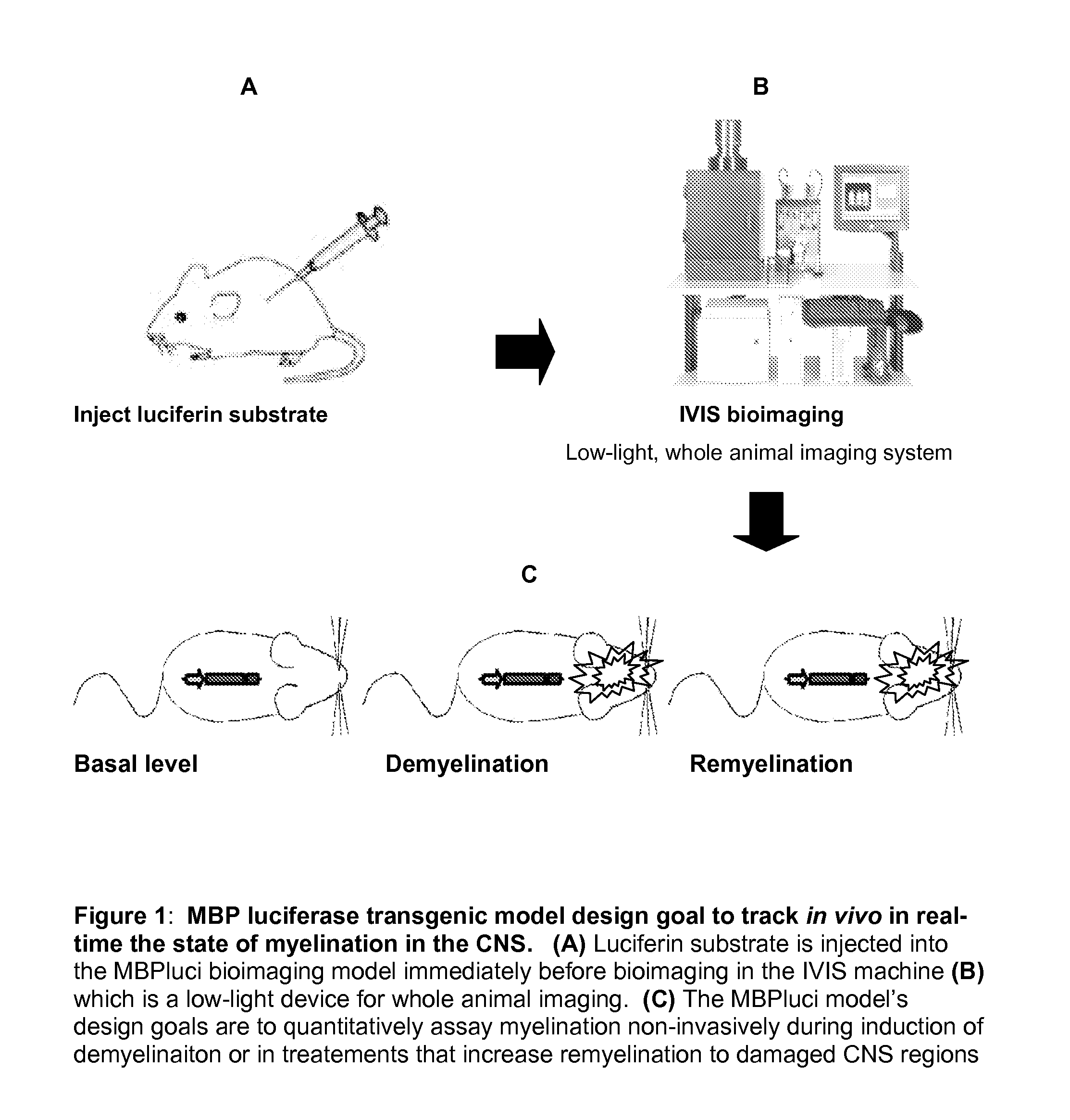
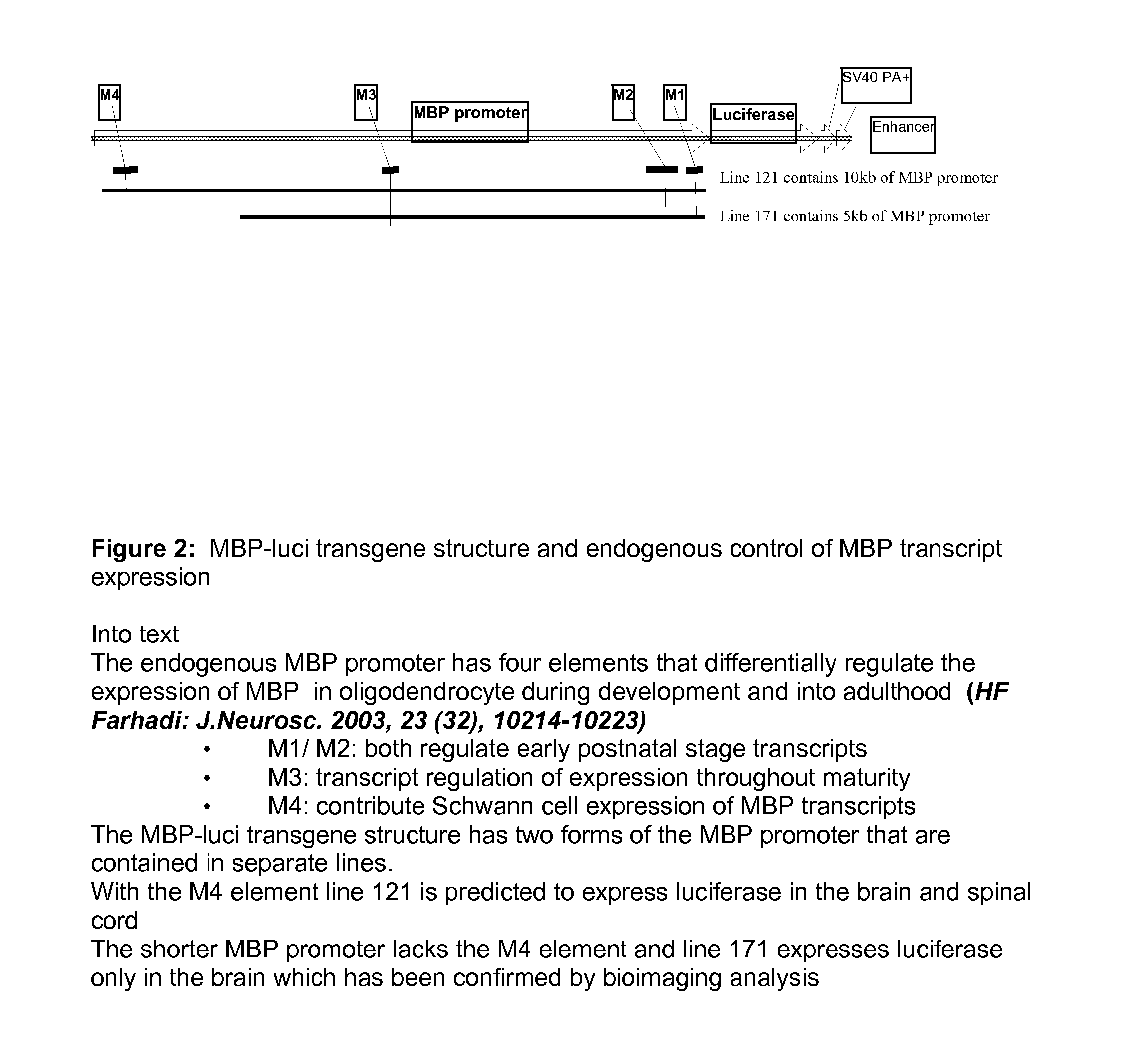
[0140]“Long” promoter is about 10 KB containing M1, M2, M3, M4 and “short” promoter is about 5 KB containing M1, M2 and M3. These were cloned with a high fidelity PCR method from a mouse Bacterial Artificial Chromosome (BAC) containing a MBP gene. Then each promoter fragment was cloned into a vector, for example into the into the poly link site of a pGL3-hygro vector (FIG. 1 and FIG. 2).

[0141]The plasmids were restricted with Not I and BamHI to release the MBP-luci transgenic expression cassettes (FIG. 3) that were used to generate transgenic mice in the FVB / Tac and in B6C3 / Tac strains using standard pronuclear microinjection techniques.

[0142]General strategies for generating transgenic (Tg) animals are well known in the art, for example as described in Pinkert, C. A. (ed.) 1994. Transgenic animal technology: A laboratory handbook. Academic Press, Inc., San Diego, Calif.; Monastersky G. M. and Robl, J. M. (ed.) (1995) Strategies in transgenic animal scienc...


PUM

PropertyMeasurementUnit
Wavelengthaaaaaaaaaa
Timeaaaaaaaaaa
Levelaaaaaaaaaa

Abstract

A Myelin Basic Protein-luciferase bioimaging noninvasive model to visualize and quantify demyelination and remyelination events in the CNS at transcriptional level in vivo is provided. Luciferase-expressing transgenic animals were generated with myelin basic protein (MBP) promoter coupled to firefly luciferase reporter. The MBP-luci bioimaging model provides a means to monitor myelination status and the efficacy of a remyelination modulating test compound. An advantage of bioimaging is that a subject in a longitudinal study can serve as its own control. The same subject can be tracked over a demyelination and remyelination process continuously over a period of at least 10 weeks. This model enables normalization of individual animal imaging response and provides quality data with considerably reduced variance. In addition, because cohorts of animals need not be sacrificed at different time points, reduction in the number necessary for a compound efficacy study is possible.

Description

FIELD OF THE INVENTION[0001]This invention relates to a manufactured Myelin Basic Protein-luciferase (MBP-luci) bioimaging model used, e.g., to visualize and quantify demyelination / remyelination events at transcriptional level in real-time with live animal bioimaging techniques.FIELD OF THE INVENTION[0002]In drug development, attrition rates are high with accounts claiming that only one in five compounds makes it through development to approval (DiMasi, J A, et al, J Health Econ 22, 151-185). Moreover, despite dramatically increased investment, the rate of introduction of novel drugs has remained relatively constant over the past 30 years, with only two to three advances in new drug classes per year eventually making it to market (Lindsay M A, Nature Rev Drug Discovery, 2, 831-838).[0003]Molecular and functional imaging applied to the initial stages of drug development can provide evidence of biological activity and confirm on-target drug effects. Accordingly, investment in molecula...

Claims


Application Information

Patent Timeline
IPC IPC(8): A01K67/027
CPCA01K67/0275A01K2217/052A01K2217/206C12N15/8509A01K2267/03A01K2267/0393A01K2227/105
Inventor CAO, JAMESCHANDROSS, KARENECONOMIDES, KYRIAKOS D.POLITES, HARRY GREGORYWEINSTOCK, DANIELYING, XIAOYOU
Owner SANOFI SA
Learn More